Magnesium »
PDB 7q7g-7qnq »
7qbm »
Magnesium in PDB 7qbm: Structure of the Activation Intermediate of Cathepsin K in Complex with the 3-Cyano-3-Aza-Beta-Amino Acid Inhibitor GU2602
Enzymatic activity of Structure of the Activation Intermediate of Cathepsin K in Complex with the 3-Cyano-3-Aza-Beta-Amino Acid Inhibitor GU2602
All present enzymatic activity of Structure of the Activation Intermediate of Cathepsin K in Complex with the 3-Cyano-3-Aza-Beta-Amino Acid Inhibitor GU2602:
3.4.22.38;
3.4.22.38;
Protein crystallography data
The structure of Structure of the Activation Intermediate of Cathepsin K in Complex with the 3-Cyano-3-Aza-Beta-Amino Acid Inhibitor GU2602, PDB code: 7qbm
was solved by
J.Benysek,
M.Busa,
M.Mares,
with X-Ray Crystallography technique. A brief refinement statistics is given in the table below:
| Resolution Low / High (Å) | 48.91 / 1.88 |
| Space group | P 43 21 2 |
| Cell size a, b, c (Å), α, β, γ (°) | 103.283, 103.283, 55.464, 90, 90, 90 |
| R / Rfree (%) | 19 / 23.4 |
Magnesium Binding Sites:
The binding sites of Magnesium atom in the Structure of the Activation Intermediate of Cathepsin K in Complex with the 3-Cyano-3-Aza-Beta-Amino Acid Inhibitor GU2602
(pdb code 7qbm). This binding sites where shown within
5.0 Angstroms radius around Magnesium atom.
In total only one binding site of Magnesium was determined in the Structure of the Activation Intermediate of Cathepsin K in Complex with the 3-Cyano-3-Aza-Beta-Amino Acid Inhibitor GU2602, PDB code: 7qbm:
In total only one binding site of Magnesium was determined in the Structure of the Activation Intermediate of Cathepsin K in Complex with the 3-Cyano-3-Aza-Beta-Amino Acid Inhibitor GU2602, PDB code: 7qbm:
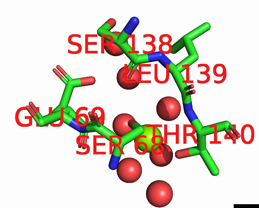
Magnesium binding site 1 out of 1 in 7qbm
Go back to
Magnesium binding site 1 out
of 1 in the Structure of the Activation Intermediate of Cathepsin K in Complex with the 3-Cyano-3-Aza-Beta-Amino Acid Inhibitor GU2602
Mono view

Stereo pair view
Mono view
Stereo pair view
A full contact list of Magnesium with other atoms in the Mg binding
site number 1 of Structure of the Activation Intermediate of Cathepsin K in Complex with the 3-Cyano-3-Aza-Beta-Amino Acid Inhibitor GU2602 within 5.0Å range:
|
Reference:
J.Benysek,
M.Busa,
P.Rubesova,
J.Fanfrlik,
M.Lepsik,
J.Brynda,
Z.Matouskova,
U.Bartz,
M.Horn,
M.Gutschow,
M.Mares.
Highly Potent Inhibitors of Cathepsin K with A Differently Positioned Cyanohydrazide Warhead: Structural Analysis of Binding Mode to Mature and Zymogen-Like Enzymes. J Enzyme Inhib Med Chem V. 37 515 2022.
ISSN: ESSN 1475-6374
PubMed: 35144520
DOI: 10.1080/14756366.2021.2024527
Page generated: Thu Oct 3 05:05:18 2024
ISSN: ESSN 1475-6374
PubMed: 35144520
DOI: 10.1080/14756366.2021.2024527
Last articles
Zn in 9JYWZn in 9IR4
Zn in 9IR3
Zn in 9GMX
Zn in 9GMW
Zn in 9JEJ
Zn in 9ERF
Zn in 9ERE
Zn in 9EGV
Zn in 9EGW