Magnesium »
PDB 7q7g-7qnq »
7qim »
Magnesium in PDB 7qim: In Situ Structure of Nebulin Bound to Actin Filament in Skeletal Sarcomere
Magnesium Binding Sites:
The binding sites of Magnesium atom in the In Situ Structure of Nebulin Bound to Actin Filament in Skeletal Sarcomere
(pdb code 7qim). This binding sites where shown within
5.0 Angstroms radius around Magnesium atom.
In total 5 binding sites of Magnesium where determined in the In Situ Structure of Nebulin Bound to Actin Filament in Skeletal Sarcomere, PDB code: 7qim:
Jump to Magnesium binding site number: 1; 2; 3; 4; 5;
In total 5 binding sites of Magnesium where determined in the In Situ Structure of Nebulin Bound to Actin Filament in Skeletal Sarcomere, PDB code: 7qim:
Jump to Magnesium binding site number: 1; 2; 3; 4; 5;
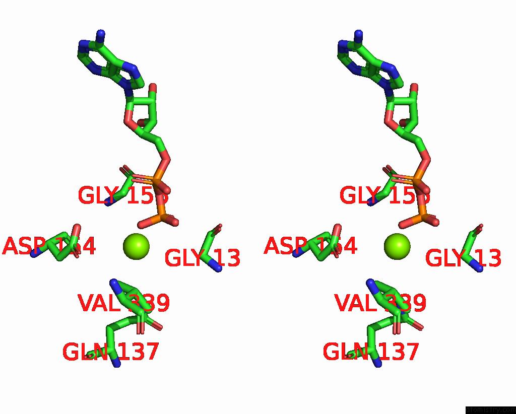
Magnesium binding site 1 out of 5 in 7qim
Go back to
Magnesium binding site 1 out
of 5 in the In Situ Structure of Nebulin Bound to Actin Filament in Skeletal Sarcomere
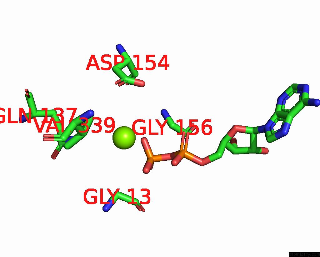
Mono view
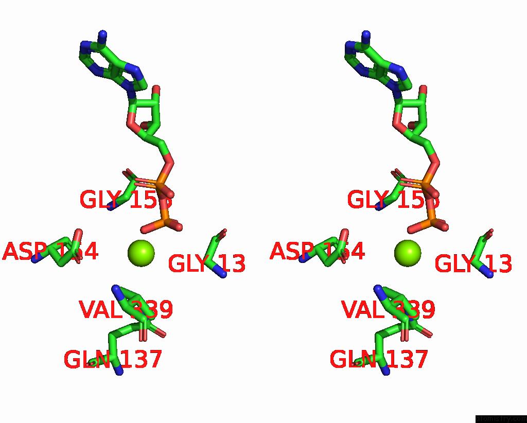
Stereo pair view
Mono view
Stereo pair view
A full contact list of Magnesium with other atoms in the Mg binding
site number 1 of In Situ Structure of Nebulin Bound to Actin Filament in Skeletal Sarcomere within 5.0Å range:
|
Magnesium binding site 2 out of 5 in 7qim
Go back to
Magnesium binding site 2 out
of 5 in the In Situ Structure of Nebulin Bound to Actin Filament in Skeletal Sarcomere
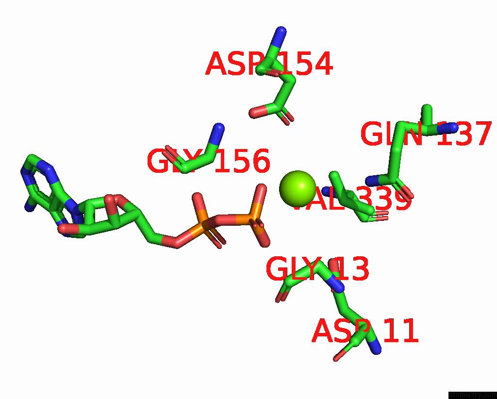
Mono view
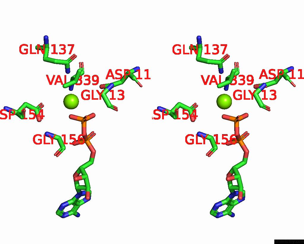
Stereo pair view
Mono view
Stereo pair view
A full contact list of Magnesium with other atoms in the Mg binding
site number 2 of In Situ Structure of Nebulin Bound to Actin Filament in Skeletal Sarcomere within 5.0Å range:
|
Magnesium binding site 3 out of 5 in 7qim
Go back to
Magnesium binding site 3 out
of 5 in the In Situ Structure of Nebulin Bound to Actin Filament in Skeletal Sarcomere

Mono view
Stereo pair view
Mono view
Stereo pair view
A full contact list of Magnesium with other atoms in the Mg binding
site number 3 of In Situ Structure of Nebulin Bound to Actin Filament in Skeletal Sarcomere within 5.0Å range:
|
Magnesium binding site 4 out of 5 in 7qim
Go back to
Magnesium binding site 4 out
of 5 in the In Situ Structure of Nebulin Bound to Actin Filament in Skeletal Sarcomere

Mono view

Stereo pair view
Mono view
Stereo pair view
A full contact list of Magnesium with other atoms in the Mg binding
site number 4 of In Situ Structure of Nebulin Bound to Actin Filament in Skeletal Sarcomere within 5.0Å range:
|
Magnesium binding site 5 out of 5 in 7qim
Go back to
Magnesium binding site 5 out
of 5 in the In Situ Structure of Nebulin Bound to Actin Filament in Skeletal Sarcomere

Mono view

Stereo pair view
Mono view
Stereo pair view
A full contact list of Magnesium with other atoms in the Mg binding
site number 5 of In Situ Structure of Nebulin Bound to Actin Filament in Skeletal Sarcomere within 5.0Å range:
|
Reference:
Z.Wang,
M.Grange,
S.Pospich,
T.Wagner,
A.L.Kho,
M.Gautel,
S.Raunser.
Structures From Intact Myofibrils Reveal Mechanism of Thin Filament Regulation Through Nebulin. Science V. 375 N1934 2022.
ISSN: ESSN 1095-9203
PubMed: 35175800
DOI: 10.1126/SCIENCE.ABN1934
Page generated: Thu Oct 3 05:07:18 2024
ISSN: ESSN 1095-9203
PubMed: 35175800
DOI: 10.1126/SCIENCE.ABN1934
Last articles
Zn in 9JYWZn in 9IR4
Zn in 9IR3
Zn in 9GMX
Zn in 9GMW
Zn in 9JEJ
Zn in 9ERF
Zn in 9ERE
Zn in 9EGV
Zn in 9EGW