Magnesium »
PDB 7s61-7sko »
7shg »
Magnesium in PDB 7shg: Polysaccharide Ribofuranosyl Transferase From Thermobacillus Composti
Protein crystallography data
The structure of Polysaccharide Ribofuranosyl Transferase From Thermobacillus Composti, PDB code: 7shg
was solved by
M.S.Kimber,
S.D.Kelly,
with X-Ray Crystallography technique. A brief refinement statistics is given in the table below:
| Resolution Low / High (Å) | 49.16 / 2.50 |
| Space group | P 2 21 21 |
| Cell size a, b, c (Å), α, β, γ (°) | 71.74, 82.5, 234.92, 90, 90, 90 |
| R / Rfree (%) | 25.1 / 29.5 |
Other elements in 7shg:
The structure of Polysaccharide Ribofuranosyl Transferase From Thermobacillus Composti also contains other interesting chemical elements:
| Chlorine | (Cl) | 2 atoms |
Magnesium Binding Sites:
The binding sites of Magnesium atom in the Polysaccharide Ribofuranosyl Transferase From Thermobacillus Composti
(pdb code 7shg). This binding sites where shown within
5.0 Angstroms radius around Magnesium atom.
In total 3 binding sites of Magnesium where determined in the Polysaccharide Ribofuranosyl Transferase From Thermobacillus Composti, PDB code: 7shg:
Jump to Magnesium binding site number: 1; 2; 3;
In total 3 binding sites of Magnesium where determined in the Polysaccharide Ribofuranosyl Transferase From Thermobacillus Composti, PDB code: 7shg:
Jump to Magnesium binding site number: 1; 2; 3;
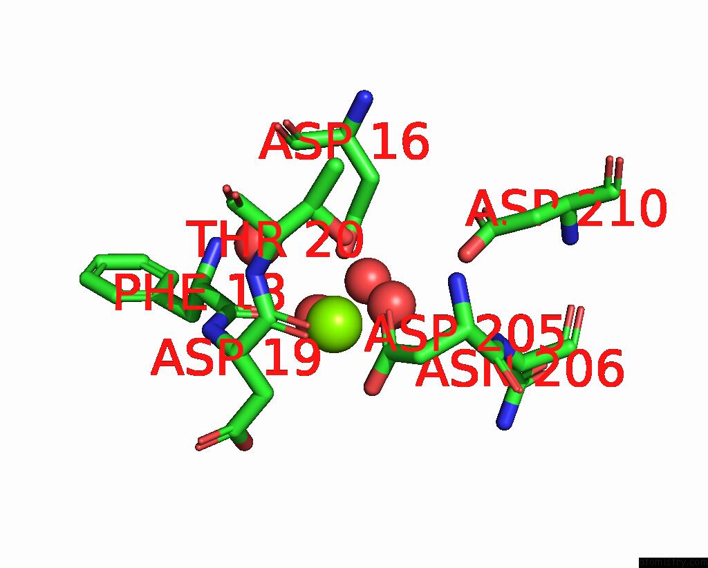
Magnesium binding site 1 out of 3 in 7shg
Go back to
Magnesium binding site 1 out
of 3 in the Polysaccharide Ribofuranosyl Transferase From Thermobacillus Composti
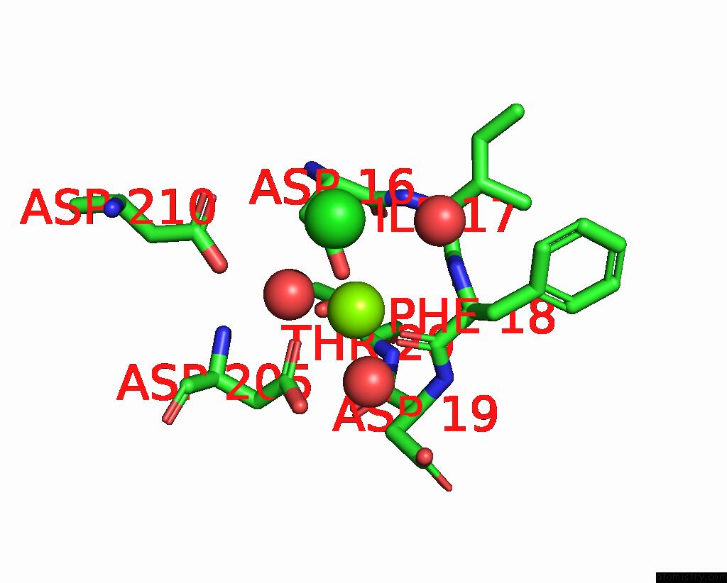
Mono view
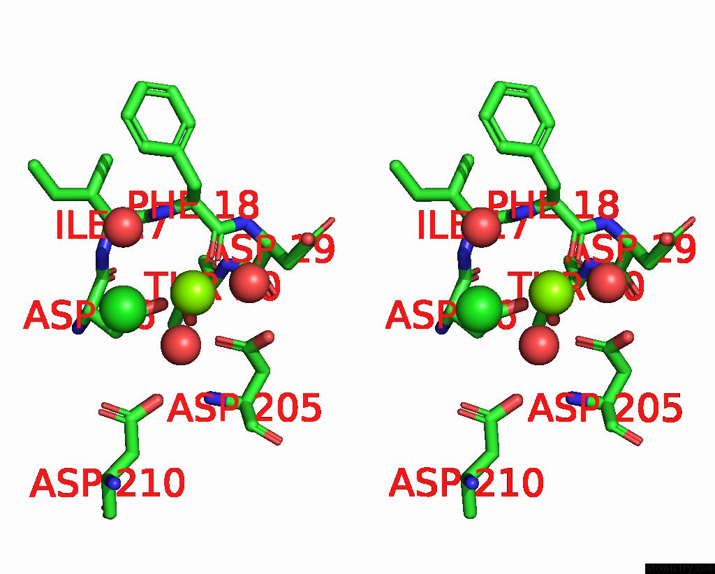
Stereo pair view
Mono view
Stereo pair view
A full contact list of Magnesium with other atoms in the Mg binding
site number 1 of Polysaccharide Ribofuranosyl Transferase From Thermobacillus Composti within 5.0Å range:
|
Magnesium binding site 2 out of 3 in 7shg
Go back to
Magnesium binding site 2 out
of 3 in the Polysaccharide Ribofuranosyl Transferase From Thermobacillus Composti
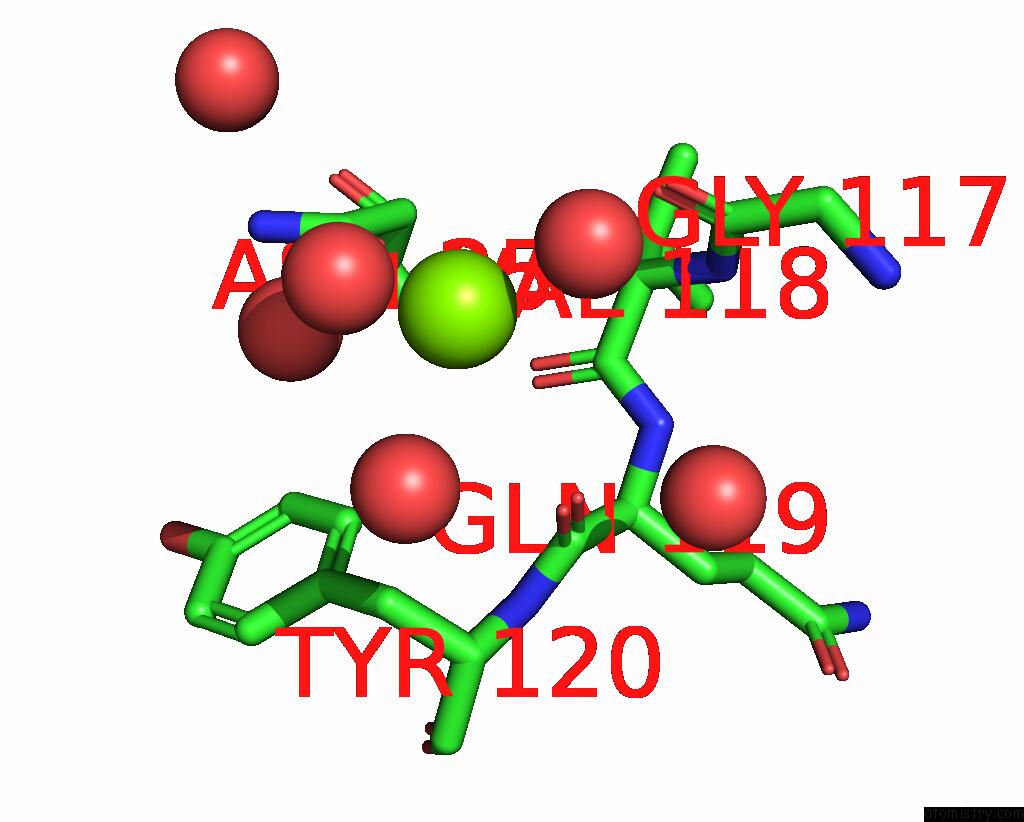
Mono view
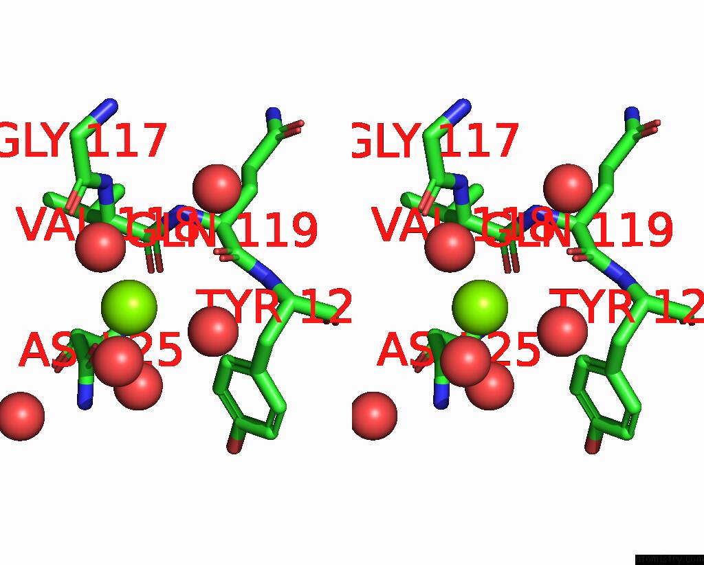
Stereo pair view
Mono view
Stereo pair view
A full contact list of Magnesium with other atoms in the Mg binding
site number 2 of Polysaccharide Ribofuranosyl Transferase From Thermobacillus Composti within 5.0Å range:
|
Magnesium binding site 3 out of 3 in 7shg
Go back to
Magnesium binding site 3 out
of 3 in the Polysaccharide Ribofuranosyl Transferase From Thermobacillus Composti
Mono view

Stereo pair view
Mono view
Stereo pair view
A full contact list of Magnesium with other atoms in the Mg binding
site number 3 of Polysaccharide Ribofuranosyl Transferase From Thermobacillus Composti within 5.0Å range:
|
Reference:
S.D.Kelly,
D.M.Williams,
J.T.Nothof,
T.Kim,
T.L.Lowary,
M.S.Kimber,
C.Whitfield.
The Biosynthetic Origin of Ribofuranose in Bacterial Polysaccharides. Nat.Chem.Biol. V. 18 530 2022.
ISSN: ESSN 1552-4469
PubMed: 35393575
DOI: 10.1038/S41589-022-01006-6
Page generated: Thu Oct 3 08:44:18 2024
ISSN: ESSN 1552-4469
PubMed: 35393575
DOI: 10.1038/S41589-022-01006-6
Last articles
Zn in 9J0NZn in 9J0O
Zn in 9J0P
Zn in 9FJX
Zn in 9EKB
Zn in 9C0F
Zn in 9CAH
Zn in 9CH0
Zn in 9CH3
Zn in 9CH1