Magnesium »
PDB 7syv-7te5 »
7tdb »
Magnesium in PDB 7tdb: Crystal Structure of the E. Coli Thim Riboswitch in Complex with Thiamine Bisphosphonate, Manganese Ions
Protein crystallography data
The structure of Crystal Structure of the E. Coli Thim Riboswitch in Complex with Thiamine Bisphosphonate, Manganese Ions, PDB code: 7tdb
was solved by
A.Nuthanakanti,
A.Serganov,
with X-Ray Crystallography technique. A brief refinement statistics is given in the table below:
| Resolution Low / High (Å) | 56.26 / 2.56 |
| Space group | P 32 1 2 |
| Cell size a, b, c (Å), α, β, γ (°) | 64.962, 64.962, 101.518, 90, 90, 120 |
| R / Rfree (%) | 21.7 / 24.6 |
Other elements in 7tdb:
The structure of Crystal Structure of the E. Coli Thim Riboswitch in Complex with Thiamine Bisphosphonate, Manganese Ions also contains other interesting chemical elements:
| Manganese | (Mn) | 3 atoms |
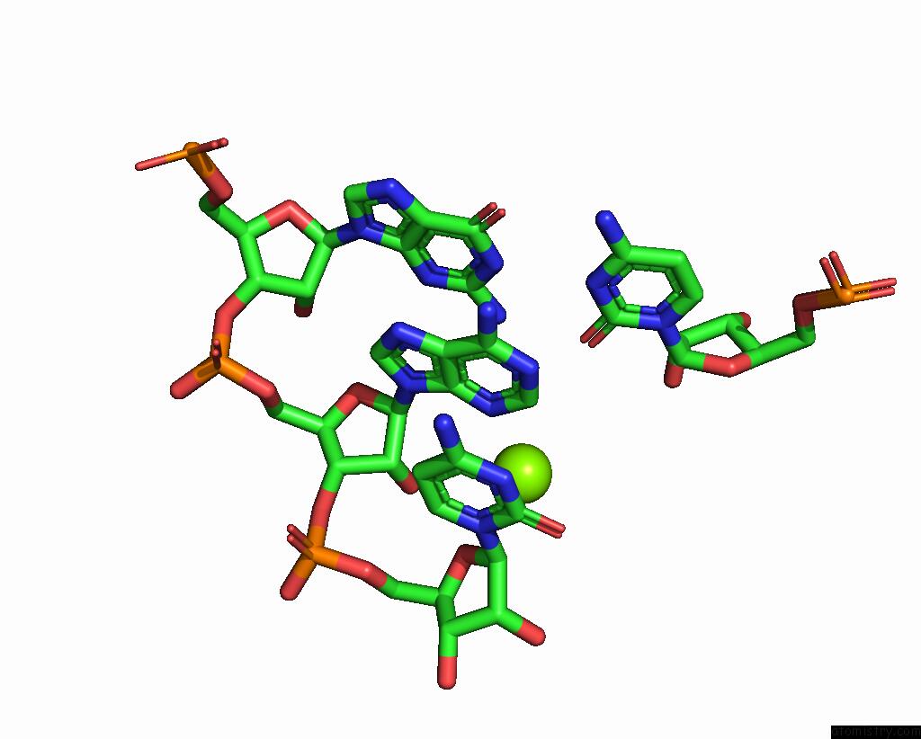
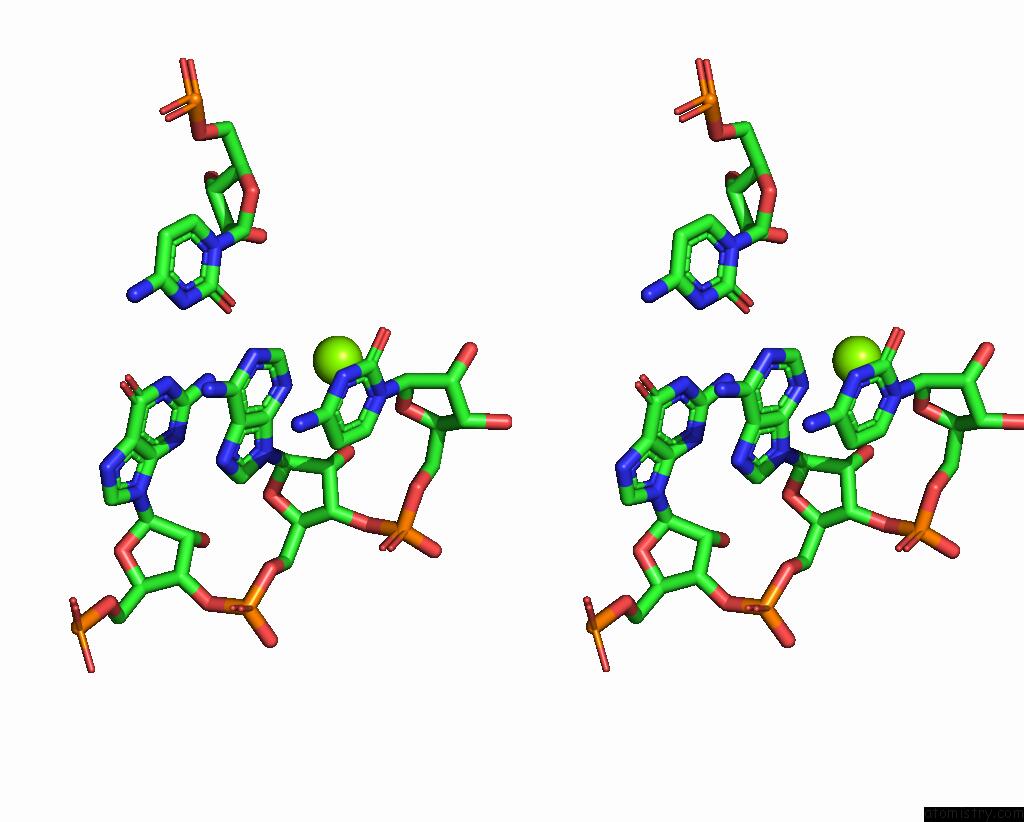
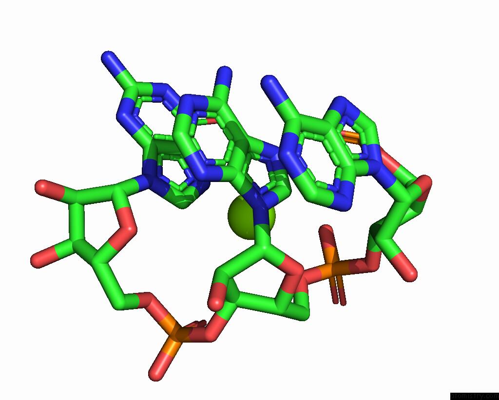
Magnesium Binding Sites:
The binding sites of Magnesium atom in the Crystal Structure of the E. Coli Thim Riboswitch in Complex with Thiamine Bisphosphonate, Manganese Ions
(pdb code 7tdb). This binding sites where shown within
5.0 Angstroms radius around Magnesium atom.
In total 6 binding sites of Magnesium where determined in the Crystal Structure of the E. Coli Thim Riboswitch in Complex with Thiamine Bisphosphonate, Manganese Ions, PDB code: 7tdb:
Jump to Magnesium binding site number: 1; 2; 3; 4; 5; 6;
In total 6 binding sites of Magnesium where determined in the Crystal Structure of the E. Coli Thim Riboswitch in Complex with Thiamine Bisphosphonate, Manganese Ions, PDB code: 7tdb:
Jump to Magnesium binding site number: 1; 2; 3; 4; 5; 6;
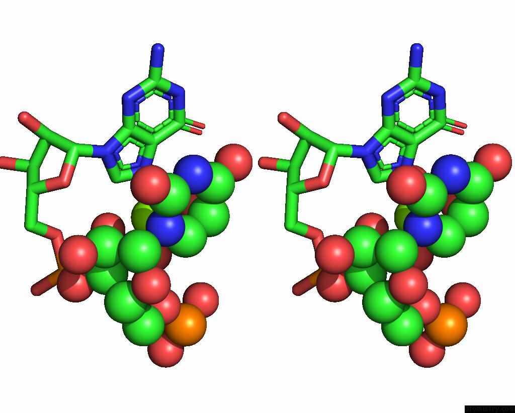
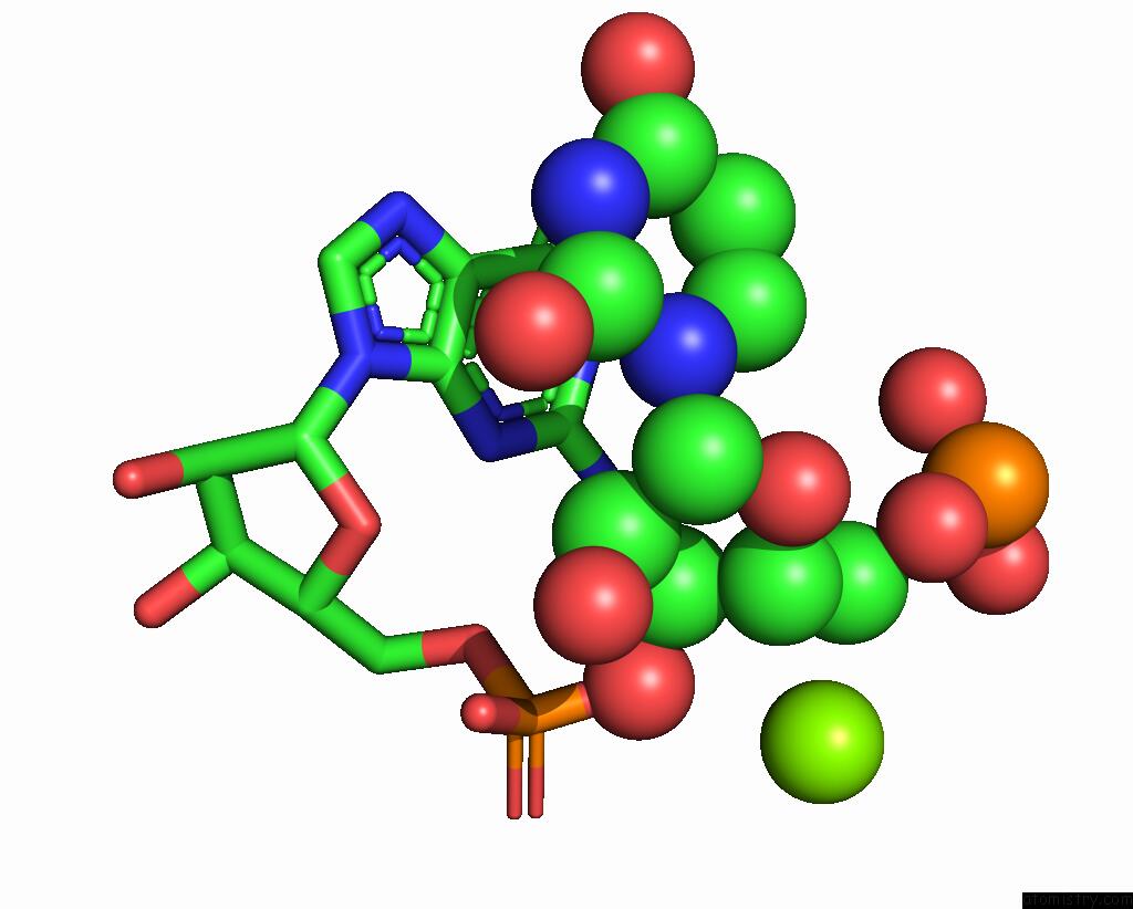
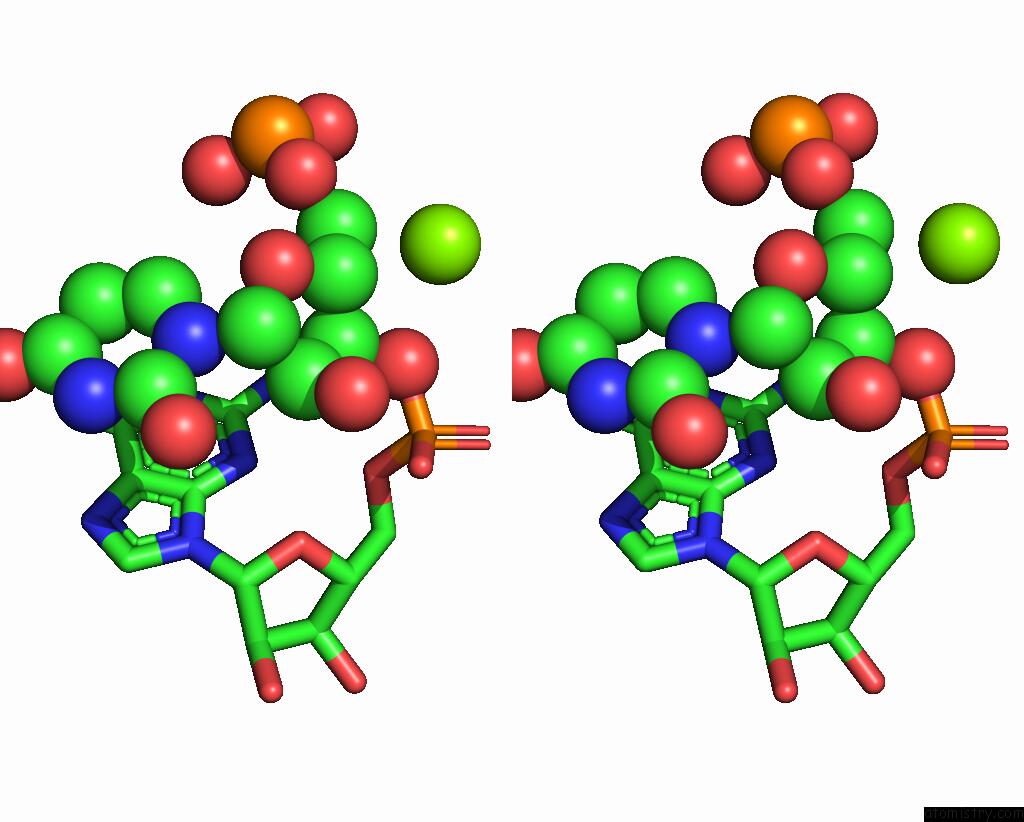
Magnesium binding site 1 out of 6 in 7tdb
Go back to
Magnesium binding site 1 out
of 6 in the Crystal Structure of the E. Coli Thim Riboswitch in Complex with Thiamine Bisphosphonate, Manganese Ions
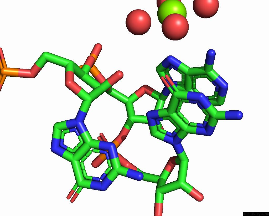
Mono view
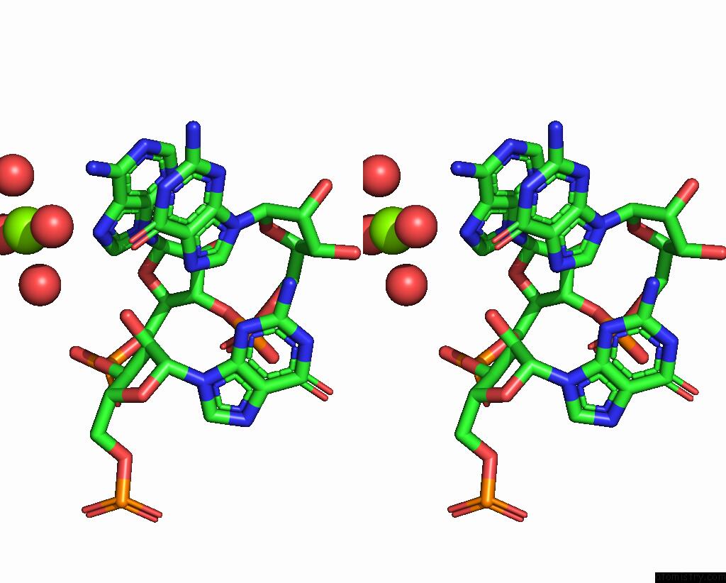
Stereo pair view
Mono view
Stereo pair view
A full contact list of Magnesium with other atoms in the Mg binding
site number 1 of Crystal Structure of the E. Coli Thim Riboswitch in Complex with Thiamine Bisphosphonate, Manganese Ions within 5.0Å range:
|
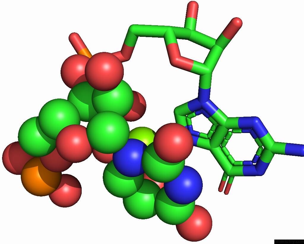
Magnesium binding site 2 out of 6 in 7tdb
Go back to
Magnesium binding site 2 out
of 6 in the Crystal Structure of the E. Coli Thim Riboswitch in Complex with Thiamine Bisphosphonate, Manganese Ions
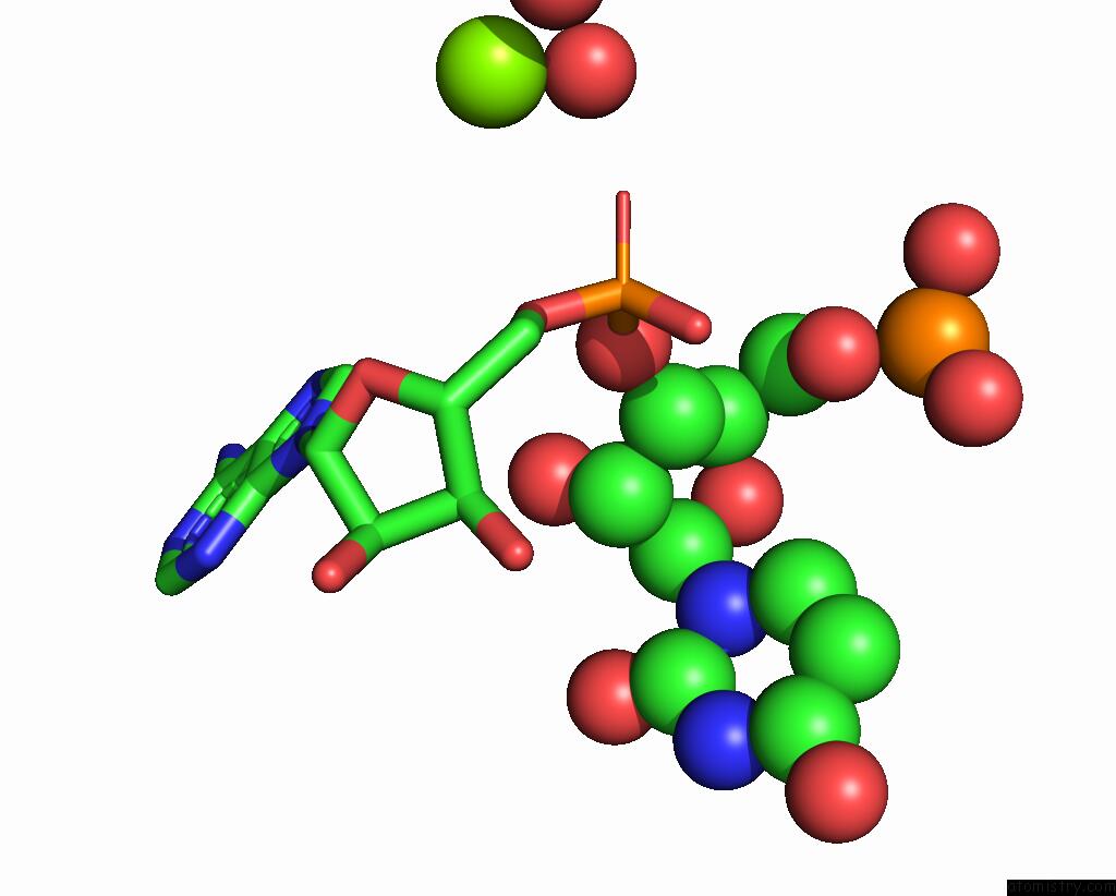
Mono view

Stereo pair view
Mono view
Stereo pair view
A full contact list of Magnesium with other atoms in the Mg binding
site number 2 of Crystal Structure of the E. Coli Thim Riboswitch in Complex with Thiamine Bisphosphonate, Manganese Ions within 5.0Å range:
|
Magnesium binding site 3 out of 6 in 7tdb
Go back to
Magnesium binding site 3 out
of 6 in the Crystal Structure of the E. Coli Thim Riboswitch in Complex with Thiamine Bisphosphonate, Manganese Ions
Mono view
Stereo pair view
Mono view
Stereo pair view
A full contact list of Magnesium with other atoms in the Mg binding
site number 3 of Crystal Structure of the E. Coli Thim Riboswitch in Complex with Thiamine Bisphosphonate, Manganese Ions within 5.0Å range:
|
Magnesium binding site 4 out of 6 in 7tdb
Go back to
Magnesium binding site 4 out
of 6 in the Crystal Structure of the E. Coli Thim Riboswitch in Complex with Thiamine Bisphosphonate, Manganese Ions
Mono view
Stereo pair view
Mono view
Stereo pair view
A full contact list of Magnesium with other atoms in the Mg binding
site number 4 of Crystal Structure of the E. Coli Thim Riboswitch in Complex with Thiamine Bisphosphonate, Manganese Ions within 5.0Å range:
|
Magnesium binding site 5 out of 6 in 7tdb
Go back to
Magnesium binding site 5 out
of 6 in the Crystal Structure of the E. Coli Thim Riboswitch in Complex with Thiamine Bisphosphonate, Manganese Ions
Mono view

Stereo pair view
Mono view
Stereo pair view
A full contact list of Magnesium with other atoms in the Mg binding
site number 5 of Crystal Structure of the E. Coli Thim Riboswitch in Complex with Thiamine Bisphosphonate, Manganese Ions within 5.0Å range:
|
Magnesium binding site 6 out of 6 in 7tdb
Go back to
Magnesium binding site 6 out
of 6 in the Crystal Structure of the E. Coli Thim Riboswitch in Complex with Thiamine Bisphosphonate, Manganese Ions
Mono view
Stereo pair view
Mono view
Stereo pair view
A full contact list of Magnesium with other atoms in the Mg binding
site number 6 of Crystal Structure of the E. Coli Thim Riboswitch in Complex with Thiamine Bisphosphonate, Manganese Ions within 5.0Å range:
|
Reference:
M.J.Zeller,
A.Nuthanakanti,
K.Li,
J.Aube,
A.Serganov,
K.M.Weeks.
Subsite Ligand Recognition and Cooperativity in the Tpp Riboswitch: Implications For Fragment-Linking in Rna Ligand Discovery. Acs Chem.Biol. V. 17 438 2022.
ISSN: ESSN 1554-8937
PubMed: 35060698
DOI: 10.1021/ACSCHEMBIO.1C00880
Page generated: Thu Oct 3 09:05:46 2024
ISSN: ESSN 1554-8937
PubMed: 35060698
DOI: 10.1021/ACSCHEMBIO.1C00880
Last articles
Ca in 5T4ZCa in 5T0X
Ca in 5T3H
Ca in 5T2N
Ca in 5T2O
Ca in 5T2H
Ca in 5SZQ
Ca in 5SZO
Ca in 5SZP
Ca in 5SZN