Magnesium »
PDB 7tr7-7u0y »
7twa »
Magnesium in PDB 7twa: Crystal Structure of Apo Besc From Streptomyces Cattleya
Protein crystallography data
The structure of Crystal Structure of Apo Besc From Streptomyces Cattleya, PDB code: 7twa
was solved by
M.E.Neugebauer,
M.J.Mcbride,
A.K.Boal,
M.C.Y.Chang,
with X-Ray Crystallography technique. A brief refinement statistics is given in the table below:
| Resolution Low / High (Å) | 126.75 / 1.70 |
| Space group | P 1 21 1 |
| Cell size a, b, c (Å), α, β, γ (°) | 60.917, 68.977, 126.748, 90, 89.99, 90 |
| R / Rfree (%) | 19.9 / 24.7 |
Other elements in 7twa:
The structure of Crystal Structure of Apo Besc From Streptomyces Cattleya also contains other interesting chemical elements:
| Sodium | (Na) | 1 atom |
Magnesium Binding Sites:
The binding sites of Magnesium atom in the Crystal Structure of Apo Besc From Streptomyces Cattleya
(pdb code 7twa). This binding sites where shown within
5.0 Angstroms radius around Magnesium atom.
In total 4 binding sites of Magnesium where determined in the Crystal Structure of Apo Besc From Streptomyces Cattleya, PDB code: 7twa:
Jump to Magnesium binding site number: 1; 2; 3; 4;
In total 4 binding sites of Magnesium where determined in the Crystal Structure of Apo Besc From Streptomyces Cattleya, PDB code: 7twa:
Jump to Magnesium binding site number: 1; 2; 3; 4;
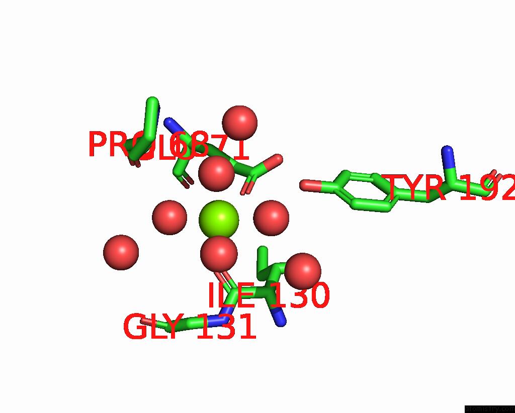
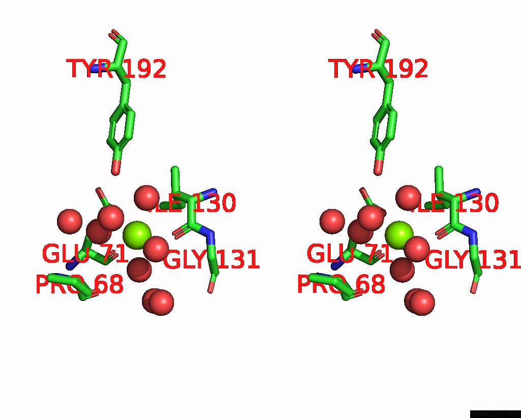
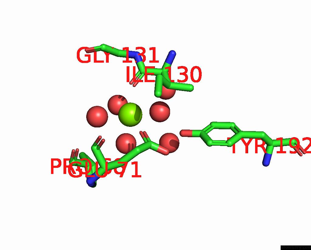
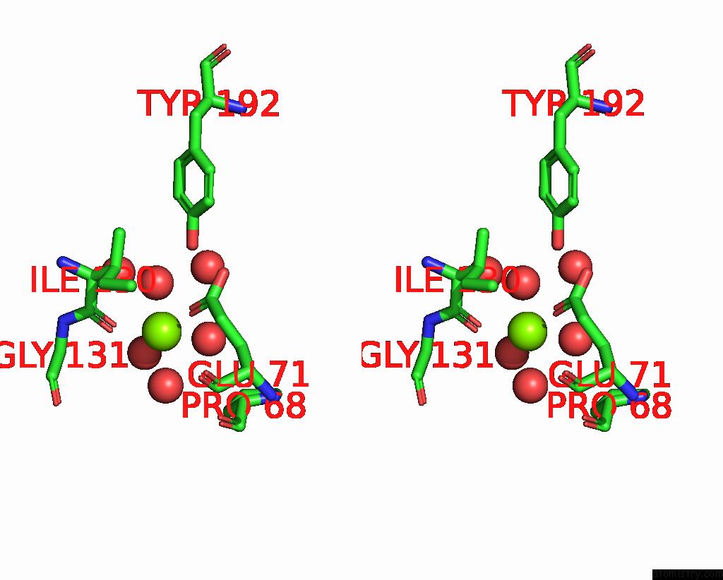
Magnesium binding site 1 out of 4 in 7twa
Go back to
Magnesium binding site 1 out
of 4 in the Crystal Structure of Apo Besc From Streptomyces Cattleya

Mono view
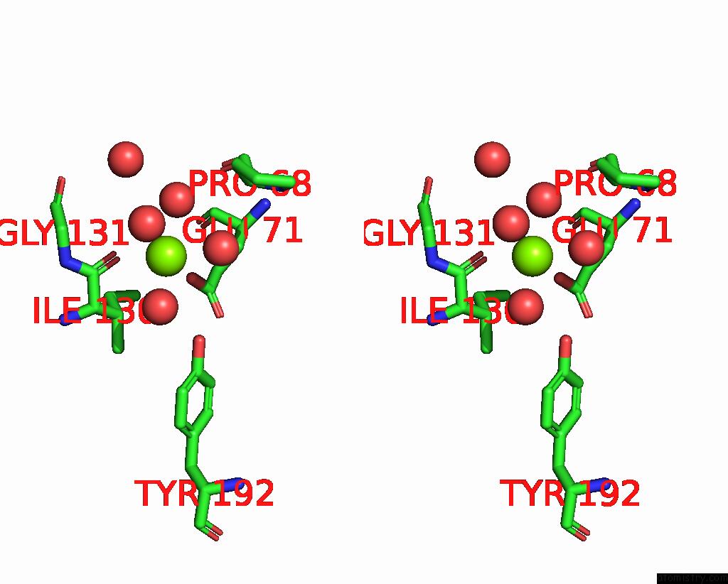
Stereo pair view
Mono view
Stereo pair view
A full contact list of Magnesium with other atoms in the Mg binding
site number 1 of Crystal Structure of Apo Besc From Streptomyces Cattleya within 5.0Å range:
|
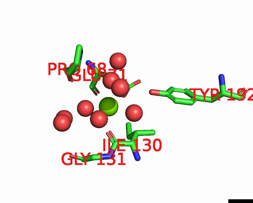
Magnesium binding site 2 out of 4 in 7twa
Go back to
Magnesium binding site 2 out
of 4 in the Crystal Structure of Apo Besc From Streptomyces Cattleya
Mono view
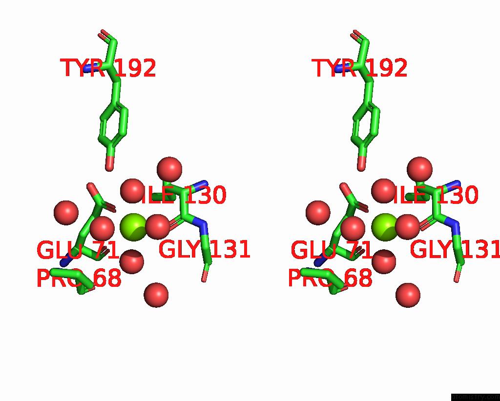
Stereo pair view
Mono view
Stereo pair view
A full contact list of Magnesium with other atoms in the Mg binding
site number 2 of Crystal Structure of Apo Besc From Streptomyces Cattleya within 5.0Å range:
|
Magnesium binding site 3 out of 4 in 7twa
Go back to
Magnesium binding site 3 out
of 4 in the Crystal Structure of Apo Besc From Streptomyces Cattleya
Mono view
Stereo pair view
Mono view
Stereo pair view
A full contact list of Magnesium with other atoms in the Mg binding
site number 3 of Crystal Structure of Apo Besc From Streptomyces Cattleya within 5.0Å range:
|
Magnesium binding site 4 out of 4 in 7twa
Go back to
Magnesium binding site 4 out
of 4 in the Crystal Structure of Apo Besc From Streptomyces Cattleya
Mono view
Stereo pair view
Mono view
Stereo pair view
A full contact list of Magnesium with other atoms in the Mg binding
site number 4 of Crystal Structure of Apo Besc From Streptomyces Cattleya within 5.0Å range:
|
Reference:
M.J.Mcbride,
M.A.Nair,
D.Sil,
J.W.Slater,
M.E.Neugebauer,
M.C.Y.Chang,
A.K.Boal,
C.Krebs,
J.M.Bollinger Jr..
Substrate-Triggered Mu-Peroxodiiron(III) Intermediate in the 4-Chloro-L-Lysine-Fragmenting Heme-Oxygenase-Like Diiron Oxidase (Hdo) Besc: Substrate Dissociation From, and C4 Targeting By, the Intermediate. Biochemistry V. 61 689 2022.
ISSN: ISSN 0006-2960
PubMed: 35380785
DOI: 10.1021/ACS.BIOCHEM.1C00774
Page generated: Thu Oct 3 09:33:12 2024
ISSN: ISSN 0006-2960
PubMed: 35380785
DOI: 10.1021/ACS.BIOCHEM.1C00774
Last articles
Fe in 2YXOFe in 2YRS
Fe in 2YXC
Fe in 2YNM
Fe in 2YVJ
Fe in 2YP1
Fe in 2YU2
Fe in 2YU1
Fe in 2YQB
Fe in 2YOO