Magnesium »
PDB 7tr7-7u0y »
7tyg »
Magnesium in PDB 7tyg: Structure of the Human Leucine Rich Repeat Protein SHOC2, Residues 80- 582
Protein crystallography data
The structure of Structure of the Human Leucine Rich Repeat Protein SHOC2, Residues 80- 582, PDB code: 7tyg
was solved by
A.Dhembi,
K.Clark,
D.A.King,
with X-Ray Crystallography technique. A brief refinement statistics is given in the table below:
| Resolution Low / High (Å) | 41.44 / 1.90 |
| Space group | C 1 2 1 |
| Cell size a, b, c (Å), α, β, γ (°) | 91.35, 102.88, 120.83, 90, 101.74, 90 |
| R / Rfree (%) | 18.8 / 22.6 |
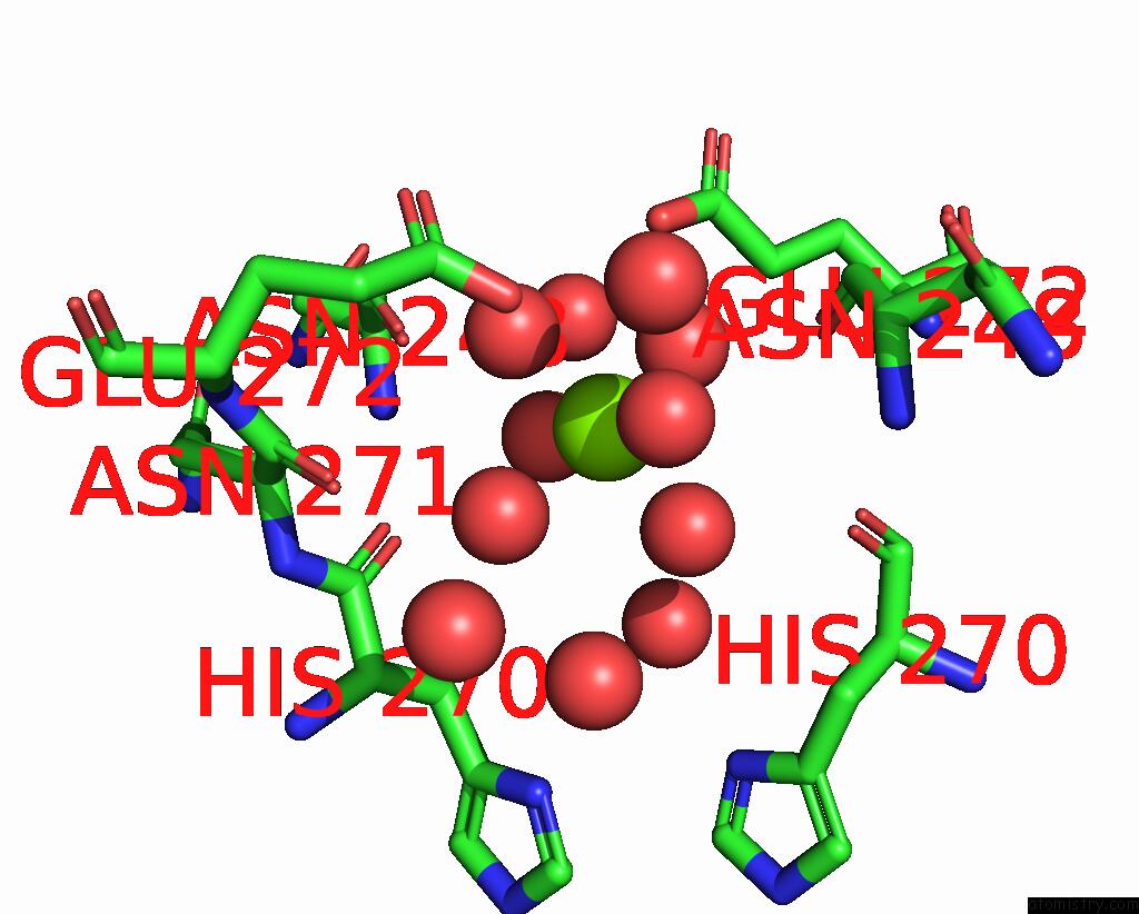
Magnesium Binding Sites:
The binding sites of Magnesium atom in the Structure of the Human Leucine Rich Repeat Protein SHOC2, Residues 80- 582
(pdb code 7tyg). This binding sites where shown within
5.0 Angstroms radius around Magnesium atom.
In total only one binding site of Magnesium was determined in the Structure of the Human Leucine Rich Repeat Protein SHOC2, Residues 80- 582, PDB code: 7tyg:
In total only one binding site of Magnesium was determined in the Structure of the Human Leucine Rich Repeat Protein SHOC2, Residues 80- 582, PDB code: 7tyg:
Magnesium binding site 1 out of 1 in 7tyg
Go back to
Magnesium binding site 1 out
of 1 in the Structure of the Human Leucine Rich Repeat Protein SHOC2, Residues 80- 582
Mono view

Stereo pair view
Mono view
Stereo pair view
A full contact list of Magnesium with other atoms in the Mg binding
site number 1 of Structure of the Human Leucine Rich Repeat Protein SHOC2, Residues 80- 582 within 5.0Å range:
|
Reference:
Z.J.Hauseman,
M.Fodor,
A.Dhembi,
J.Viscomi,
D.Egli,
M.Bleu,
S.Katz,
E.Park,
D.M.Jang,
K.A.Porter,
F.Meili,
H.Guo,
G.Kerr,
S.Molle,
C.Velez-Vega,
K.S.Beyer,
G.G.Galli,
S.M.Maira,
T.Stams,
K.Clark,
M.J.Eck,
L.Tordella,
C.R.Thoma,
D.A.King.
Structure of the Mras-SHOC2-PP1C Phosphatase Complex. Nature V. 609 416 2022.
ISSN: ESSN 1476-4687
PubMed: 35830882
DOI: 10.1038/S41586-022-05086-1
Page generated: Thu Oct 3 09:34:55 2024
ISSN: ESSN 1476-4687
PubMed: 35830882
DOI: 10.1038/S41586-022-05086-1
Last articles
Zn in 9MJ5Zn in 9HNW
Zn in 9G0L
Zn in 9FNE
Zn in 9DZN
Zn in 9E0I
Zn in 9D32
Zn in 9DAK
Zn in 8ZXC
Zn in 8ZUF