Magnesium »
PDB 7tr7-7u0y »
7tzs »
Magnesium in PDB 7tzs: Crystal Structure of the E. Coli Thim Riboswitch in Complex with Quinoxalin-6-Ylmethanamine (Compound 17)
Protein crystallography data
The structure of Crystal Structure of the E. Coli Thim Riboswitch in Complex with Quinoxalin-6-Ylmethanamine (Compound 17), PDB code: 7tzs
was solved by
A.Nuthanakanti,
A.Serganov,
with X-Ray Crystallography technique. A brief refinement statistics is given in the table below:
| Resolution Low / High (Å) | 29.78 / 2.21 |
| Space group | C 1 2 1 |
| Cell size a, b, c (Å), α, β, γ (°) | 148.734, 30.4, 95.57, 90, 93.51, 90 |
| R / Rfree (%) | 20.4 / 24.5 |
Other elements in 7tzs:
The structure of Crystal Structure of the E. Coli Thim Riboswitch in Complex with Quinoxalin-6-Ylmethanamine (Compound 17) also contains other interesting chemical elements:
| Potassium | (K) | 5 atoms |
| Sodium | (Na) | 1 atom |
Magnesium Binding Sites:
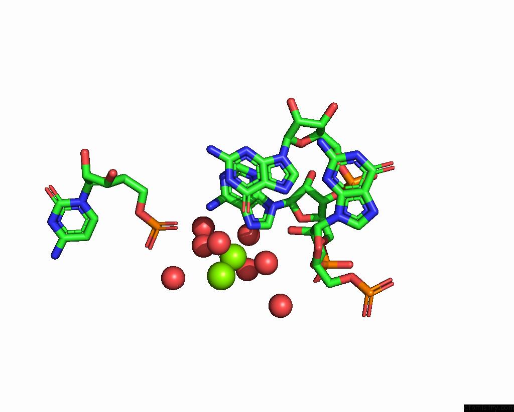
The binding sites of Magnesium atom in the Crystal Structure of the E. Coli Thim Riboswitch in Complex with Quinoxalin-6-Ylmethanamine (Compound 17)
(pdb code 7tzs). This binding sites where shown within
5.0 Angstroms radius around Magnesium atom.
In total 5 binding sites of Magnesium where determined in the Crystal Structure of the E. Coli Thim Riboswitch in Complex with Quinoxalin-6-Ylmethanamine (Compound 17), PDB code: 7tzs:
Jump to Magnesium binding site number: 1; 2; 3; 4; 5;
In total 5 binding sites of Magnesium where determined in the Crystal Structure of the E. Coli Thim Riboswitch in Complex with Quinoxalin-6-Ylmethanamine (Compound 17), PDB code: 7tzs:
Jump to Magnesium binding site number: 1; 2; 3; 4; 5;
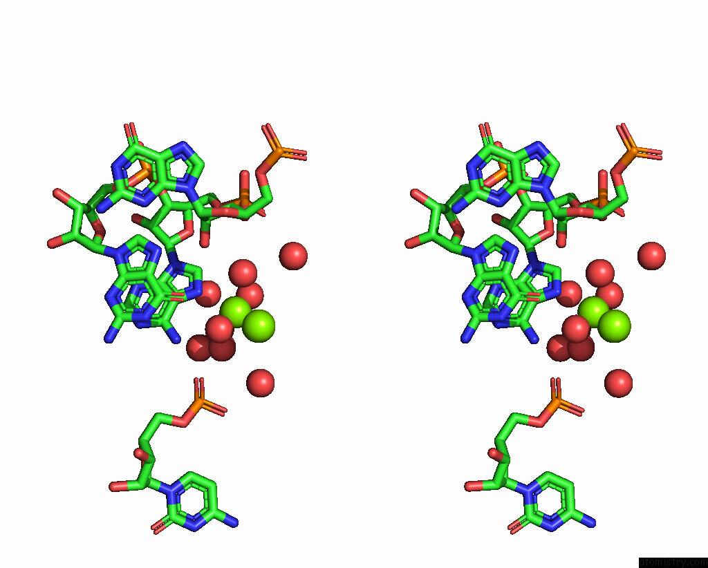
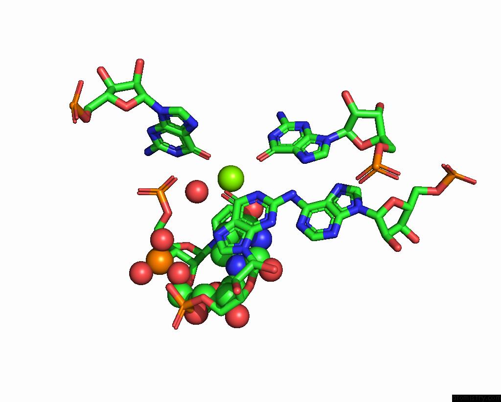
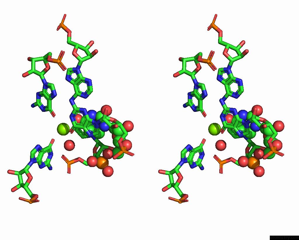
Magnesium binding site 1 out of 5 in 7tzs
Go back to
Magnesium binding site 1 out
of 5 in the Crystal Structure of the E. Coli Thim Riboswitch in Complex with Quinoxalin-6-Ylmethanamine (Compound 17)

Mono view

Stereo pair view
Mono view
Stereo pair view
A full contact list of Magnesium with other atoms in the Mg binding
site number 1 of Crystal Structure of the E. Coli Thim Riboswitch in Complex with Quinoxalin-6-Ylmethanamine (Compound 17) within 5.0Å range:
|
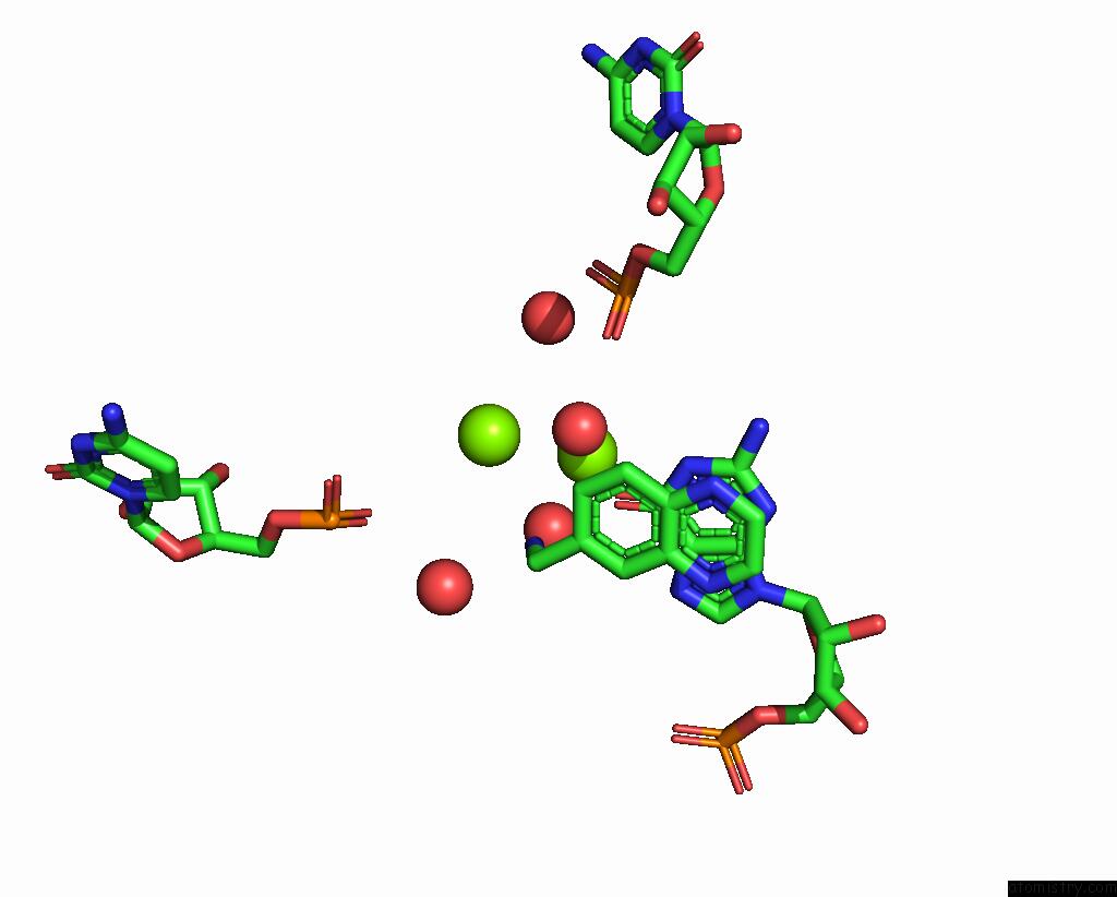
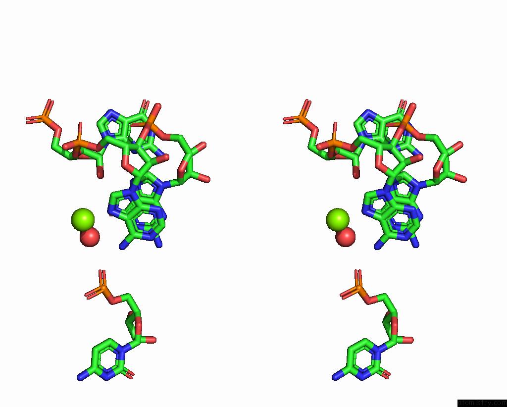
Magnesium binding site 2 out of 5 in 7tzs
Go back to
Magnesium binding site 2 out
of 5 in the Crystal Structure of the E. Coli Thim Riboswitch in Complex with Quinoxalin-6-Ylmethanamine (Compound 17)
Mono view
Stereo pair view
Mono view
Stereo pair view
A full contact list of Magnesium with other atoms in the Mg binding
site number 2 of Crystal Structure of the E. Coli Thim Riboswitch in Complex with Quinoxalin-6-Ylmethanamine (Compound 17) within 5.0Å range:
|
Magnesium binding site 3 out of 5 in 7tzs
Go back to
Magnesium binding site 3 out
of 5 in the Crystal Structure of the E. Coli Thim Riboswitch in Complex with Quinoxalin-6-Ylmethanamine (Compound 17)
Mono view
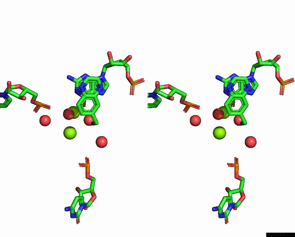
Stereo pair view
Mono view
Stereo pair view
A full contact list of Magnesium with other atoms in the Mg binding
site number 3 of Crystal Structure of the E. Coli Thim Riboswitch in Complex with Quinoxalin-6-Ylmethanamine (Compound 17) within 5.0Å range:
|
Magnesium binding site 4 out of 5 in 7tzs
Go back to
Magnesium binding site 4 out
of 5 in the Crystal Structure of the E. Coli Thim Riboswitch in Complex with Quinoxalin-6-Ylmethanamine (Compound 17)
Mono view
Stereo pair view
Mono view
Stereo pair view
A full contact list of Magnesium with other atoms in the Mg binding
site number 4 of Crystal Structure of the E. Coli Thim Riboswitch in Complex with Quinoxalin-6-Ylmethanamine (Compound 17) within 5.0Å range:
|
Magnesium binding site 5 out of 5 in 7tzs
Go back to
Magnesium binding site 5 out
of 5 in the Crystal Structure of the E. Coli Thim Riboswitch in Complex with Quinoxalin-6-Ylmethanamine (Compound 17)

Mono view
Stereo pair view
Mono view
Stereo pair view
A full contact list of Magnesium with other atoms in the Mg binding
site number 5 of Crystal Structure of the E. Coli Thim Riboswitch in Complex with Quinoxalin-6-Ylmethanamine (Compound 17) within 5.0Å range:
|
Reference:
M.J.Zeller,
O.Favorov,
K.Li,
A.Nuthanakanti,
D.Hussein,
A.Michaud,
D.A.Lafontaine,
S.Busan,
A.Serganov,
J.Aube,
K.M.Weeks.
Shape-Enabled Fragment-Based Ligand Discovery For Rna. Proc.Natl.Acad.Sci.Usa V. 119 60119 2022.
ISSN: ESSN 1091-6490
PubMed: 35561226
DOI: 10.1073/PNAS.2122660119
Page generated: Thu Oct 3 09:35:26 2024
ISSN: ESSN 1091-6490
PubMed: 35561226
DOI: 10.1073/PNAS.2122660119
Last articles
Zn in 9MJ5Zn in 9HNW
Zn in 9G0L
Zn in 9FNE
Zn in 9DZN
Zn in 9E0I
Zn in 9D32
Zn in 9DAK
Zn in 8ZXC
Zn in 8ZUF