Magnesium »
PDB 7u0y-7udu »
7u67 »
Magnesium in PDB 7u67: Structure of E. Coli Dgtpase Bound to T7 Bacteriophage Protein GP1.2 and Gtp
Enzymatic activity of Structure of E. Coli Dgtpase Bound to T7 Bacteriophage Protein GP1.2 and Gtp
All present enzymatic activity of Structure of E. Coli Dgtpase Bound to T7 Bacteriophage Protein GP1.2 and Gtp:
3.1.5.1;
3.1.5.1;
Magnesium Binding Sites:
The binding sites of Magnesium atom in the Structure of E. Coli Dgtpase Bound to T7 Bacteriophage Protein GP1.2 and Gtp
(pdb code 7u67). This binding sites where shown within
5.0 Angstroms radius around Magnesium atom.
In total 6 binding sites of Magnesium where determined in the Structure of E. Coli Dgtpase Bound to T7 Bacteriophage Protein GP1.2 and Gtp, PDB code: 7u67:
Jump to Magnesium binding site number: 1; 2; 3; 4; 5; 6;
In total 6 binding sites of Magnesium where determined in the Structure of E. Coli Dgtpase Bound to T7 Bacteriophage Protein GP1.2 and Gtp, PDB code: 7u67:
Jump to Magnesium binding site number: 1; 2; 3; 4; 5; 6;
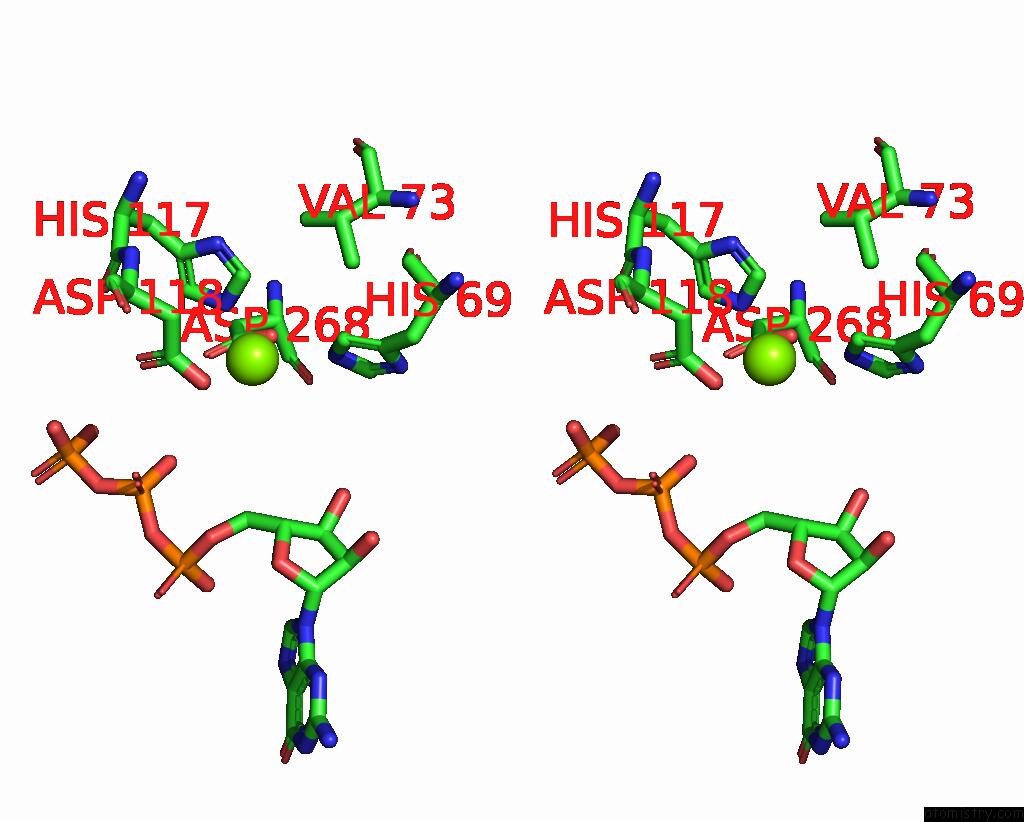
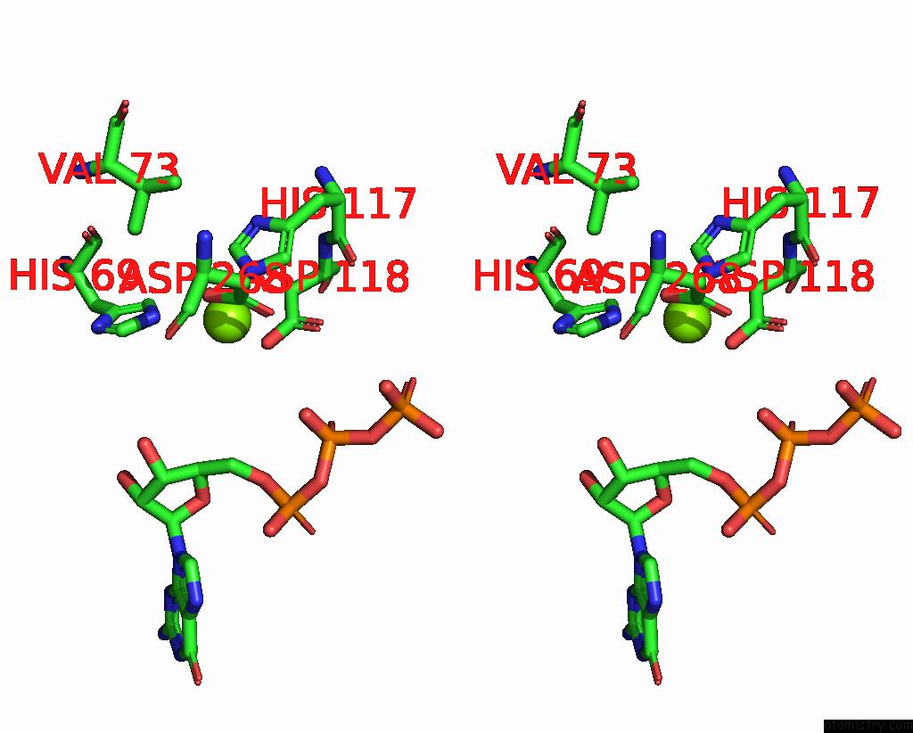
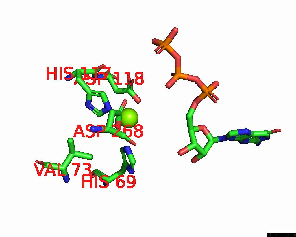
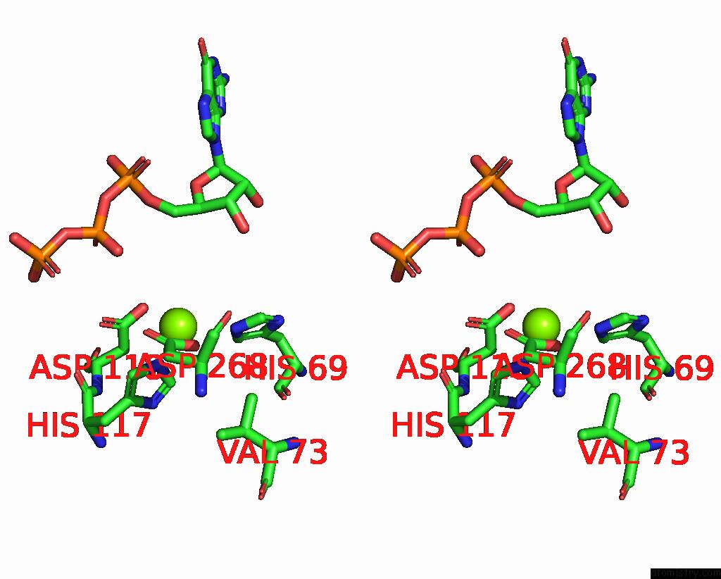
Magnesium binding site 1 out of 6 in 7u67
Go back to
Magnesium binding site 1 out
of 6 in the Structure of E. Coli Dgtpase Bound to T7 Bacteriophage Protein GP1.2 and Gtp
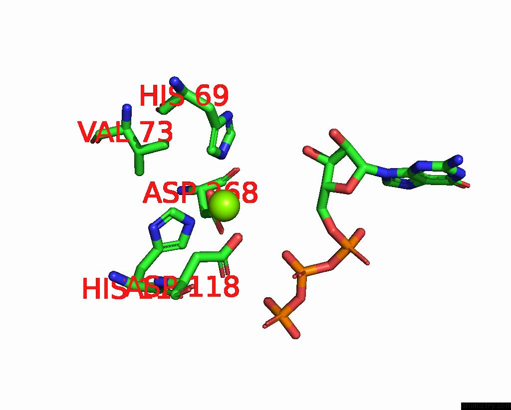
Mono view

Stereo pair view
Mono view
Stereo pair view
A full contact list of Magnesium with other atoms in the Mg binding
site number 1 of Structure of E. Coli Dgtpase Bound to T7 Bacteriophage Protein GP1.2 and Gtp within 5.0Å range:
|
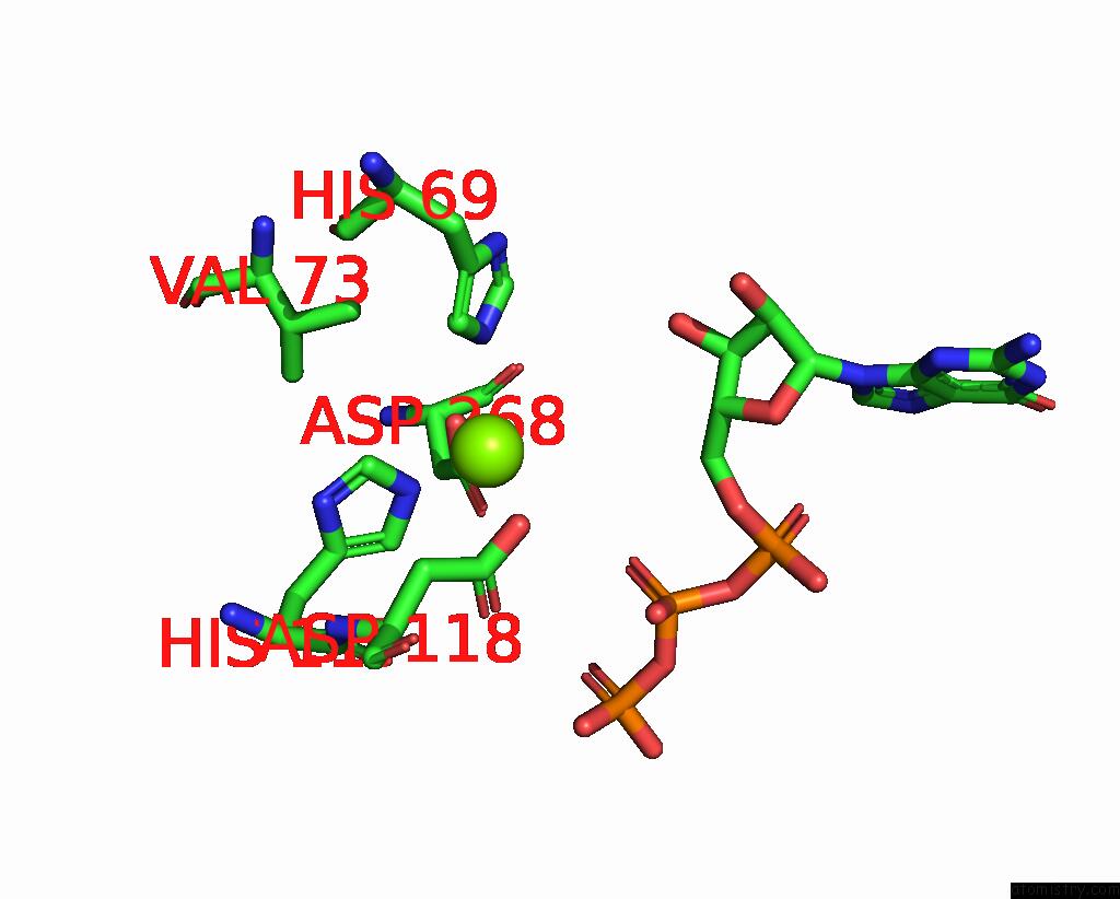
Magnesium binding site 2 out of 6 in 7u67
Go back to
Magnesium binding site 2 out
of 6 in the Structure of E. Coli Dgtpase Bound to T7 Bacteriophage Protein GP1.2 and Gtp
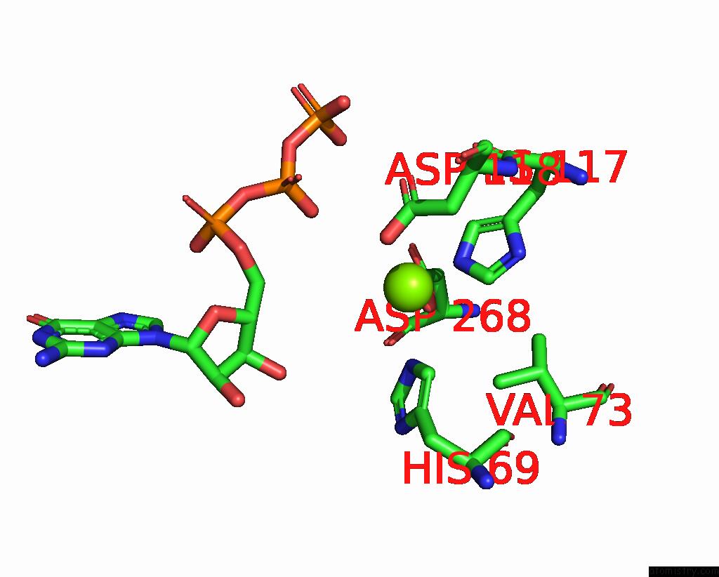
Mono view
Stereo pair view
Mono view
Stereo pair view
A full contact list of Magnesium with other atoms in the Mg binding
site number 2 of Structure of E. Coli Dgtpase Bound to T7 Bacteriophage Protein GP1.2 and Gtp within 5.0Å range:
|
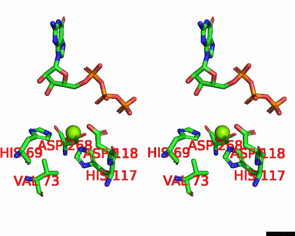
Magnesium binding site 3 out of 6 in 7u67
Go back to
Magnesium binding site 3 out
of 6 in the Structure of E. Coli Dgtpase Bound to T7 Bacteriophage Protein GP1.2 and Gtp
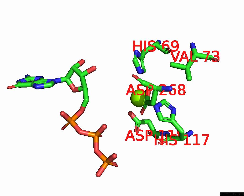
Mono view
Stereo pair view
Mono view
Stereo pair view
A full contact list of Magnesium with other atoms in the Mg binding
site number 3 of Structure of E. Coli Dgtpase Bound to T7 Bacteriophage Protein GP1.2 and Gtp within 5.0Å range:
|
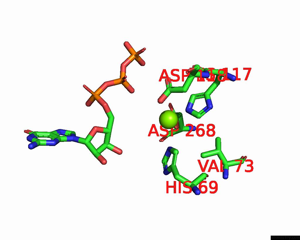
Magnesium binding site 4 out of 6 in 7u67
Go back to
Magnesium binding site 4 out
of 6 in the Structure of E. Coli Dgtpase Bound to T7 Bacteriophage Protein GP1.2 and Gtp
Mono view
Stereo pair view
Mono view
Stereo pair view
A full contact list of Magnesium with other atoms in the Mg binding
site number 4 of Structure of E. Coli Dgtpase Bound to T7 Bacteriophage Protein GP1.2 and Gtp within 5.0Å range:
|
Magnesium binding site 5 out of 6 in 7u67
Go back to
Magnesium binding site 5 out
of 6 in the Structure of E. Coli Dgtpase Bound to T7 Bacteriophage Protein GP1.2 and Gtp
Mono view

Stereo pair view
Mono view
Stereo pair view
A full contact list of Magnesium with other atoms in the Mg binding
site number 5 of Structure of E. Coli Dgtpase Bound to T7 Bacteriophage Protein GP1.2 and Gtp within 5.0Å range:
|
Magnesium binding site 6 out of 6 in 7u67
Go back to
Magnesium binding site 6 out
of 6 in the Structure of E. Coli Dgtpase Bound to T7 Bacteriophage Protein GP1.2 and Gtp
Mono view
Stereo pair view
Mono view
Stereo pair view
A full contact list of Magnesium with other atoms in the Mg binding
site number 6 of Structure of E. Coli Dgtpase Bound to T7 Bacteriophage Protein GP1.2 and Gtp within 5.0Å range:
|
Reference:
B.P.Klemm,
D.Singh,
C.E.Smith,
A.L.Hsu,
L.B.Dillard,
J.M.Krahn,
R.E.London,
G.A.Mueller,
M.J.Borgnia,
R.M.Schaaper.
Mechanism By Which T7 Bacteriophage Protein GP1.2 Inhibits Escherichia Coli Dgtpase. Proc.Natl.Acad.Sci.Usa V. 119 92119 2022.
ISSN: ESSN 1091-6490
PubMed: 36067314
DOI: 10.1073/PNAS.2123092119
Page generated: Thu Oct 3 09:48:23 2024
ISSN: ESSN 1091-6490
PubMed: 36067314
DOI: 10.1073/PNAS.2123092119
Last articles
Zn in 9J0NZn in 9J0O
Zn in 9J0P
Zn in 9FJX
Zn in 9EKB
Zn in 9C0F
Zn in 9CAH
Zn in 9CH0
Zn in 9CH3
Zn in 9CH1