Magnesium »
PDB 7ue3-7uo7 »
7ujp »
Magnesium in PDB 7ujp: Cocrystal Structure of Human Camkii-Alpha (CAMK2A)Kinase Domain and GLUN2B
Enzymatic activity of Cocrystal Structure of Human Camkii-Alpha (CAMK2A)Kinase Domain and GLUN2B
All present enzymatic activity of Cocrystal Structure of Human Camkii-Alpha (CAMK2A)Kinase Domain and GLUN2B:
2.7.11.17;
2.7.11.17;
Protein crystallography data
The structure of Cocrystal Structure of Human Camkii-Alpha (CAMK2A)Kinase Domain and GLUN2B, PDB code: 7ujp
was solved by
C.Ozden,
J.C.Foster,
M.M.Stratton,
S.C.Garman,
with X-Ray Crystallography technique. A brief refinement statistics is given in the table below:
| Resolution Low / High (Å) | 48.56 / 2.56 |
| Space group | P 21 21 21 |
| Cell size a, b, c (Å), α, β, γ (°) | 73.143, 91.423, 91.922, 90, 90, 90 |
| R / Rfree (%) | 23.2 / 28.1 |
Magnesium Binding Sites:
The binding sites of Magnesium atom in the Cocrystal Structure of Human Camkii-Alpha (CAMK2A)Kinase Domain and GLUN2B
(pdb code 7ujp). This binding sites where shown within
5.0 Angstroms radius around Magnesium atom.
In total 3 binding sites of Magnesium where determined in the Cocrystal Structure of Human Camkii-Alpha (CAMK2A)Kinase Domain and GLUN2B, PDB code: 7ujp:
Jump to Magnesium binding site number: 1; 2; 3;
In total 3 binding sites of Magnesium where determined in the Cocrystal Structure of Human Camkii-Alpha (CAMK2A)Kinase Domain and GLUN2B, PDB code: 7ujp:
Jump to Magnesium binding site number: 1; 2; 3;
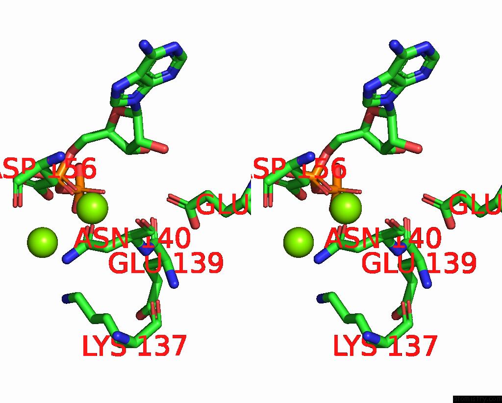
Magnesium binding site 1 out of 3 in 7ujp
Go back to
Magnesium binding site 1 out
of 3 in the Cocrystal Structure of Human Camkii-Alpha (CAMK2A)Kinase Domain and GLUN2B

Mono view
Stereo pair view
Mono view
Stereo pair view
A full contact list of Magnesium with other atoms in the Mg binding
site number 1 of Cocrystal Structure of Human Camkii-Alpha (CAMK2A)Kinase Domain and GLUN2B within 5.0Å range:
|
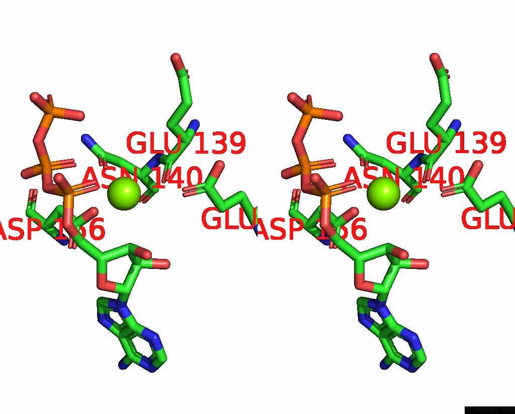
Magnesium binding site 2 out of 3 in 7ujp
Go back to
Magnesium binding site 2 out
of 3 in the Cocrystal Structure of Human Camkii-Alpha (CAMK2A)Kinase Domain and GLUN2B

Mono view
Stereo pair view
Mono view
Stereo pair view
A full contact list of Magnesium with other atoms in the Mg binding
site number 2 of Cocrystal Structure of Human Camkii-Alpha (CAMK2A)Kinase Domain and GLUN2B within 5.0Å range:
|
Magnesium binding site 3 out of 3 in 7ujp
Go back to
Magnesium binding site 3 out
of 3 in the Cocrystal Structure of Human Camkii-Alpha (CAMK2A)Kinase Domain and GLUN2B

Mono view

Stereo pair view
Mono view
Stereo pair view
A full contact list of Magnesium with other atoms in the Mg binding
site number 3 of Cocrystal Structure of Human Camkii-Alpha (CAMK2A)Kinase Domain and GLUN2B within 5.0Å range:
|
Reference:
C.Ozden,
R.Sloutsky,
T.Mitsugi,
N.Santos,
E.Agnello,
C.Gaubitz,
J.Foster,
E.Lapinskas,
E.A.Esposito,
T.Saneyoshi,
B.A.Kelch,
S.C.Garman,
Y.Hayashi,
M.M.Stratton.
Camkii Binds Both Substrates and Activators at the Active Site. Cell Rep V. 40 11064 2022.
ISSN: ESSN 2211-1247
PubMed: 35830796
DOI: 10.1016/J.CELREP.2022.111064
Page generated: Thu Oct 3 10:06:38 2024
ISSN: ESSN 2211-1247
PubMed: 35830796
DOI: 10.1016/J.CELREP.2022.111064
Last articles
Zn in 9MJ5Zn in 9HNW
Zn in 9G0L
Zn in 9FNE
Zn in 9DZN
Zn in 9E0I
Zn in 9D32
Zn in 9DAK
Zn in 8ZXC
Zn in 8ZUF