Magnesium »
PDB 7ue3-7uo7 »
7ujx »
Magnesium in PDB 7ujx: Structure of Camp-Dependent Protein Kinase Using A Md-Mx Procedure, Produced Using 2.4 Angstrom Data
Enzymatic activity of Structure of Camp-Dependent Protein Kinase Using A Md-Mx Procedure, Produced Using 2.4 Angstrom Data
All present enzymatic activity of Structure of Camp-Dependent Protein Kinase Using A Md-Mx Procedure, Produced Using 2.4 Angstrom Data:
2.7.11.11;
2.7.11.11;
Protein crystallography data
The structure of Structure of Camp-Dependent Protein Kinase Using A Md-Mx Procedure, Produced Using 2.4 Angstrom Data, PDB code: 7ujx
was solved by
D.C.Wych,
P.C.Aoto,
M.E.Wall,
with X-Ray Crystallography technique. A brief refinement statistics is given in the table below:
| Resolution Low / High (Å) | 49.53 / 2.40 |
| Space group | P 21 21 21 |
| Cell size a, b, c (Å), α, β, γ (°) | 58.976, 79.744, 99.058, 90, 90, 90 |
| R / Rfree (%) | 12.8 / 17.7 |
Magnesium Binding Sites:
The binding sites of Magnesium atom in the Structure of Camp-Dependent Protein Kinase Using A Md-Mx Procedure, Produced Using 2.4 Angstrom Data
(pdb code 7ujx). This binding sites where shown within
5.0 Angstroms radius around Magnesium atom.
In total 2 binding sites of Magnesium where determined in the Structure of Camp-Dependent Protein Kinase Using A Md-Mx Procedure, Produced Using 2.4 Angstrom Data, PDB code: 7ujx:
Jump to Magnesium binding site number: 1; 2;
In total 2 binding sites of Magnesium where determined in the Structure of Camp-Dependent Protein Kinase Using A Md-Mx Procedure, Produced Using 2.4 Angstrom Data, PDB code: 7ujx:
Jump to Magnesium binding site number: 1; 2;
Magnesium binding site 1 out of 2 in 7ujx
Go back to
Magnesium binding site 1 out
of 2 in the Structure of Camp-Dependent Protein Kinase Using A Md-Mx Procedure, Produced Using 2.4 Angstrom Data
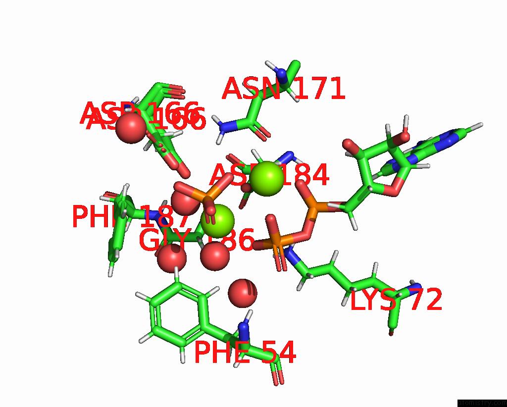
Mono view
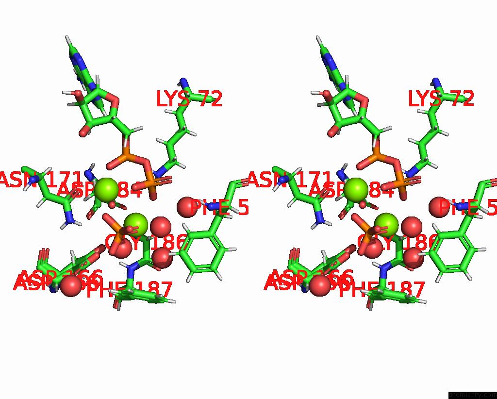
Stereo pair view
Mono view
Stereo pair view
A full contact list of Magnesium with other atoms in the Mg binding
site number 1 of Structure of Camp-Dependent Protein Kinase Using A Md-Mx Procedure, Produced Using 2.4 Angstrom Data within 5.0Å range:
|
Magnesium binding site 2 out of 2 in 7ujx
Go back to
Magnesium binding site 2 out
of 2 in the Structure of Camp-Dependent Protein Kinase Using A Md-Mx Procedure, Produced Using 2.4 Angstrom Data
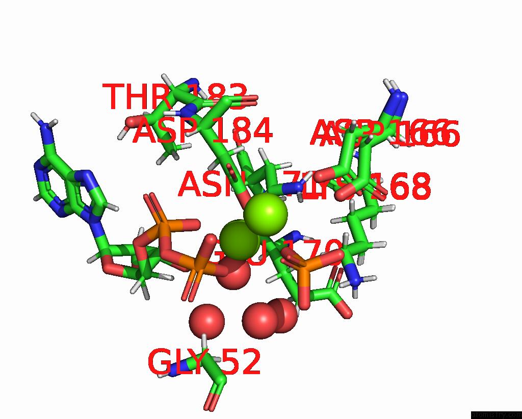
Mono view
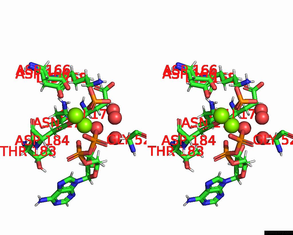
Stereo pair view
Mono view
Stereo pair view
A full contact list of Magnesium with other atoms in the Mg binding
site number 2 of Structure of Camp-Dependent Protein Kinase Using A Md-Mx Procedure, Produced Using 2.4 Angstrom Data within 5.0Å range:
|
Reference:
D.C.Wych,
P.C.Aoto,
L.T.Vu,
A.M.Wolff,
D.L.Mobley,
J.S.Fraser,
S.S.Taylor,
M.E.Wall.
Molecular Dynamics Simulation Methods For Macromolecular Crystallography To Be Published.
Page generated: Thu Oct 3 10:07:14 2024
Last articles
Fe in 2YXOFe in 2YRS
Fe in 2YXC
Fe in 2YNM
Fe in 2YVJ
Fe in 2YP1
Fe in 2YU2
Fe in 2YU1
Fe in 2YQB
Fe in 2YOO