Magnesium »
PDB 7vdo-7vpi »
7vhv »
Magnesium in PDB 7vhv: Crystal Structure of S. Aureus D-Alanine Alanyl Carrier Protein Ligase
Enzymatic activity of Crystal Structure of S. Aureus D-Alanine Alanyl Carrier Protein Ligase
All present enzymatic activity of Crystal Structure of S. Aureus D-Alanine Alanyl Carrier Protein Ligase:
6.2.1.54;
6.2.1.54;
Protein crystallography data
The structure of Crystal Structure of S. Aureus D-Alanine Alanyl Carrier Protein Ligase, PDB code: 7vhv
was solved by
B.J.Lee,
I.-G.Lee,
H.G.Im,
H.J.Yoon,
with X-Ray Crystallography technique. A brief refinement statistics is given in the table below:
| Resolution Low / High (Å) | 39.91 / 2.55 |
| Space group | P 1 21 1 |
| Cell size a, b, c (Å), α, β, γ (°) | 89.468, 88.51, 130.85, 90, 91.67, 90 |
| R / Rfree (%) | 18.9 / 24 |
Magnesium Binding Sites:
The binding sites of Magnesium atom in the Crystal Structure of S. Aureus D-Alanine Alanyl Carrier Protein Ligase
(pdb code 7vhv). This binding sites where shown within
5.0 Angstroms radius around Magnesium atom.
In total 4 binding sites of Magnesium where determined in the Crystal Structure of S. Aureus D-Alanine Alanyl Carrier Protein Ligase, PDB code: 7vhv:
Jump to Magnesium binding site number: 1; 2; 3; 4;
In total 4 binding sites of Magnesium where determined in the Crystal Structure of S. Aureus D-Alanine Alanyl Carrier Protein Ligase, PDB code: 7vhv:
Jump to Magnesium binding site number: 1; 2; 3; 4;
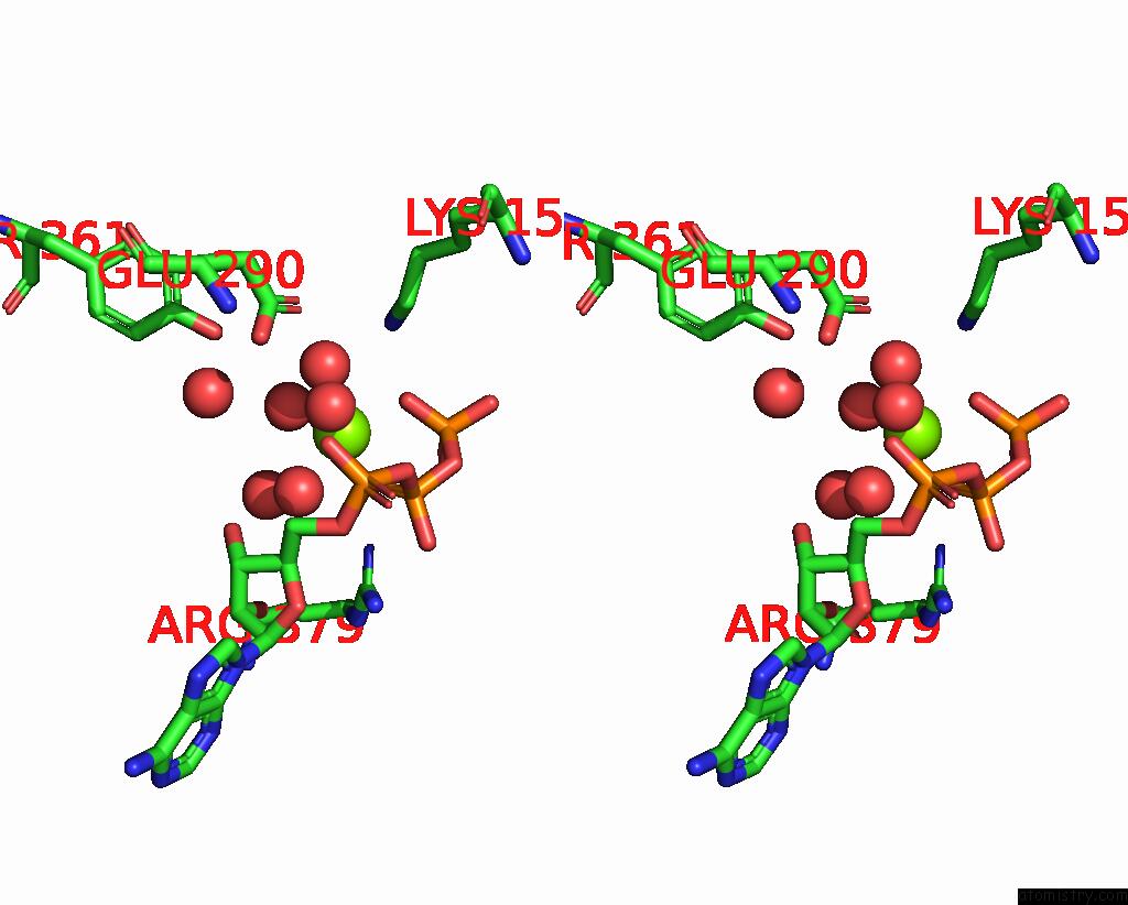
Magnesium binding site 1 out of 4 in 7vhv
Go back to
Magnesium binding site 1 out
of 4 in the Crystal Structure of S. Aureus D-Alanine Alanyl Carrier Protein Ligase
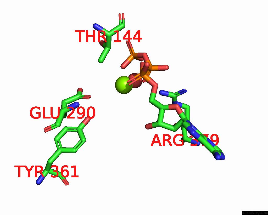
Mono view
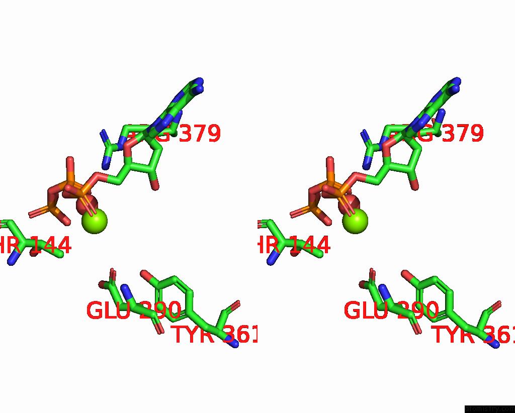
Stereo pair view
Mono view
Stereo pair view
A full contact list of Magnesium with other atoms in the Mg binding
site number 1 of Crystal Structure of S. Aureus D-Alanine Alanyl Carrier Protein Ligase within 5.0Å range:
|
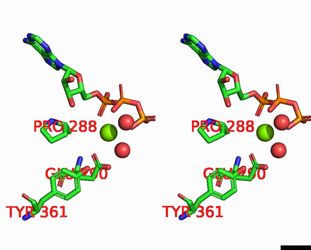
Magnesium binding site 2 out of 4 in 7vhv
Go back to
Magnesium binding site 2 out
of 4 in the Crystal Structure of S. Aureus D-Alanine Alanyl Carrier Protein Ligase

Mono view
Stereo pair view
Mono view
Stereo pair view
A full contact list of Magnesium with other atoms in the Mg binding
site number 2 of Crystal Structure of S. Aureus D-Alanine Alanyl Carrier Protein Ligase within 5.0Å range:
|
Magnesium binding site 3 out of 4 in 7vhv
Go back to
Magnesium binding site 3 out
of 4 in the Crystal Structure of S. Aureus D-Alanine Alanyl Carrier Protein Ligase
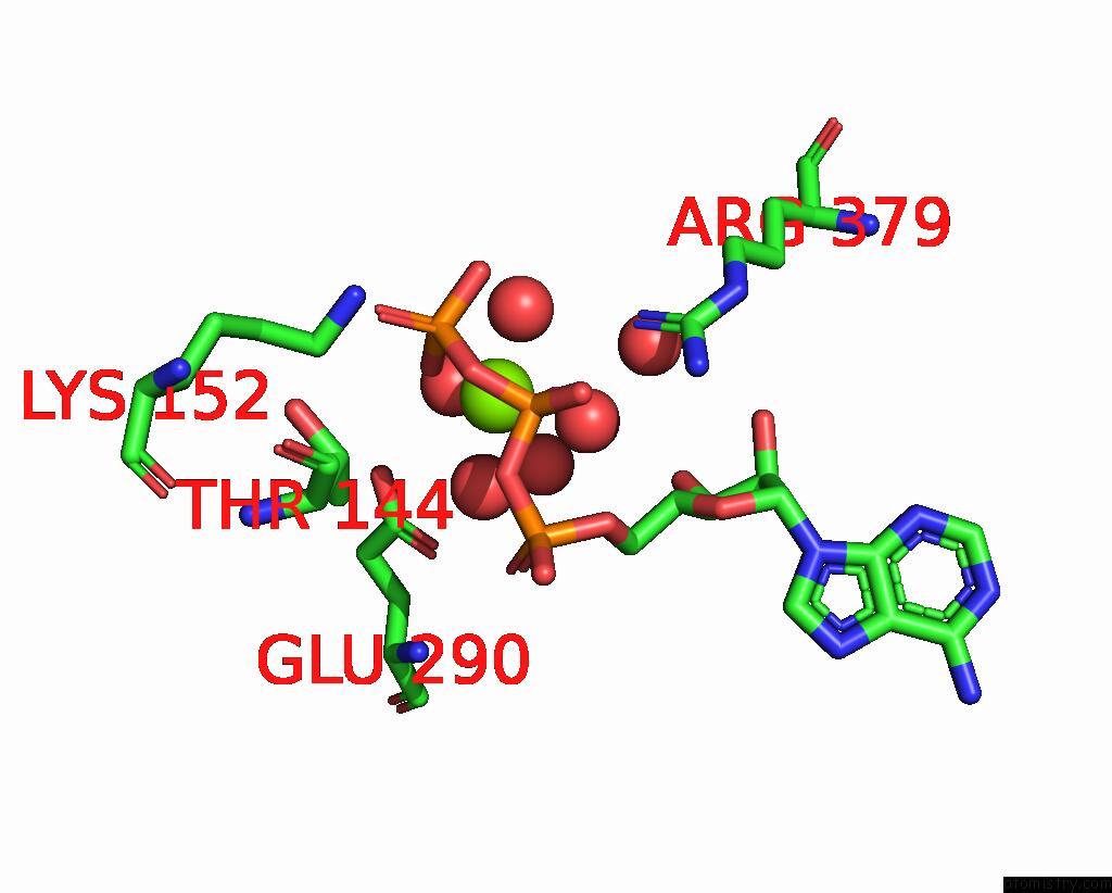
Mono view

Stereo pair view
Mono view
Stereo pair view
A full contact list of Magnesium with other atoms in the Mg binding
site number 3 of Crystal Structure of S. Aureus D-Alanine Alanyl Carrier Protein Ligase within 5.0Å range:
|
Magnesium binding site 4 out of 4 in 7vhv
Go back to
Magnesium binding site 4 out
of 4 in the Crystal Structure of S. Aureus D-Alanine Alanyl Carrier Protein Ligase

Mono view
Stereo pair view
Mono view
Stereo pair view
A full contact list of Magnesium with other atoms in the Mg binding
site number 4 of Crystal Structure of S. Aureus D-Alanine Alanyl Carrier Protein Ligase within 5.0Å range:
|
Reference:
I.G.Lee,
C.Song,
S.Yang,
H.Jeon,
J.Park,
H.J.Yoon,
H.Im,
S.M.Kang,
H.J.Eun,
B.J.Lee.
Structural and Functional Analysis of the D-Alanyl Carrier Protein Ligase Dlta From Staphylococcus Aureus MU50. Acta Crystallogr D Struct V. 78 424 2022BIOL.
ISSN: ISSN 2059-7983
PubMed: 35362466
DOI: 10.1107/S2059798322000547
Page generated: Thu Oct 3 10:31:13 2024
ISSN: ISSN 2059-7983
PubMed: 35362466
DOI: 10.1107/S2059798322000547
Last articles
Zn in 9J0NZn in 9J0O
Zn in 9J0P
Zn in 9FJX
Zn in 9EKB
Zn in 9C0F
Zn in 9CAH
Zn in 9CH0
Zn in 9CH3
Zn in 9CH1