Magnesium »
PDB 7vdt-7vpj »
7vmj »
Magnesium in PDB 7vmj: Crystal Structure of Tubulin with 17A
Protein crystallography data
The structure of Crystal Structure of Tubulin with 17A, PDB code: 7vmj
was solved by
Z.Jifa,
T.Lun,
with X-Ray Crystallography technique. A brief refinement statistics is given in the table below:
| Resolution Low / High (Å) | 87.43 / 2.90 |
| Space group | P 21 21 21 |
| Cell size a, b, c (Å), α, β, γ (°) | 104.976, 157.934, 182.334, 90, 90, 90 |
| R / Rfree (%) | 19.9 / 24.3 |
Other elements in 7vmj:
The structure of Crystal Structure of Tubulin with 17A also contains other interesting chemical elements:
| Fluorine | (F) | 2 atoms |
| Calcium | (Ca) | 2 atoms |
| Chlorine | (Cl) | 1 atom |
Magnesium Binding Sites:
The binding sites of Magnesium atom in the Crystal Structure of Tubulin with 17A
(pdb code 7vmj). This binding sites where shown within
5.0 Angstroms radius around Magnesium atom.
In total 3 binding sites of Magnesium where determined in the Crystal Structure of Tubulin with 17A, PDB code: 7vmj:
Jump to Magnesium binding site number: 1; 2; 3;
In total 3 binding sites of Magnesium where determined in the Crystal Structure of Tubulin with 17A, PDB code: 7vmj:
Jump to Magnesium binding site number: 1; 2; 3;
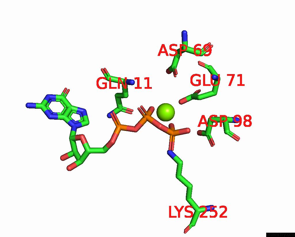
Magnesium binding site 1 out of 3 in 7vmj
Go back to
Magnesium binding site 1 out
of 3 in the Crystal Structure of Tubulin with 17A
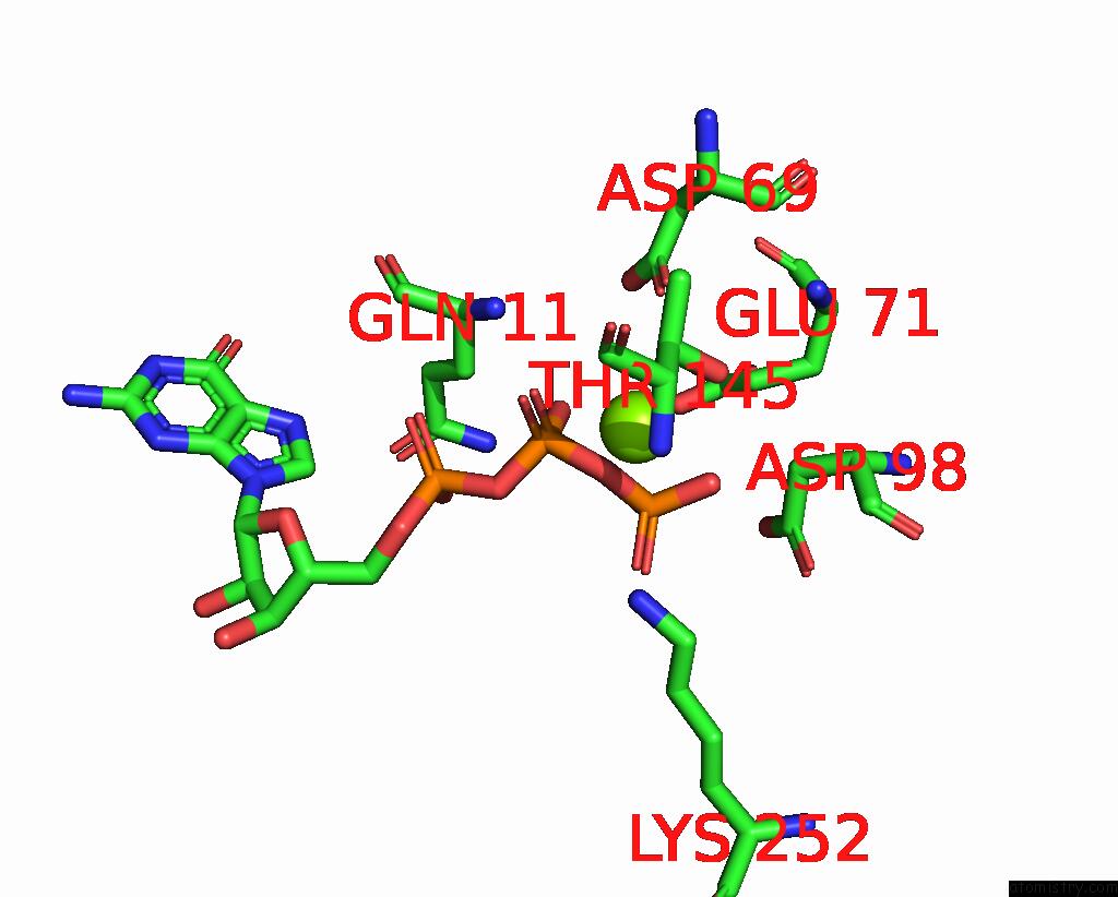
Mono view
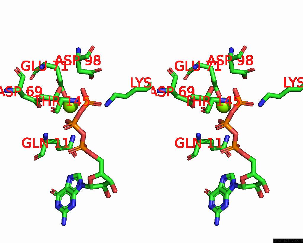
Stereo pair view
Mono view
Stereo pair view
A full contact list of Magnesium with other atoms in the Mg binding
site number 1 of Crystal Structure of Tubulin with 17A within 5.0Å range:
|
Magnesium binding site 2 out of 3 in 7vmj
Go back to
Magnesium binding site 2 out
of 3 in the Crystal Structure of Tubulin with 17A
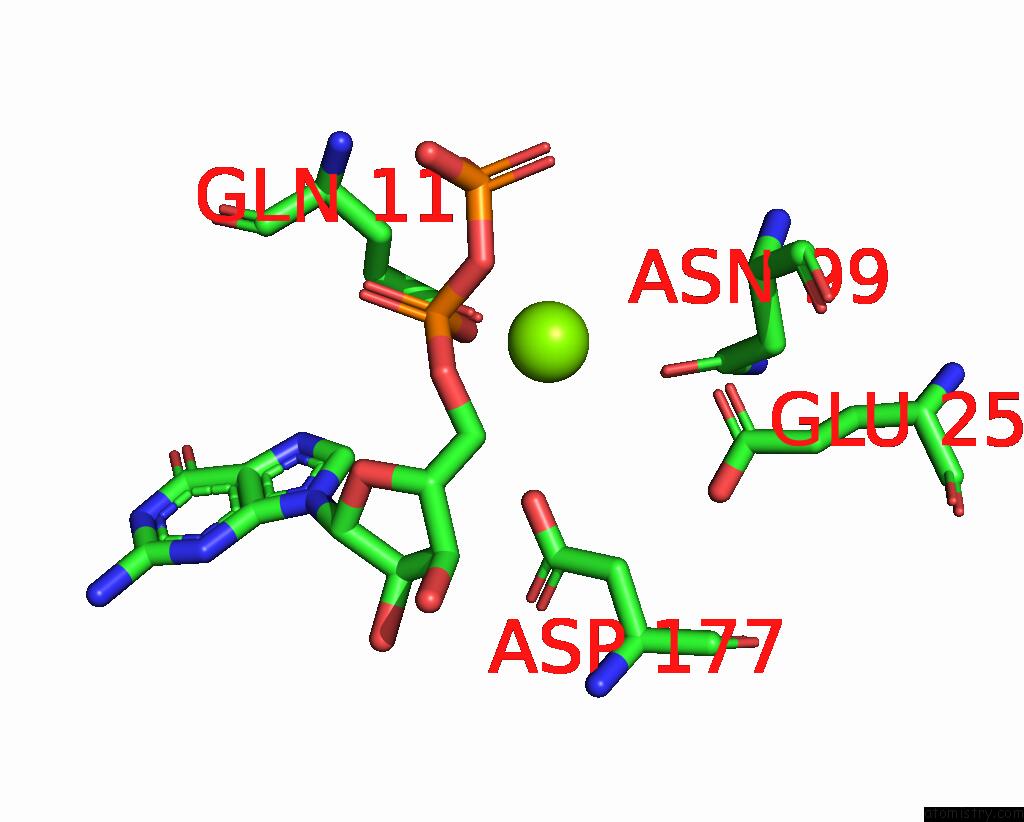
Mono view
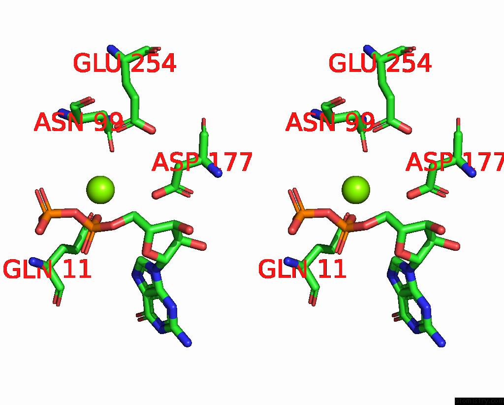
Stereo pair view
Mono view
Stereo pair view
A full contact list of Magnesium with other atoms in the Mg binding
site number 2 of Crystal Structure of Tubulin with 17A within 5.0Å range:
|
Magnesium binding site 3 out of 3 in 7vmj
Go back to
Magnesium binding site 3 out
of 3 in the Crystal Structure of Tubulin with 17A
Mono view

Stereo pair view
Mono view
Stereo pair view
A full contact list of Magnesium with other atoms in the Mg binding
site number 3 of Crystal Structure of Tubulin with 17A within 5.0Å range:
|
Reference:
Z.Jifa,
T.Lun.
Crystal Structure of Tubulin with 17J To Be Published.
Page generated: Thu Oct 3 10:33:11 2024
Last articles
F in 7LG8F in 7LD3
F in 7LCR
F in 7LCM
F in 7LCO
F in 7LCK
F in 7LCJ
F in 7LCI
F in 7L9Y
F in 7LCD