Magnesium »
PDB 7w8d-7wmb »
7wjv »
Magnesium in PDB 7wjv: Crystal Structure of Human Liver Fbpase Complexed with An Covalent Inhibitor
Protein crystallography data
The structure of Crystal Structure of Human Liver Fbpase Complexed with An Covalent Inhibitor, PDB code: 7wjv
was solved by
H.Cao,
Y.Huang,
Y.Ren,
J.Wan,
with X-Ray Crystallography technique. A brief refinement statistics is given in the table below:
| Resolution Low / High (Å) | 46.45 / 1.72 |
| Space group | P 21 21 21 |
| Cell size a, b, c (Å), α, β, γ (°) | 67.641, 83.617, 278.717, 90, 90, 90 |
| R / Rfree (%) | 17.4 / 19.8 |
Magnesium Binding Sites:
The binding sites of Magnesium atom in the Crystal Structure of Human Liver Fbpase Complexed with An Covalent Inhibitor
(pdb code 7wjv). This binding sites where shown within
5.0 Angstroms radius around Magnesium atom.
In total 2 binding sites of Magnesium where determined in the Crystal Structure of Human Liver Fbpase Complexed with An Covalent Inhibitor, PDB code: 7wjv:
Jump to Magnesium binding site number: 1; 2;
In total 2 binding sites of Magnesium where determined in the Crystal Structure of Human Liver Fbpase Complexed with An Covalent Inhibitor, PDB code: 7wjv:
Jump to Magnesium binding site number: 1; 2;
Magnesium binding site 1 out of 2 in 7wjv
Go back to
Magnesium binding site 1 out
of 2 in the Crystal Structure of Human Liver Fbpase Complexed with An Covalent Inhibitor
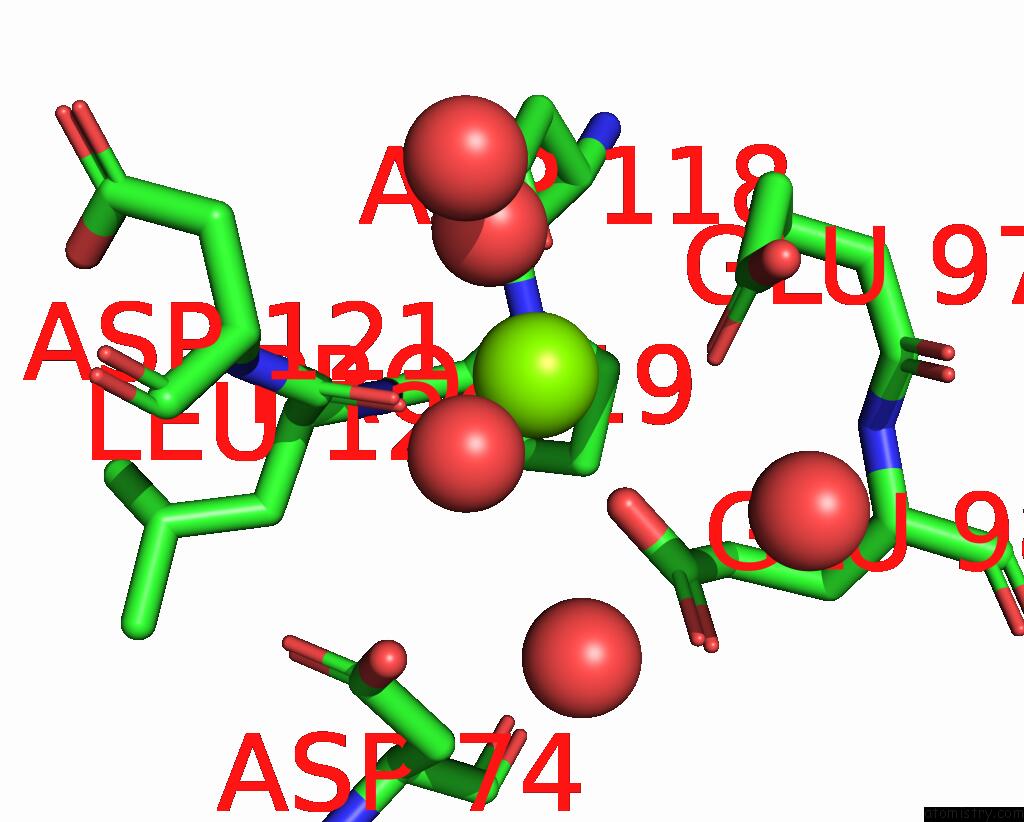
Mono view
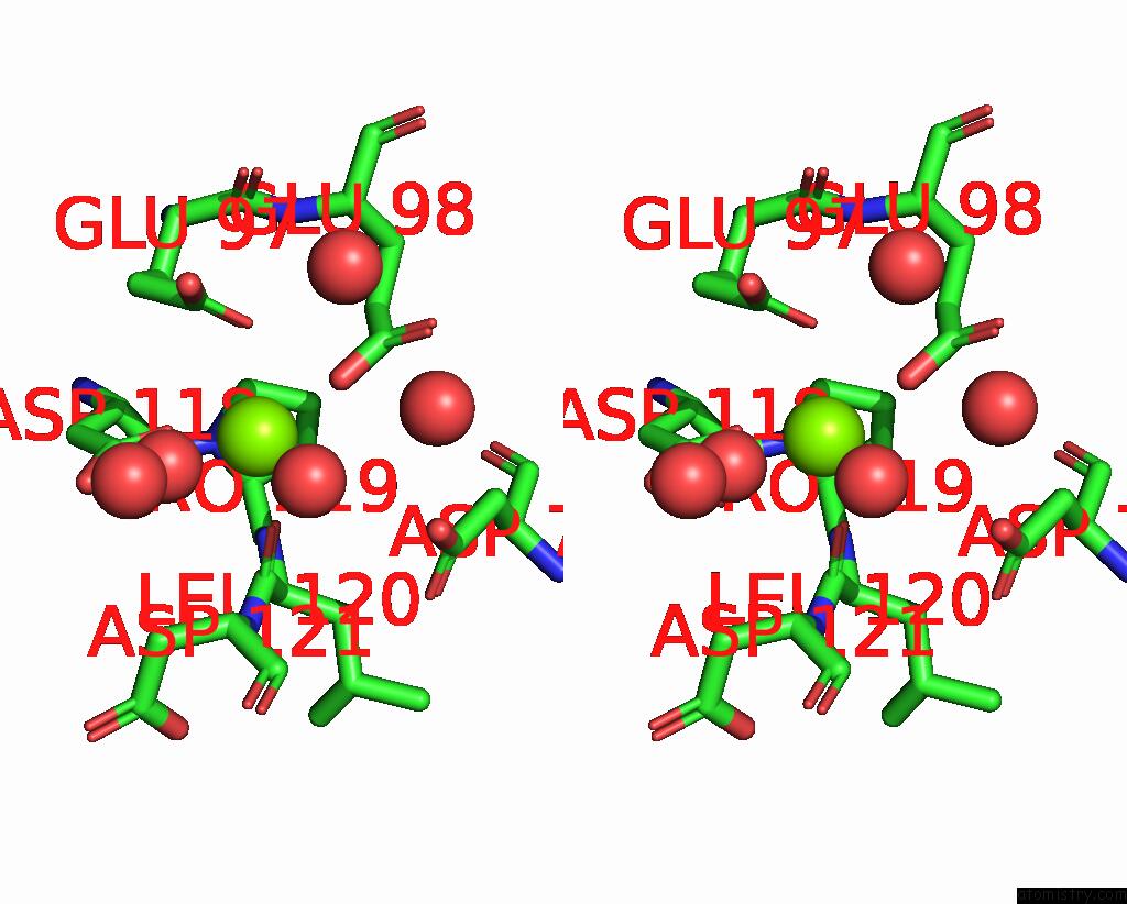
Stereo pair view
Mono view
Stereo pair view
A full contact list of Magnesium with other atoms in the Mg binding
site number 1 of Crystal Structure of Human Liver Fbpase Complexed with An Covalent Inhibitor within 5.0Å range:
|
Magnesium binding site 2 out of 2 in 7wjv
Go back to
Magnesium binding site 2 out
of 2 in the Crystal Structure of Human Liver Fbpase Complexed with An Covalent Inhibitor
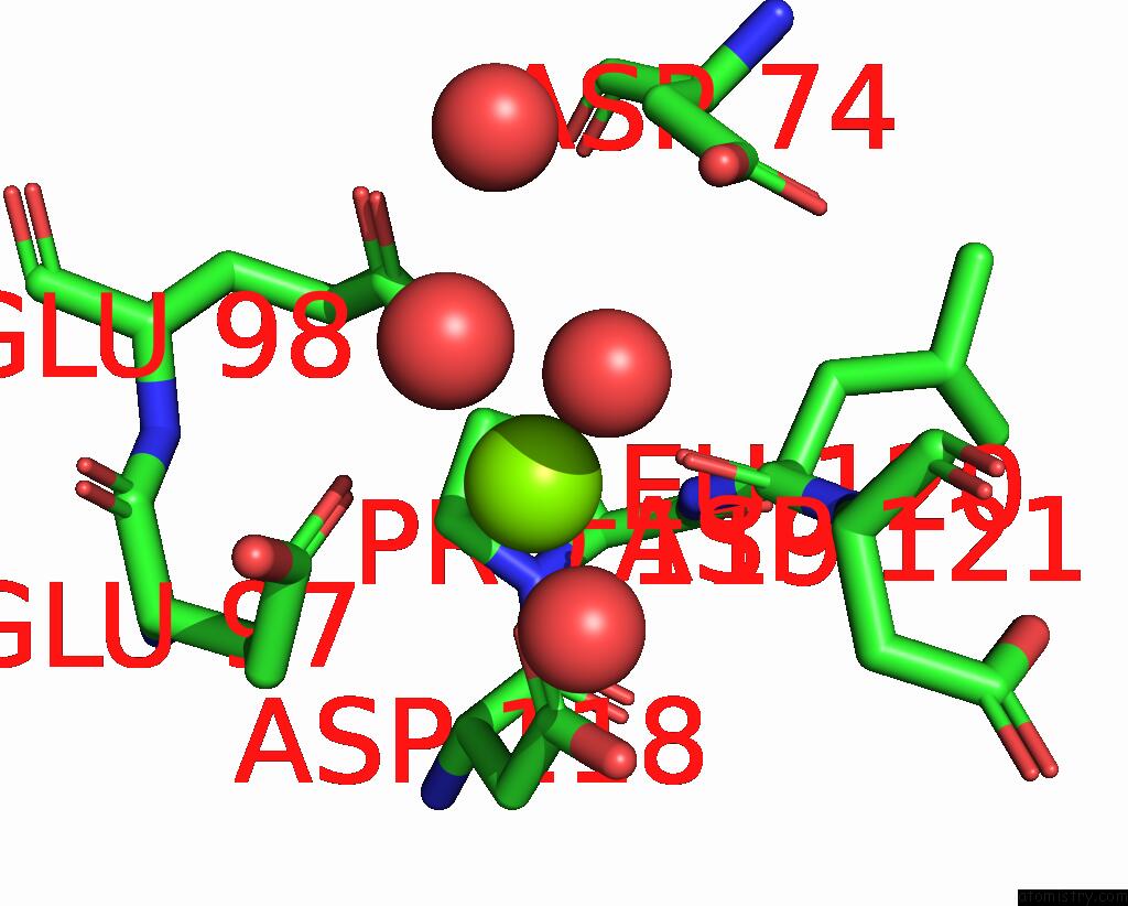
Mono view
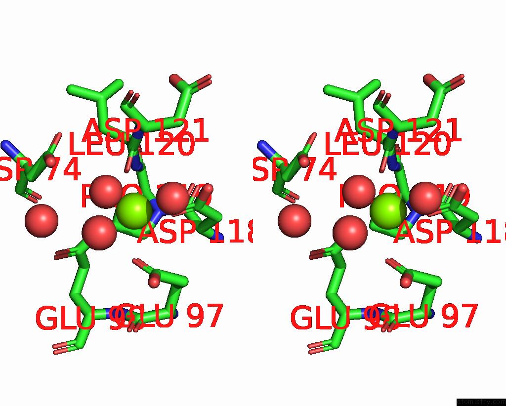
Stereo pair view
Mono view
Stereo pair view
A full contact list of Magnesium with other atoms in the Mg binding
site number 2 of Crystal Structure of Human Liver Fbpase Complexed with An Covalent Inhibitor within 5.0Å range:
|
Reference:
W.Wen,
H.Cao,
Y.Xu,
Y.Ren,
L.Rao,
X.Shao,
H.Chen,
L.Wu,
J.Liu,
C.Su,
C.Peng,
Y.Huang,
J.Wan.
N -Acylamino Saccharin As An Emerging Cysteine-Directed Covalent Warhead and Its Application in the Identification of Novel Fbpase Inhibitors Toward Glucose Reduction. J.Med.Chem. V. 65 9126 2022.
ISSN: ISSN 0022-2623
PubMed: 35786925
DOI: 10.1021/ACS.JMEDCHEM.2C00336
Page generated: Thu Oct 3 11:19:21 2024
ISSN: ISSN 0022-2623
PubMed: 35786925
DOI: 10.1021/ACS.JMEDCHEM.2C00336
Last articles
Zn in 9MJ5Zn in 9HNW
Zn in 9G0L
Zn in 9FNE
Zn in 9DZN
Zn in 9E0I
Zn in 9D32
Zn in 9DAK
Zn in 8ZXC
Zn in 8ZUF