Magnesium in PDB 7x1z: Structure of the Phosphorylation-Site Double Mutant S431E/T432E of the Kaic Circadian Clock Protein

Magnesium Binding Sites:

Pages:

>>> Page 1 <<< Page 2, Binding sites: 11 - 12;

Binding sites:

The binding sites of Magnesium atom in the Structure of the Phosphorylation-Site Double Mutant S431E/T432E of the Kaic Circadian Clock Protein (pdb code 7x1z). This binding sites where shown within 5.0 Angstroms radius around Magnesium atom.
In total 12 binding sites of Magnesium where determined in the Structure of the Phosphorylation-Site Double Mutant S431E/T432E of the Kaic Circadian Clock Protein, PDB code: 7x1z:
Jump to Magnesium binding site number: 1; 2; 3; 4; 5; 6; 7; 8; 9; 10;

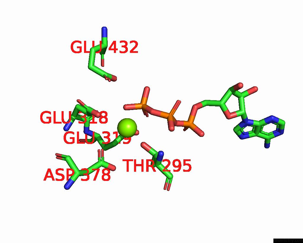
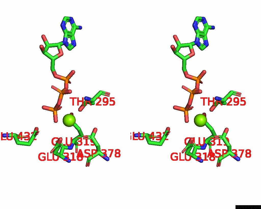
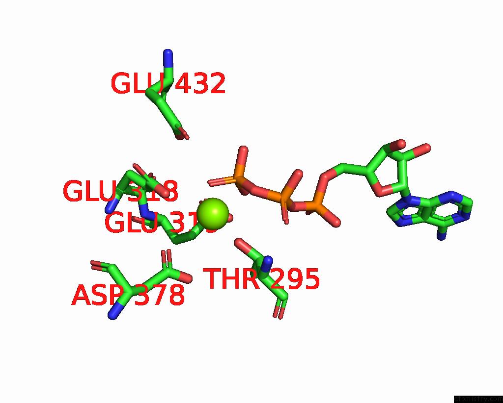
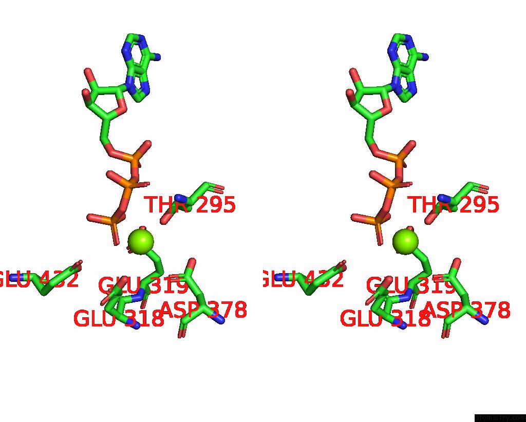
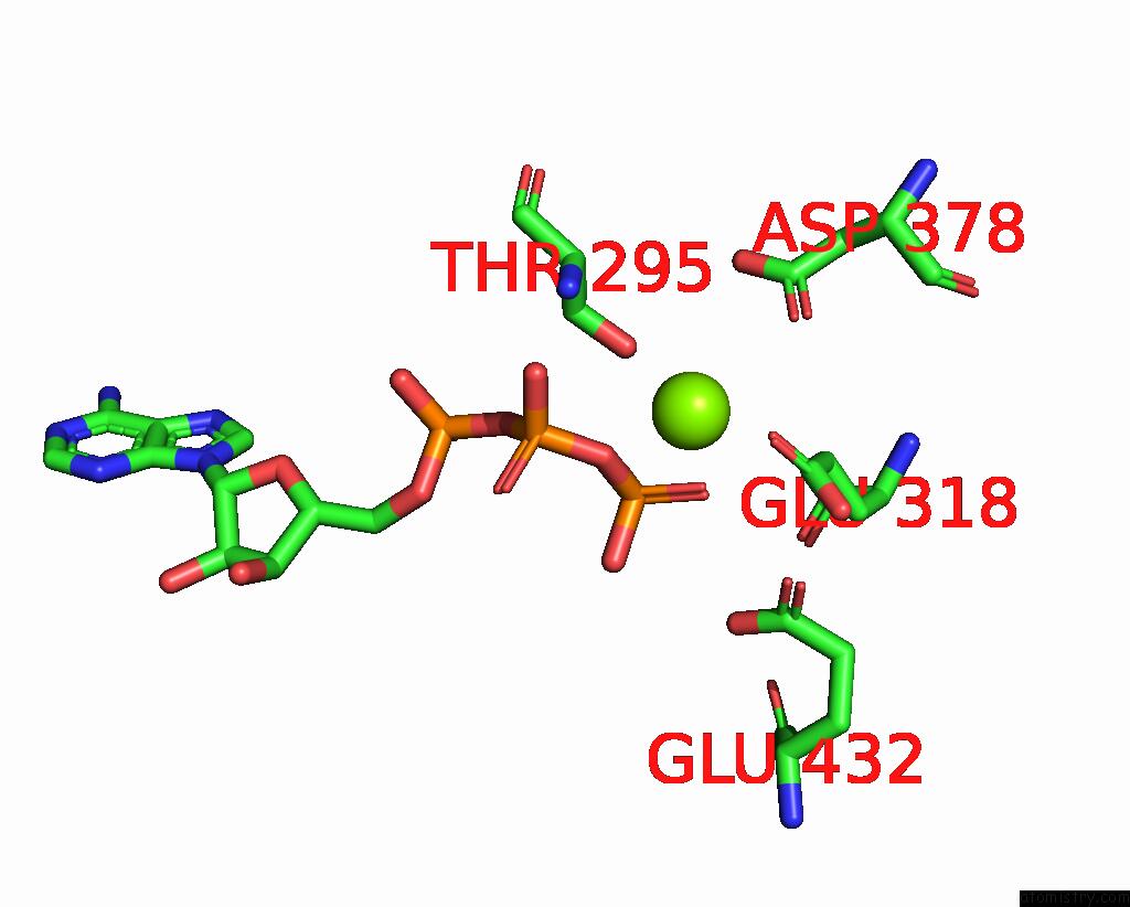
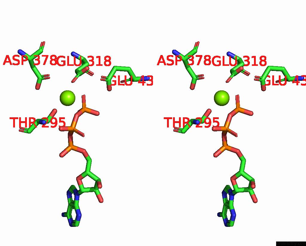
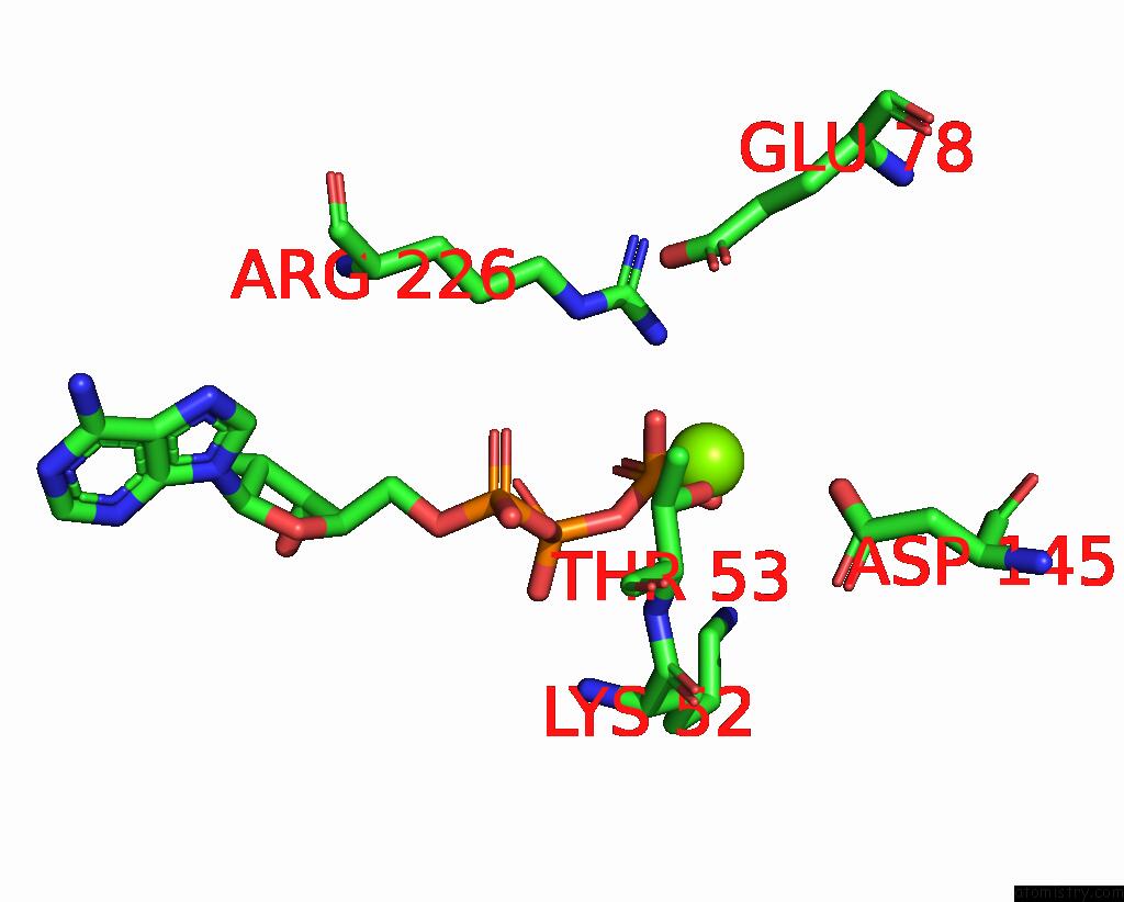
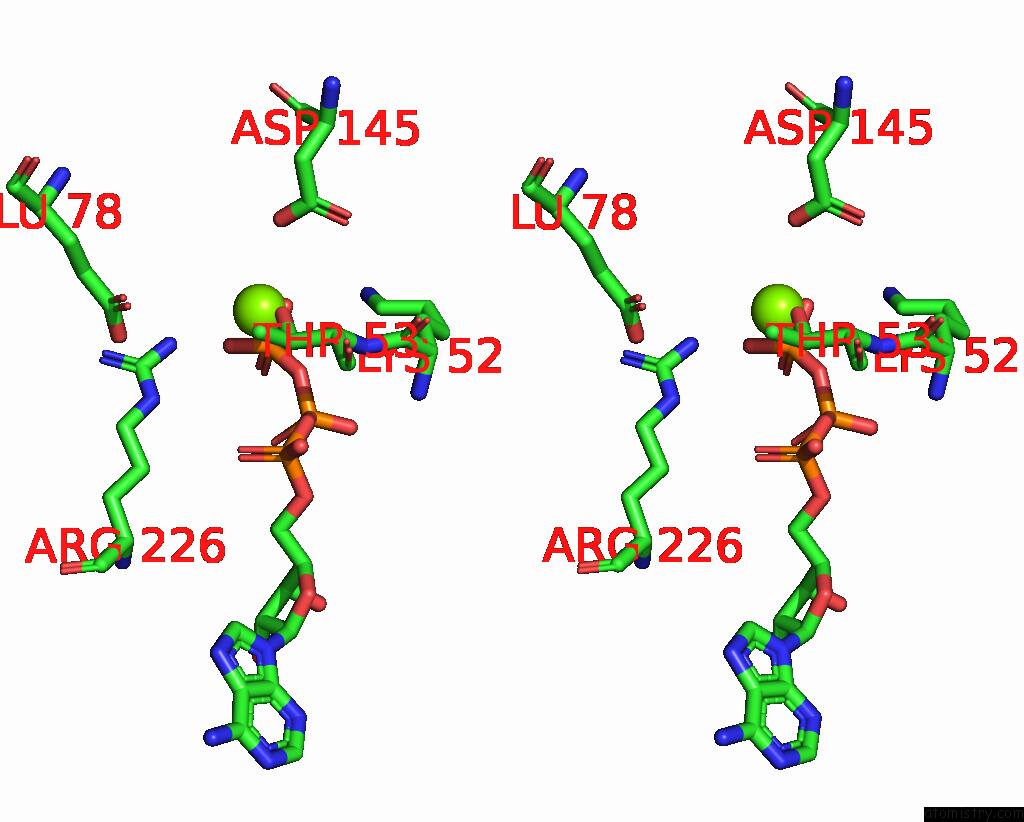
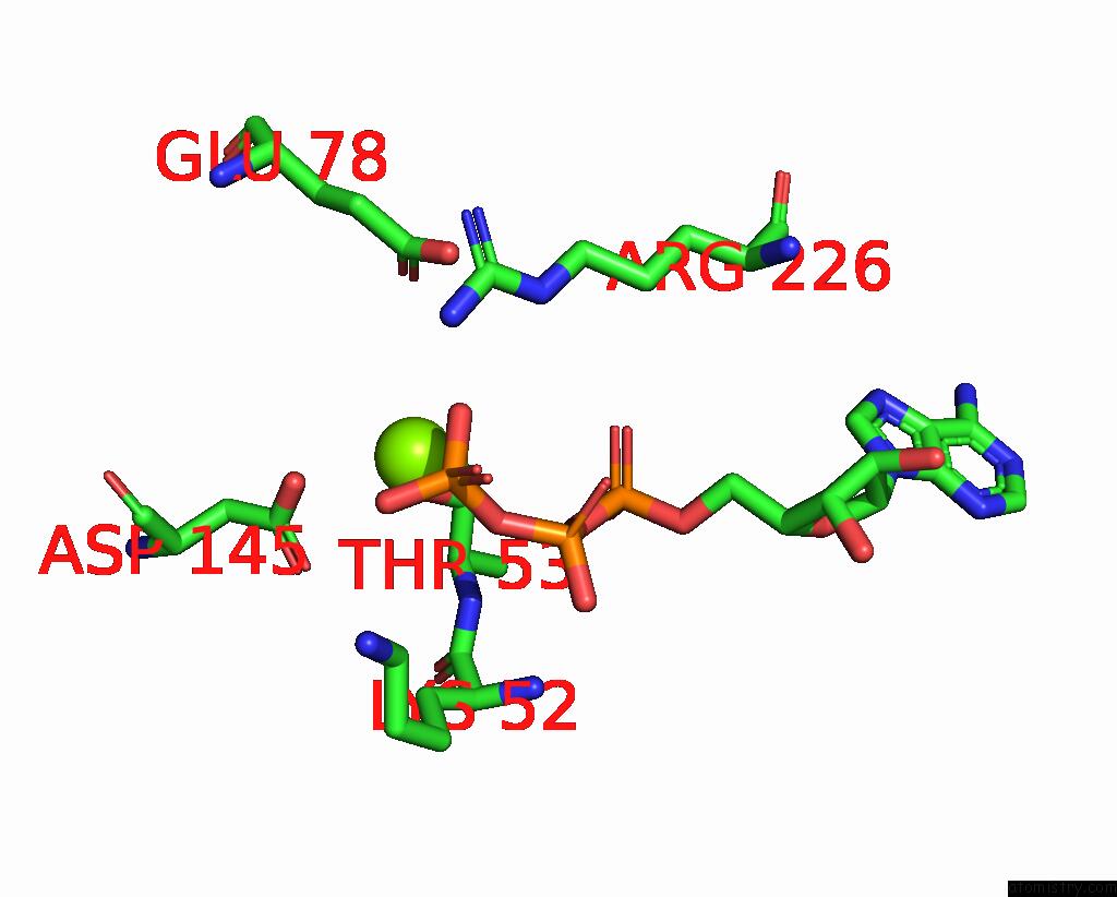
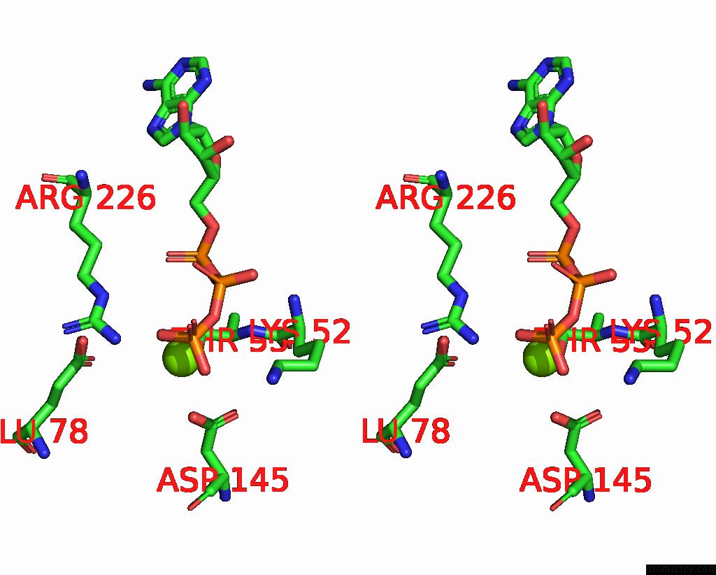
Magnesium binding site 1 out of 12 in 7x1z

Go back to Magnesium Binding Sites List in 7x1z
Magnesium binding site 1 out of 12 in the Structure of the Phosphorylation-Site Double Mutant S431E/T432E of the Kaic Circadian Clock Protein


Mono view


Stereo pair view

A full contact list of Magnesium with other atoms in the Mg binding site number 1 of Structure of the Phosphorylation-Site Double Mutant S431E/T432E of the Kaic Circadian Clock Protein within 5.0Å range:
probe atom residue distance (Å) B Occ
A:Mg903

b:64.0
occ:1.00
OG1 A:THR295 2.1 59.0 1.0
O3G A:ATP901 2.1 63.0 1.0
O1G A:ATP901 2.2 63.0 1.0
PG A:ATP901 2.5 63.0 1.0
OE2 A:GLU318 2.9 67.9 1.0
OD1 A:ASP378 3.2 56.6 1.0
O3B A:ATP901 3.2 63.0 1.0
CB A:THR295 3.5 59.0 1.0
OD2 A:ASP378 3.8 56.6 1.0
CG A:ASP378 3.9 56.6 1.0
O2G A:ATP901 3.9 63.0 1.0
CD A:GLU318 3.9 67.9 1.0
CG2 A:THR295 4.3 59.0 1.0
CG A:GLU318 4.3 67.9 1.0
O3A A:ATP901 4.3 63.0 1.0
CA A:THR295 4.4 59.0 1.0
N A:THR295 4.4 59.0 1.0
PB A:ATP901 4.5 63.0 1.0
OE1 B:GLU432 4.7 66.2 1.0
OE2 B:GLU432 4.7 66.2 1.0
OE2 A:GLU319 4.9 71.3 1.0

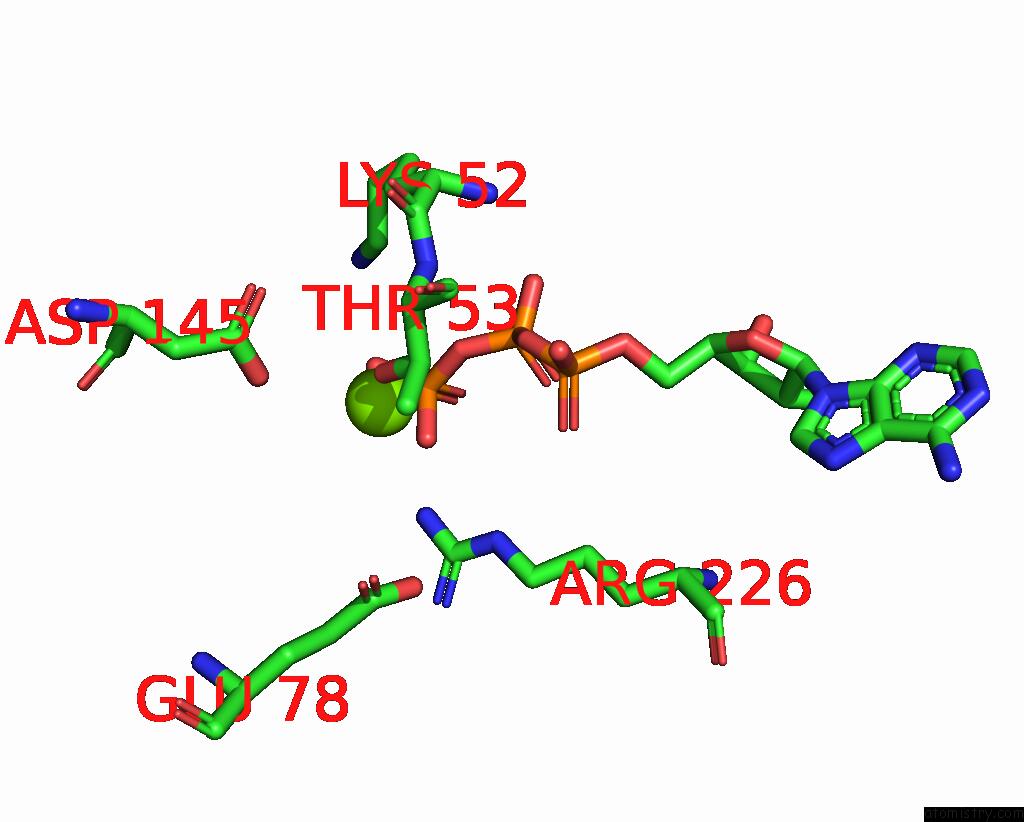
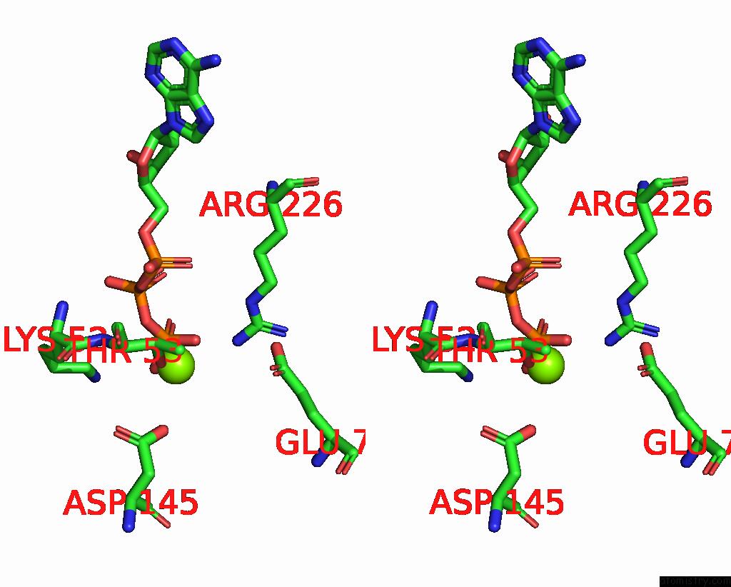
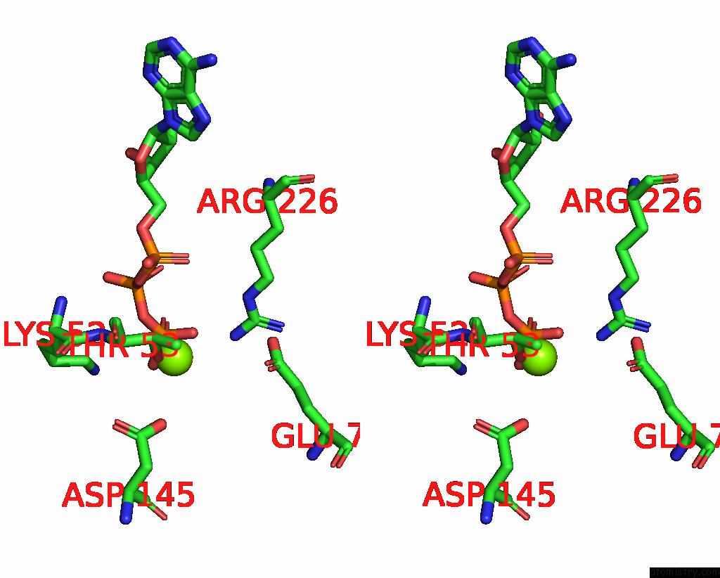
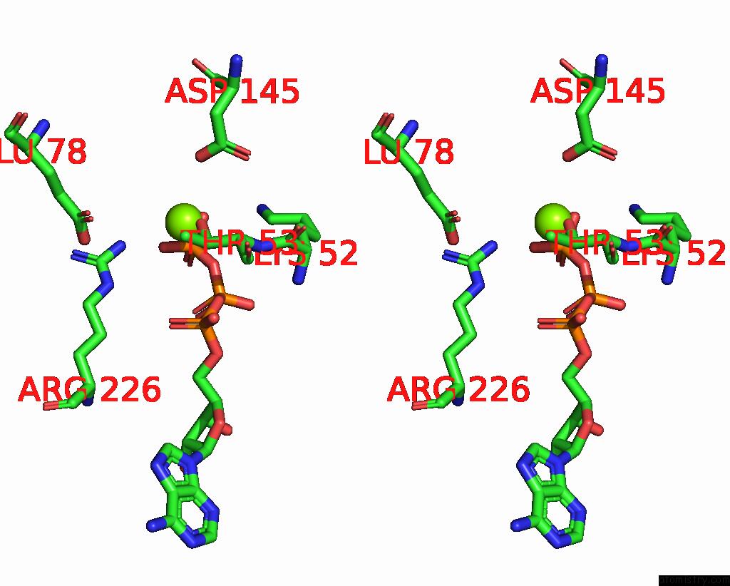
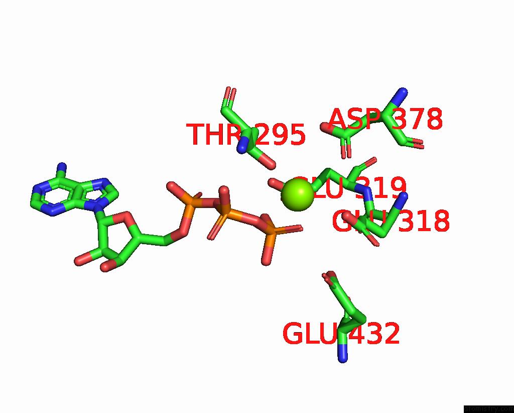
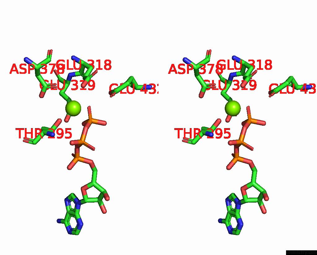
Magnesium binding site 2 out of 12 in 7x1z

Go back to Magnesium Binding Sites List in 7x1z
Magnesium binding site 2 out of 12 in the Structure of the Phosphorylation-Site Double Mutant S431E/T432E of the Kaic Circadian Clock Protein


Mono view


Stereo pair view

A full contact list of Magnesium with other atoms in the Mg binding site number 2 of Structure of the Phosphorylation-Site Double Mutant S431E/T432E of the Kaic Circadian Clock Protein within 5.0Å range:
probe atom residue distance (Å) B Occ
A:Mg904

b:53.8
occ:1.00
OG1 A:THR53 2.1 49.3 1.0
O2G A:ATP902 2.1 52.5 1.0
PG A:ATP902 2.6 52.5 1.0
O1G A:ATP902 2.7 52.5 1.0
O3B A:ATP902 2.9 52.5 1.0
CB A:THR53 3.4 49.3 1.0
OD2 A:ASP145 3.9 64.6 1.0
O3A A:ATP902 4.0 52.5 1.0
O3G A:ATP902 4.1 52.5 1.0
PB A:ATP902 4.1 52.5 1.0
NH2 B:ARG226 4.2 53.4 1.0
CG2 A:THR53 4.2 49.3 1.0
N A:THR53 4.3 49.3 1.0
CA A:THR53 4.4 49.3 1.0
OE2 A:GLU78 4.8 75.3 1.0
O2B A:ATP902 4.8 52.5 1.0
CE A:LYS52 4.8 46.7 1.0
CZ B:ARG226 4.9 53.4 1.0
NE B:ARG226 4.9 53.4 1.0
O2A A:ATP902 4.9 52.5 1.0
CG A:ASP145 4.9 64.6 1.0
PA A:ATP902 5.0 52.5 1.0

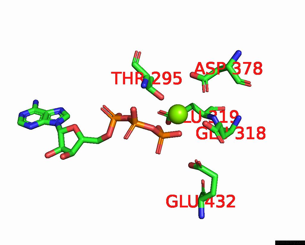
Magnesium binding site 3 out of 12 in 7x1z

Go back to Magnesium Binding Sites List in 7x1z
Magnesium binding site 3 out of 12 in the Structure of the Phosphorylation-Site Double Mutant S431E/T432E of the Kaic Circadian Clock Protein


Mono view


Stereo pair view

A full contact list of Magnesium with other atoms in the Mg binding site number 3 of Structure of the Phosphorylation-Site Double Mutant S431E/T432E of the Kaic Circadian Clock Protein within 5.0Å range:
probe atom residue distance (Å) B Occ
B:Mg903

b:68.5
occ:1.00
OG1 B:THR295 2.1 61.4 1.0
O3G B:ATP901 2.1 64.2 1.0
O1G B:ATP901 2.2 64.2 1.0
PG B:ATP901 2.5 64.2 1.0
OE2 B:GLU318 2.8 69.2 1.0
O3B B:ATP901 3.3 64.2 1.0
OD1 B:ASP378 3.3 58.3 1.0
CB B:THR295 3.5 61.4 1.0
OD2 B:ASP378 3.7 58.3 1.0
CD B:GLU318 3.9 69.2 1.0
O2G B:ATP901 3.9 64.2 1.0
CG B:ASP378 3.9 58.3 1.0
CG2 B:THR295 4.2 61.4 1.0
O3A B:ATP901 4.3 64.2 1.0
CG B:GLU318 4.4 69.2 1.0
CA B:THR295 4.4 61.4 1.0
N B:THR295 4.5 61.4 1.0
PB B:ATP901 4.5 64.2 1.0
OE2 C:GLU432 4.6 67.0 1.0
OE2 B:GLU319 4.7 72.8 1.0
OE1 C:GLU432 4.8 67.0 1.0
OE1 B:GLU318 4.9 69.2 1.0

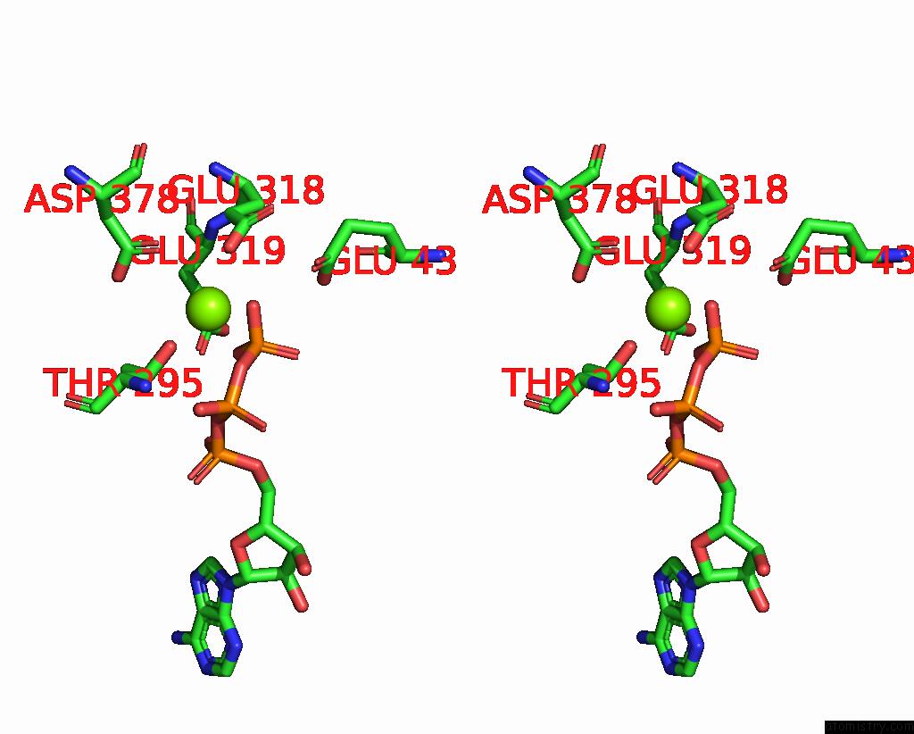
Magnesium binding site 4 out of 12 in 7x1z

Go back to Magnesium Binding Sites List in 7x1z
Magnesium binding site 4 out of 12 in the Structure of the Phosphorylation-Site Double Mutant S431E/T432E of the Kaic Circadian Clock Protein


Mono view


Stereo pair view

A full contact list of Magnesium with other atoms in the Mg binding site number 4 of Structure of the Phosphorylation-Site Double Mutant S431E/T432E of the Kaic Circadian Clock Protein within 5.0Å range:
probe atom residue distance (Å) B Occ
B:Mg904

b:53.5
occ:1.00
OG1 B:THR53 2.1 51.3 1.0
O2G B:ATP902 2.1 53.7 1.0
PG B:ATP902 2.5 53.7 1.0
O1G B:ATP902 2.5 53.7 1.0
O3B B:ATP902 2.8 53.7 1.0
CB B:THR53 3.4 51.3 1.0
O3A B:ATP902 4.0 53.7 1.0
OD2 B:ASP145 4.0 64.2 1.0
O3G B:ATP902 4.0 53.7 1.0
PB B:ATP902 4.1 53.7 1.0
NH2 C:ARG226 4.2 54.1 1.0
CG2 B:THR53 4.2 51.3 1.0
N B:THR53 4.3 51.3 1.0
CA B:THR53 4.4 51.3 1.0
O2B B:ATP902 4.7 53.7 1.0
OE2 B:GLU78 4.7 73.9 1.0
O2A B:ATP902 4.8 53.7 1.0
NE C:ARG226 4.8 54.1 1.0
CZ C:ARG226 4.8 54.1 1.0
CE B:LYS52 4.9 47.8 1.0
PA B:ATP902 4.9 53.7 1.0

Magnesium binding site 5 out of 12 in 7x1z

Go back to Magnesium Binding Sites List in 7x1z
Magnesium binding site 5 out of 12 in the Structure of the Phosphorylation-Site Double Mutant S431E/T432E of the Kaic Circadian Clock Protein


Mono view


Stereo pair view

A full contact list of Magnesium with other atoms in the Mg binding site number 5 of Structure of the Phosphorylation-Site Double Mutant S431E/T432E of the Kaic Circadian Clock Protein within 5.0Å range:
probe atom residue distance (Å) B Occ
C:Mg903

b:63.2
occ:1.00
OG1 C:THR295 2.1 57.9 1.0
OE1 C:GLU318 2.1 66.8 1.0
O1G C:ATP901 2.2 63.1 1.0
O3G C:ATP901 2.2 63.1 1.0
PG C:ATP901 2.5 63.1 1.0
O3B C:ATP901 3.0 63.1 1.0
OD1 C:ASP378 3.3 57.6 1.0
CD C:GLU318 3.3 66.8 1.0
CB C:THR295 3.5 57.9 1.0
OD2 C:ASP378 3.9 57.6 1.0
O2G C:ATP901 3.9 63.1 1.0
CG C:ASP378 4.0 57.6 1.0
OE2 C:GLU318 4.1 66.8 1.0
N C:THR295 4.2 57.9 1.0
O3A C:ATP901 4.2 63.1 1.0
PB C:ATP901 4.3 63.1 1.0
CA C:THR295 4.3 57.9 1.0
CG C:GLU318 4.3 66.8 1.0
CG2 C:THR295 4.4 57.9 1.0
OE1 D:GLU432 4.7 67.4 1.0
O1B C:ATP901 4.8 63.1 1.0
OE2 D:GLU432 4.9 67.4 1.0

Magnesium binding site 6 out of 12 in 7x1z

Go back to Magnesium Binding Sites List in 7x1z
Magnesium binding site 6 out of 12 in the Structure of the Phosphorylation-Site Double Mutant S431E/T432E of the Kaic Circadian Clock Protein


Mono view


Stereo pair view

A full contact list of Magnesium with other atoms in the Mg binding site number 6 of Structure of the Phosphorylation-Site Double Mutant S431E/T432E of the Kaic Circadian Clock Protein within 5.0Å range:
probe atom residue distance (Å) B Occ
C:Mg904

b:53.2
occ:1.00
OG1 C:THR53 2.1 50.1 1.0
O2G C:ATP902 2.1 53.5 1.0
PG C:ATP902 2.6 53.5 1.0
O1G C:ATP902 2.7 53.5 1.0
O3B C:ATP902 2.8 53.5 1.0
CB C:THR53 3.4 50.1 1.0
OD2 C:ASP145 3.9 64.1 1.0
O3A C:ATP902 4.0 53.5 1.0
O3G C:ATP902 4.1 53.5 1.0
PB C:ATP902 4.1 53.5 1.0
NH2 D:ARG226 4.2 53.6 1.0
CG2 C:THR53 4.2 50.1 1.0
N C:THR53 4.3 50.1 1.0
CA C:THR53 4.4 50.1 1.0
O2B C:ATP902 4.7 53.5 1.0
OE2 C:GLU78 4.7 77.0 1.0
O2A C:ATP902 4.8 53.5 1.0
CZ D:ARG226 4.8 53.6 1.0
NE D:ARG226 4.8 53.6 1.0
CE C:LYS52 4.9 47.9 1.0
PA C:ATP902 4.9 53.5 1.0
CG C:ASP145 5.0 64.1 1.0

Magnesium binding site 7 out of 12 in 7x1z

Go back to Magnesium Binding Sites List in 7x1z
Magnesium binding site 7 out of 12 in the Structure of the Phosphorylation-Site Double Mutant S431E/T432E of the Kaic Circadian Clock Protein


Mono view


Stereo pair view

A full contact list of Magnesium with other atoms in the Mg binding site number 7 of Structure of the Phosphorylation-Site Double Mutant S431E/T432E of the Kaic Circadian Clock Protein within 5.0Å range:
probe atom residue distance (Å) B Occ
D:Mg903

b:63.6
occ:1.00
OG1 D:THR295 2.1 59.9 1.0
O3G D:ATP901 2.1 63.3 1.0
O1G D:ATP901 2.2 63.3 1.0
PG D:ATP901 2.5 63.3 1.0
OE2 D:GLU318 2.6 67.5 1.0
OD1 D:ASP378 3.2 57.2 1.0
O3B D:ATP901 3.3 63.3 1.0
CB D:THR295 3.5 59.9 1.0
CD D:GLU318 3.7 67.5 1.0
OD2 D:ASP378 3.8 57.2 1.0
CG D:ASP378 3.9 57.2 1.0
O2G D:ATP901 3.9 63.3 1.0
CG2 D:THR295 4.3 59.9 1.0
CG D:GLU318 4.3 67.5 1.0
O3A D:ATP901 4.3 63.3 1.0
CA D:THR295 4.4 59.9 1.0
N D:THR295 4.4 59.9 1.0
PB D:ATP901 4.5 63.3 1.0
OE1 D:GLU318 4.6 67.5 1.0
OE1 E:GLU432 4.7 66.3 1.0
OE2 E:GLU432 4.7 66.3 1.0
OE1 D:GLU319 4.9 71.3 1.0

Magnesium binding site 8 out of 12 in 7x1z

Go back to Magnesium Binding Sites List in 7x1z
Magnesium binding site 8 out of 12 in the Structure of the Phosphorylation-Site Double Mutant S431E/T432E of the Kaic Circadian Clock Protein


Mono view


Stereo pair view

A full contact list of Magnesium with other atoms in the Mg binding site number 8 of Structure of the Phosphorylation-Site Double Mutant S431E/T432E of the Kaic Circadian Clock Protein within 5.0Å range:
probe atom residue distance (Å) B Occ
D:Mg904

b:53.5
occ:1.00
OG1 D:THR53 2.1 49.8 1.0
O2G D:ATP902 2.1 52.4 1.0
PG D:ATP902 2.6 52.4 1.0
O1G D:ATP902 2.7 52.4 1.0
O3B D:ATP902 2.9 52.4 1.0
CB D:THR53 3.4 49.8 1.0
OD2 D:ASP145 3.9 65.5 1.0
O3A D:ATP902 4.0 52.4 1.0
O3G D:ATP902 4.1 52.4 1.0
PB D:ATP902 4.1 52.4 1.0
NH2 E:ARG226 4.2 54.2 1.0
CG2 D:THR53 4.2 49.8 1.0
N D:THR53 4.3 49.8 1.0
CA D:THR53 4.4 49.8 1.0
O2B D:ATP902 4.8 52.4 1.0
OE2 D:GLU78 4.8 76.3 1.0
CE D:LYS52 4.8 47.3 1.0
CZ E:ARG226 4.9 54.2 1.0
O2A D:ATP902 4.9 52.4 1.0
NE E:ARG226 4.9 54.2 1.0
CG D:ASP145 5.0 65.5 1.0
PA D:ATP902 5.0 52.4 1.0

Magnesium binding site 9 out of 12 in 7x1z

Go back to Magnesium Binding Sites List in 7x1z
Magnesium binding site 9 out of 12 in the Structure of the Phosphorylation-Site Double Mutant S431E/T432E of the Kaic Circadian Clock Protein


Mono view


Stereo pair view

A full contact list of Magnesium with other atoms in the Mg binding site number 9 of Structure of the Phosphorylation-Site Double Mutant S431E/T432E of the Kaic Circadian Clock Protein within 5.0Å range:
probe atom residue distance (Å) B Occ
E:Mg903

b:68.0
occ:1.00
OG1 E:THR295 2.1 61.2 1.0
O3G E:ATP901 2.1 64.6 1.0
O1G E:ATP901 2.2 64.6 1.0
PG E:ATP901 2.5 64.6 1.0
OE2 E:GLU318 2.8 69.2 1.0
O3B E:ATP901 3.3 64.6 1.0
OD1 E:ASP378 3.4 59.3 1.0
CB E:THR295 3.5 61.2 1.0
OD2 E:ASP378 3.8 59.3 1.0
CD E:GLU318 3.9 69.2 1.0
O2G E:ATP901 3.9 64.6 1.0
CG E:ASP378 4.0 59.3 1.0
CG2 E:THR295 4.2 61.2 1.0
O3A E:ATP901 4.3 64.6 1.0
CA E:THR295 4.4 61.2 1.0
CG E:GLU318 4.4 69.2 1.0
N E:THR295 4.4 61.2 1.0
PB E:ATP901 4.5 64.6 1.0
OE2 F:GLU432 4.6 68.4 1.0
OE1 F:GLU432 4.8 68.4 1.0
OE2 E:GLU319 4.9 72.0 1.0
OE1 E:GLU318 4.9 69.2 1.0

Magnesium binding site 10 out of 12 in 7x1z

Go back to Magnesium Binding Sites List in 7x1z
Magnesium binding site 10 out of 12 in the Structure of the Phosphorylation-Site Double Mutant S431E/T432E of the Kaic Circadian Clock Protein


Mono view


Stereo pair view

A full contact list of Magnesium with other atoms in the Mg binding site number 10 of Structure of the Phosphorylation-Site Double Mutant S431E/T432E of the Kaic Circadian Clock Protein within 5.0Å range:
probe atom residue distance (Å) B Occ
E:Mg904

b:53.4
occ:1.00
OG1 E:THR53 2.1 51.4 1.0
O2G E:ATP902 2.1 54.2 1.0
PG E:ATP902 2.5 54.2 1.0
O1G E:ATP902 2.6 54.2 1.0
O3B E:ATP902 2.8 54.2 1.0
CB E:THR53 3.4 51.4 1.0
O3A E:ATP902 4.0 54.2 1.0
OD2 E:ASP145 4.0 64.9 1.0
O3G E:ATP902 4.0 54.2 1.0
PB E:ATP902 4.1 54.2 1.0
NH2 F:ARG226 4.2 54.7 1.0
CG2 E:THR53 4.2 51.4 1.0
N E:THR53 4.3 51.4 1.0
CA E:THR53 4.4 51.4 1.0
O2B E:ATP902 4.7 54.2 1.0
OE2 E:GLU78 4.7 75.1 1.0
O2A E:ATP902 4.8 54.2 1.0
NE F:ARG226 4.8 54.7 1.0
CZ F:ARG226 4.8 54.7 1.0
CE E:LYS52 4.9 48.2 1.0
PA E:ATP902 4.9 54.2 1.0

Reference:

X.Han, D.L.Zhang, L.Hong, D.Q.Yu, Z.L.Wu, T.Yang, M.J.Rust, Y.H.Tu, Q.Ouyang. A Cooperative Switch Within the Kaic Hexamer Revealed By Cryo-Em To Be Published.
Page generated: Tue Apr 25 22:25:19 2023

Last articles

Zn in 8WB0
Zn in 8WAX
Zn in 8WAU
Zn in 8WAZ
Zn in 8WAY
Zn in 8WAV
Zn in 8WAW
Zn in 8WAT
Zn in 8W7M
Zn in 8WD3
© Copyright 2008-2020 by atomistry.com
Home   |    Site Map   |    Copyright   |    Contact us   |    Privacy