Magnesium »
PDB 7ygb-7yv1 »
7yqa »
Magnesium in PDB 7yqa: Crystal Structure of D-Threonine Aldolase From Chlamydomonas Reinhardtii
Enzymatic activity of Crystal Structure of D-Threonine Aldolase From Chlamydomonas Reinhardtii
All present enzymatic activity of Crystal Structure of D-Threonine Aldolase From Chlamydomonas Reinhardtii:
4.1.2.42;
4.1.2.42;
Protein crystallography data
The structure of Crystal Structure of D-Threonine Aldolase From Chlamydomonas Reinhardtii, PDB code: 7yqa
was solved by
Y.Hirato,
M.Goto,
T.Mizobuchi,
H.Muramatsu,
M.Tanigawa,
K.Nishimura,
with X-Ray Crystallography technique. A brief refinement statistics is given in the table below:
| Resolution Low / High (Å) | 83.46 / 1.85 |
| Space group | P 1 |
| Cell size a, b, c (Å), α, β, γ (°) | 64.792, 74.096, 89.939, 77.07, 69.34, 71.93 |
| R / Rfree (%) | 21.7 / 24.9 |
Magnesium Binding Sites:
The binding sites of Magnesium atom in the Crystal Structure of D-Threonine Aldolase From Chlamydomonas Reinhardtii
(pdb code 7yqa). This binding sites where shown within
5.0 Angstroms radius around Magnesium atom.
In total 4 binding sites of Magnesium where determined in the Crystal Structure of D-Threonine Aldolase From Chlamydomonas Reinhardtii, PDB code: 7yqa:
Jump to Magnesium binding site number: 1; 2; 3; 4;
In total 4 binding sites of Magnesium where determined in the Crystal Structure of D-Threonine Aldolase From Chlamydomonas Reinhardtii, PDB code: 7yqa:
Jump to Magnesium binding site number: 1; 2; 3; 4;
Magnesium binding site 1 out of 4 in 7yqa
Go back to
Magnesium binding site 1 out
of 4 in the Crystal Structure of D-Threonine Aldolase From Chlamydomonas Reinhardtii

Mono view
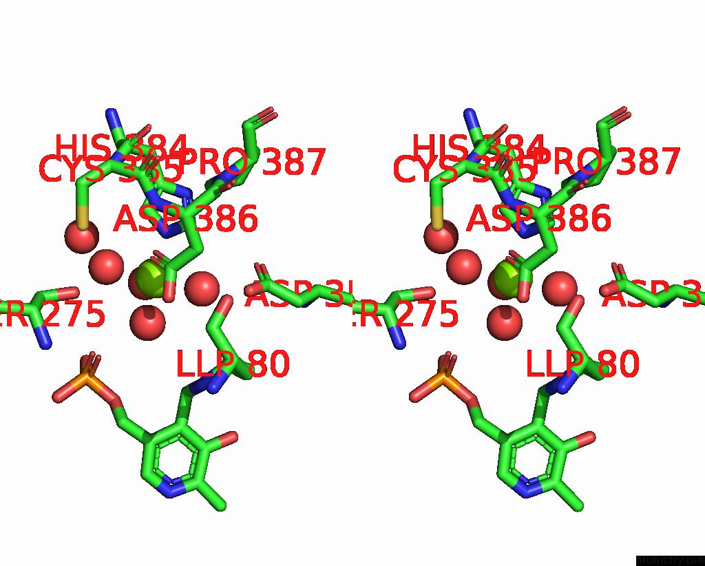
Stereo pair view
Mono view
Stereo pair view
A full contact list of Magnesium with other atoms in the Mg binding
site number 1 of Crystal Structure of D-Threonine Aldolase From Chlamydomonas Reinhardtii within 5.0Å range:
|
Magnesium binding site 2 out of 4 in 7yqa
Go back to
Magnesium binding site 2 out
of 4 in the Crystal Structure of D-Threonine Aldolase From Chlamydomonas Reinhardtii

Mono view
Stereo pair view
Mono view
Stereo pair view
A full contact list of Magnesium with other atoms in the Mg binding
site number 2 of Crystal Structure of D-Threonine Aldolase From Chlamydomonas Reinhardtii within 5.0Å range:
|
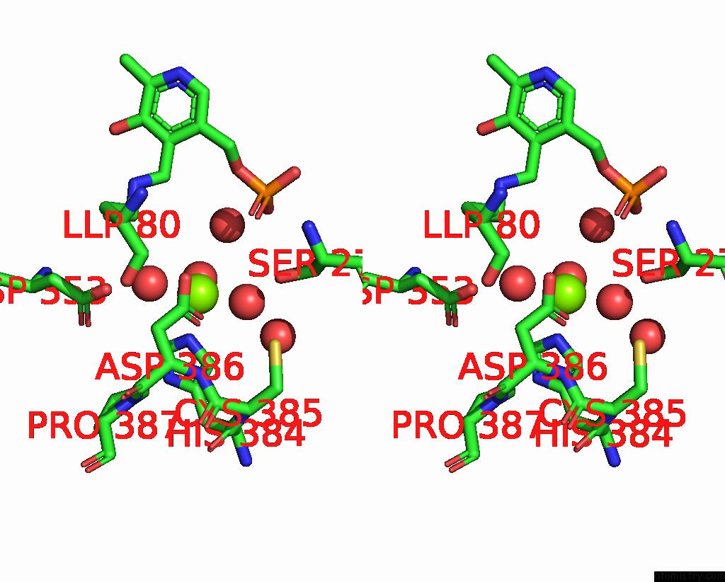
Magnesium binding site 3 out of 4 in 7yqa
Go back to
Magnesium binding site 3 out
of 4 in the Crystal Structure of D-Threonine Aldolase From Chlamydomonas Reinhardtii

Mono view

Stereo pair view
Mono view
Stereo pair view
A full contact list of Magnesium with other atoms in the Mg binding
site number 3 of Crystal Structure of D-Threonine Aldolase From Chlamydomonas Reinhardtii within 5.0Å range:
|
Magnesium binding site 4 out of 4 in 7yqa
Go back to
Magnesium binding site 4 out
of 4 in the Crystal Structure of D-Threonine Aldolase From Chlamydomonas Reinhardtii

Mono view

Stereo pair view
Mono view
Stereo pair view
A full contact list of Magnesium with other atoms in the Mg binding
site number 4 of Crystal Structure of D-Threonine Aldolase From Chlamydomonas Reinhardtii within 5.0Å range:
|
Reference:
Y.Hirato,
M.Goto,
T.Mizobuchi,
H.Muramatsu,
M.Tanigawa,
K.Nishimura.
Structure of Pyridoxal 5'-Phosphate-Bound D-Threonine Aldolase From Chlamydomonas Reinhardtii. Acta Crystallogr.,Sect.F V. 79 31 2023.
ISSN: ESSN 2053-230X
PubMed: 36748339
DOI: 10.1107/S2053230X23000304
Page generated: Thu Oct 3 15:38:55 2024
ISSN: ESSN 2053-230X
PubMed: 36748339
DOI: 10.1107/S2053230X23000304
Last articles
Cl in 7SS8Cl in 7SR6
Cl in 7SS7
Cl in 7SS6
Cl in 7SQB
Cl in 7SQA
Cl in 7SPP
Cl in 7SQ3
Cl in 7SPG
Cl in 7SP3