Magnesium »
PDB 7zmj-7zz1 »
7zvn »
Magnesium in PDB 7zvn: Crystal Structure of Human Annexin A2 in Complex with Full Phosphorothioate 5-10 2'-Methoxyethyl Dna Gapmer Antisense Oligonucleotide Solved at 1.87 A Resolution
Protein crystallography data
The structure of Crystal Structure of Human Annexin A2 in Complex with Full Phosphorothioate 5-10 2'-Methoxyethyl Dna Gapmer Antisense Oligonucleotide Solved at 1.87 A Resolution, PDB code: 7zvn
was solved by
M.Hyjek-Skladanowska,
B.Anderson,
V.Mykhaylyk,
C.Orr,
A.Wagner,
K.Skowronek,
P.Seth,
M.Nowotny,
with X-Ray Crystallography technique. A brief refinement statistics is given in the table below:
| Resolution Low / High (Å) | 43.75 / 1.87 |
| Space group | P 1 21 1 |
| Cell size a, b, c (Å), α, β, γ (°) | 55.621, 57.502, 70.478, 90, 90.24, 90 |
| R / Rfree (%) | 16.7 / 19.8 |
Other elements in 7zvn:
The structure of Crystal Structure of Human Annexin A2 in Complex with Full Phosphorothioate 5-10 2'-Methoxyethyl Dna Gapmer Antisense Oligonucleotide Solved at 1.87 A Resolution also contains other interesting chemical elements:
| Calcium | (Ca) | 7 atoms |
Magnesium Binding Sites:
The binding sites of Magnesium atom in the Crystal Structure of Human Annexin A2 in Complex with Full Phosphorothioate 5-10 2'-Methoxyethyl Dna Gapmer Antisense Oligonucleotide Solved at 1.87 A Resolution
(pdb code 7zvn). This binding sites where shown within
5.0 Angstroms radius around Magnesium atom.
In total only one binding site of Magnesium was determined in the Crystal Structure of Human Annexin A2 in Complex with Full Phosphorothioate 5-10 2'-Methoxyethyl Dna Gapmer Antisense Oligonucleotide Solved at 1.87 A Resolution, PDB code: 7zvn:
In total only one binding site of Magnesium was determined in the Crystal Structure of Human Annexin A2 in Complex with Full Phosphorothioate 5-10 2'-Methoxyethyl Dna Gapmer Antisense Oligonucleotide Solved at 1.87 A Resolution, PDB code: 7zvn:
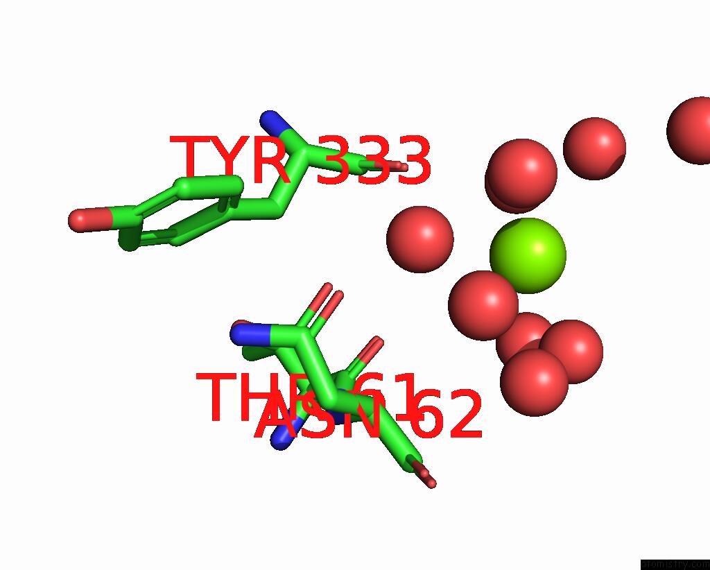
Magnesium binding site 1 out of 1 in 7zvn
Go back to
Magnesium binding site 1 out
of 1 in the Crystal Structure of Human Annexin A2 in Complex with Full Phosphorothioate 5-10 2'-Methoxyethyl Dna Gapmer Antisense Oligonucleotide Solved at 1.87 A Resolution
Mono view

Stereo pair view
Mono view
Stereo pair view
A full contact list of Magnesium with other atoms in the Mg binding
site number 1 of Crystal Structure of Human Annexin A2 in Complex with Full Phosphorothioate 5-10 2'-Methoxyethyl Dna Gapmer Antisense Oligonucleotide Solved at 1.87 A Resolution within 5.0Å range:
|
Reference:
M.Hyjek-Skladanowska,
B.A.Anderson,
V.Mykhaylyk,
C.Orr,
A.Wagner,
J.T.Poznanski,
K.Skowronek,
P.Seth,
M.Nowotny.
Structures of Annexin A2-Ps Dna Complexes Show Dominance of Hydrophobic Interactions in Phosphorothioate Binding. Nucleic Acids Res. 2022.
ISSN: ESSN 1362-4962
PubMed: 36124719
DOI: 10.1093/NAR/GKAC774
Page generated: Thu Oct 3 17:06:04 2024
ISSN: ESSN 1362-4962
PubMed: 36124719
DOI: 10.1093/NAR/GKAC774
Last articles
Zn in 9J0NZn in 9J0O
Zn in 9J0P
Zn in 9FJX
Zn in 9EKB
Zn in 9C0F
Zn in 9CAH
Zn in 9CH0
Zn in 9CH3
Zn in 9CH1