Magnesium in PDB 8bz6: Crystal Structure of the L. Monocytogenes Rmlt in Complex with Udp- Glucose
Protein crystallography data
The structure of Crystal Structure of the L. Monocytogenes Rmlt in Complex with Udp- Glucose, PDB code: 8bz6
was solved by
T.B.Cereija,
J.H.Morais-Cabral,
with X-Ray Crystallography technique. A brief refinement statistics is given in the table below:
Resolution Low / High (Å)
|
44.22 /
2.40
|
Space group
|
P 1 21 1
|
Cell size a, b, c (Å), α, β, γ (°)
|
85.523,
291.057,
89.824,
90,
100.08,
90
|
R / Rfree (%)
|
19.5 /
23.5
|
Other elements in 8bz6:
The structure of Crystal Structure of the L. Monocytogenes Rmlt in Complex with Udp- Glucose also contains other interesting chemical elements:
Magnesium Binding Sites:
The binding sites of Magnesium atom in the Crystal Structure of the L. Monocytogenes Rmlt in Complex with Udp- Glucose
(pdb code 8bz6). This binding sites where shown within
5.0 Angstroms radius around Magnesium atom.
In total 6 binding sites of Magnesium where determined in the
Crystal Structure of the L. Monocytogenes Rmlt in Complex with Udp- Glucose, PDB code: 8bz6:
Jump to Magnesium binding site number:
1;
2;
3;
4;
5;
6;
Magnesium binding site 1 out
of 6 in 8bz6
Go back to
Magnesium Binding Sites List in 8bz6
Magnesium binding site 1 out
of 6 in the Crystal Structure of the L. Monocytogenes Rmlt in Complex with Udp- Glucose
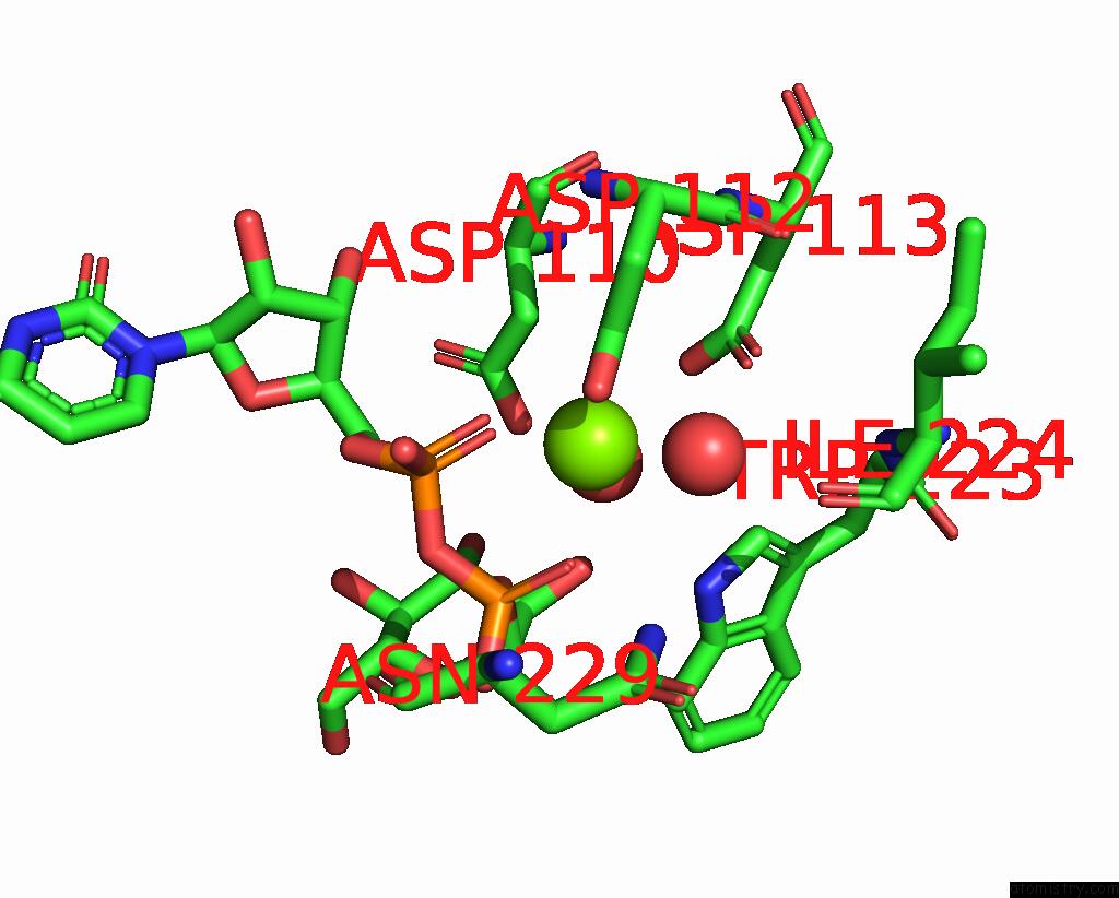
Mono view
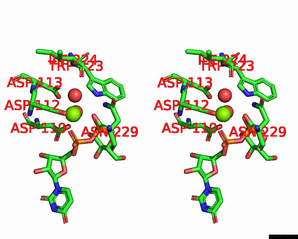
Stereo pair view
|
A full contact list of Magnesium with other atoms in the Mg binding
site number 1 of Crystal Structure of the L. Monocytogenes Rmlt in Complex with Udp- Glucose within 5.0Å range:
probe
|
atom
|
residue
|
distance (Å)
|
B
|
Occ
|
A:Mg703
b:65.5
occ:1.00
|
O2A
|
A:UPG701
|
2.0
|
70.5
|
1.0
|
OD2
|
A:ASP112
|
2.1
|
65.8
|
1.0
|
O
|
A:HOH809
|
2.1
|
59.4
|
1.0
|
O
|
A:HOH891
|
2.2
|
61.9
|
1.0
|
OD1
|
A:ASP112
|
2.2
|
60.0
|
1.0
|
O2B
|
A:UPG701
|
2.3
|
70.2
|
1.0
|
CG
|
A:ASP112
|
2.4
|
64.9
|
1.0
|
PA
|
A:UPG701
|
3.1
|
75.5
|
1.0
|
PB
|
A:UPG701
|
3.3
|
80.1
|
1.0
|
O3A
|
A:UPG701
|
3.5
|
80.5
|
1.0
|
O3B
|
A:UPG701
|
3.7
|
78.5
|
1.0
|
OD1
|
A:ASP113
|
3.7
|
58.4
|
1.0
|
O1A
|
A:UPG701
|
3.9
|
82.4
|
1.0
|
CB
|
A:ASP112
|
3.9
|
58.2
|
1.0
|
NE1
|
A:TRP223
|
4.1
|
60.9
|
1.0
|
OD2
|
A:ASP113
|
4.2
|
55.4
|
1.0
|
CG
|
A:ASP113
|
4.3
|
56.7
|
1.0
|
CB
|
A:ASP110
|
4.3
|
61.6
|
1.0
|
CD1
|
A:TRP223
|
4.4
|
56.9
|
1.0
|
O5C
|
A:UPG701
|
4.4
|
75.4
|
1.0
|
ND2
|
A:ASN229
|
4.5
|
84.8
|
1.0
|
O
|
A:ILE224
|
4.6
|
56.7
|
1.0
|
OD2
|
A:ASP110
|
4.6
|
65.8
|
1.0
|
C5C
|
A:UPG701
|
4.6
|
74.3
|
1.0
|
O2'
|
A:UPG701
|
4.7
|
72.5
|
1.0
|
O1B
|
A:UPG701
|
4.7
|
85.7
|
1.0
|
CA
|
A:ASP112
|
4.8
|
59.5
|
1.0
|
N
|
A:ASP112
|
4.8
|
63.3
|
1.0
|
CG
|
A:ASP110
|
4.9
|
64.5
|
1.0
|
C1'
|
A:UPG701
|
4.9
|
76.6
|
1.0
|
C
|
A:ASP112
|
4.9
|
58.0
|
1.0
|
|
Magnesium binding site 2 out
of 6 in 8bz6
Go back to
Magnesium Binding Sites List in 8bz6
Magnesium binding site 2 out
of 6 in the Crystal Structure of the L. Monocytogenes Rmlt in Complex with Udp- Glucose
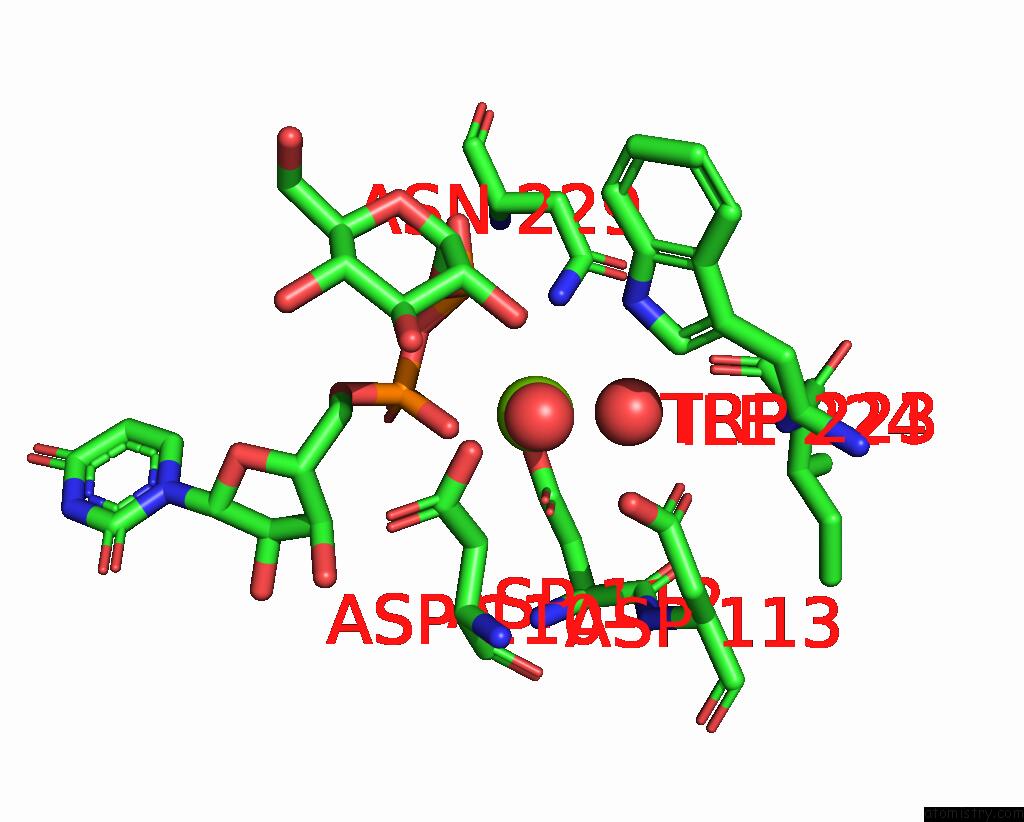
Mono view
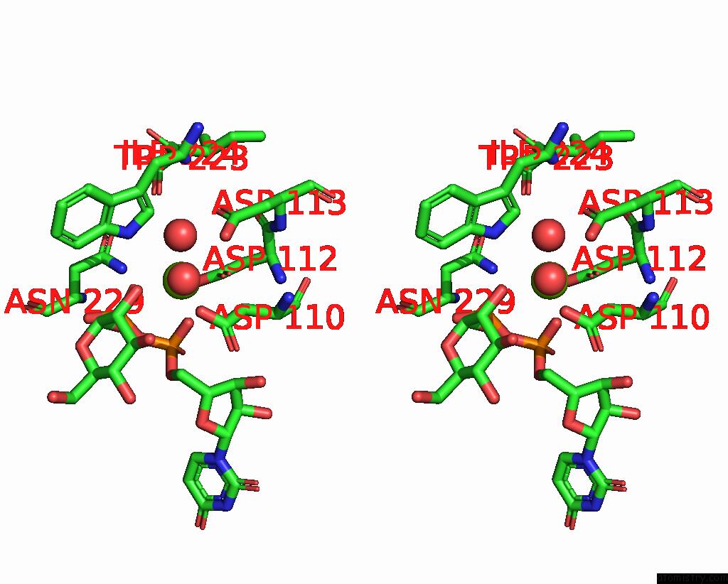
Stereo pair view
|
A full contact list of Magnesium with other atoms in the Mg binding
site number 2 of Crystal Structure of the L. Monocytogenes Rmlt in Complex with Udp- Glucose within 5.0Å range:
probe
|
atom
|
residue
|
distance (Å)
|
B
|
Occ
|
B:Mg702
b:66.6
occ:1.00
|
O1A
|
B:UPG701
|
2.0
|
66.4
|
1.0
|
OD2
|
B:ASP112
|
2.1
|
69.7
|
1.0
|
O
|
B:HOH831
|
2.1
|
60.4
|
1.0
|
OD1
|
B:ASP112
|
2.1
|
65.4
|
1.0
|
O
|
B:HOH806
|
2.1
|
61.8
|
1.0
|
O2B
|
B:UPG701
|
2.3
|
68.7
|
1.0
|
CG
|
B:ASP112
|
2.4
|
65.9
|
1.0
|
PA
|
B:UPG701
|
3.1
|
65.4
|
1.0
|
PB
|
B:UPG701
|
3.4
|
69.4
|
1.0
|
O3A
|
B:UPG701
|
3.4
|
74.5
|
1.0
|
O2A
|
B:UPG701
|
3.9
|
72.0
|
1.0
|
CB
|
B:ASP112
|
3.9
|
60.6
|
1.0
|
OD1
|
B:ASP113
|
4.0
|
58.9
|
1.0
|
O3B
|
B:UPG701
|
4.0
|
69.0
|
1.0
|
ND2
|
B:ASN229
|
4.0
|
96.0
|
1.0
|
NE1
|
B:TRP223
|
4.1
|
61.7
|
1.0
|
OD2
|
B:ASP113
|
4.3
|
58.6
|
1.0
|
CB
|
B:ASP110
|
4.3
|
63.3
|
1.0
|
CD1
|
B:TRP223
|
4.4
|
60.0
|
1.0
|
CG
|
B:ASP113
|
4.4
|
60.8
|
1.0
|
O5C
|
B:UPG701
|
4.5
|
67.5
|
1.0
|
O
|
B:ILE224
|
4.5
|
69.1
|
1.0
|
O2'
|
B:UPG701
|
4.5
|
71.1
|
1.0
|
OD2
|
B:ASP110
|
4.7
|
64.7
|
1.0
|
O1B
|
B:UPG701
|
4.7
|
79.2
|
1.0
|
C5C
|
B:UPG701
|
4.7
|
69.8
|
1.0
|
CA
|
B:ASP112
|
4.7
|
65.2
|
1.0
|
N
|
B:ASP112
|
4.8
|
67.8
|
1.0
|
C
|
B:ASP112
|
4.9
|
65.0
|
1.0
|
CG
|
B:ASP110
|
4.9
|
63.5
|
1.0
|
|
Magnesium binding site 3 out
of 6 in 8bz6
Go back to
Magnesium Binding Sites List in 8bz6
Magnesium binding site 3 out
of 6 in the Crystal Structure of the L. Monocytogenes Rmlt in Complex with Udp- Glucose
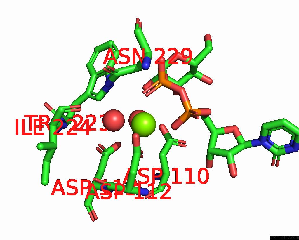
Mono view
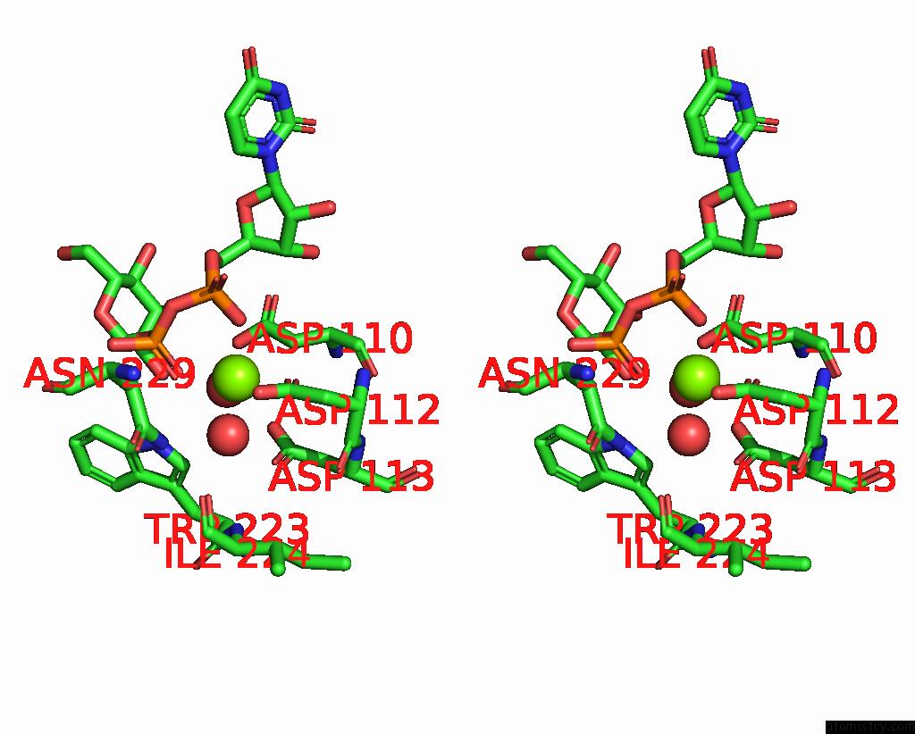
Stereo pair view
|
A full contact list of Magnesium with other atoms in the Mg binding
site number 3 of Crystal Structure of the L. Monocytogenes Rmlt in Complex with Udp- Glucose within 5.0Å range:
probe
|
atom
|
residue
|
distance (Å)
|
B
|
Occ
|
C:Mg703
b:58.6
occ:1.00
|
OD1
|
C:ASP112
|
2.1
|
58.9
|
1.0
|
OD2
|
C:ASP112
|
2.1
|
60.9
|
1.0
|
O
|
C:HOH818
|
2.1
|
54.6
|
1.0
|
O2B
|
C:UPG701
|
2.2
|
64.0
|
1.0
|
O2A
|
C:UPG701
|
2.2
|
59.9
|
1.0
|
O
|
C:HOH817
|
2.2
|
58.2
|
1.0
|
CG
|
C:ASP112
|
2.4
|
58.1
|
1.0
|
PB
|
C:UPG701
|
3.3
|
65.0
|
1.0
|
PA
|
C:UPG701
|
3.4
|
64.8
|
1.0
|
O3A
|
C:UPG701
|
3.6
|
69.1
|
1.0
|
O3B
|
C:UPG701
|
3.7
|
68.6
|
1.0
|
OD1
|
C:ASP113
|
3.8
|
54.8
|
1.0
|
CB
|
C:ASP112
|
3.9
|
56.9
|
1.0
|
OD2
|
C:ASP113
|
4.0
|
53.4
|
1.0
|
CB
|
C:ASP110
|
4.1
|
59.2
|
1.0
|
CG
|
C:ASP113
|
4.2
|
54.1
|
1.0
|
O1A
|
C:UPG701
|
4.3
|
68.3
|
1.0
|
NE1
|
C:TRP223
|
4.3
|
61.5
|
1.0
|
ND2
|
C:ASN229
|
4.5
|
81.9
|
1.0
|
CD1
|
C:TRP223
|
4.5
|
59.7
|
1.0
|
OD2
|
C:ASP110
|
4.5
|
64.2
|
1.0
|
O5C
|
C:UPG701
|
4.6
|
63.3
|
1.0
|
O1B
|
C:UPG701
|
4.6
|
67.5
|
1.0
|
C5C
|
C:UPG701
|
4.7
|
63.7
|
1.0
|
N
|
C:ASP112
|
4.7
|
61.8
|
1.0
|
O
|
C:ILE224
|
4.7
|
61.9
|
1.0
|
CA
|
C:ASP112
|
4.7
|
56.1
|
1.0
|
CG
|
C:ASP110
|
4.7
|
59.4
|
1.0
|
C
|
C:ASP112
|
4.8
|
57.9
|
1.0
|
O2'
|
C:UPG701
|
4.8
|
67.5
|
1.0
|
C1'
|
C:UPG701
|
5.0
|
70.3
|
1.0
|
|
Magnesium binding site 4 out
of 6 in 8bz6
Go back to
Magnesium Binding Sites List in 8bz6
Magnesium binding site 4 out
of 6 in the Crystal Structure of the L. Monocytogenes Rmlt in Complex with Udp- Glucose
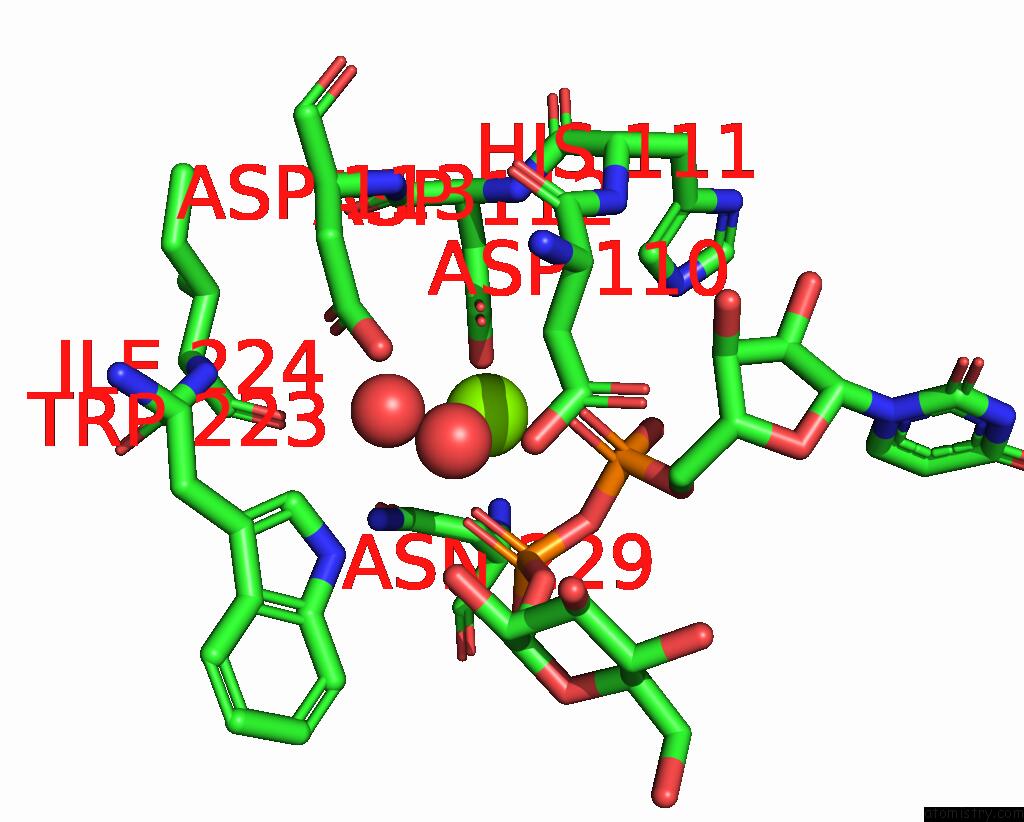
Mono view
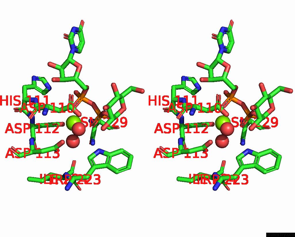
Stereo pair view
|
A full contact list of Magnesium with other atoms in the Mg binding
site number 4 of Crystal Structure of the L. Monocytogenes Rmlt in Complex with Udp- Glucose within 5.0Å range:
probe
|
atom
|
residue
|
distance (Å)
|
B
|
Occ
|
D:Mg703
b:65.2
occ:1.00
|
O2A
|
D:UPG701
|
2.0
|
56.4
|
1.0
|
O
|
D:HOH814
|
2.1
|
55.0
|
1.0
|
OD2
|
D:ASP112
|
2.1
|
68.2
|
1.0
|
OD1
|
D:ASP112
|
2.1
|
57.0
|
1.0
|
O2B
|
D:UPG701
|
2.2
|
61.3
|
1.0
|
O
|
D:HOH827
|
2.3
|
56.1
|
1.0
|
CG
|
D:ASP112
|
2.4
|
64.2
|
1.0
|
PA
|
D:UPG701
|
3.1
|
63.0
|
1.0
|
PB
|
D:UPG701
|
3.2
|
59.5
|
1.0
|
O3A
|
D:UPG701
|
3.2
|
59.5
|
1.0
|
O3B
|
D:UPG701
|
3.8
|
63.4
|
1.0
|
O1A
|
D:UPG701
|
3.9
|
70.0
|
1.0
|
CB
|
D:ASP112
|
3.9
|
57.0
|
1.0
|
OD1
|
D:ASP113
|
4.0
|
57.2
|
1.0
|
ND2
|
D:ASN229
|
4.2
|
78.4
|
1.0
|
OD2
|
D:ASP113
|
4.2
|
53.9
|
1.0
|
O5C
|
D:UPG701
|
4.3
|
60.5
|
1.0
|
CB
|
D:ASP110
|
4.3
|
57.3
|
1.0
|
CG
|
D:ASP113
|
4.4
|
55.8
|
1.0
|
NE1
|
D:TRP223
|
4.5
|
55.0
|
1.0
|
CB
|
D:ASN229
|
4.6
|
86.4
|
1.0
|
O1B
|
D:UPG701
|
4.6
|
62.1
|
1.0
|
C5C
|
D:UPG701
|
4.6
|
61.0
|
1.0
|
O
|
D:ILE224
|
4.7
|
59.9
|
1.0
|
O2'
|
D:UPG701
|
4.7
|
62.3
|
1.0
|
CD1
|
D:TRP223
|
4.7
|
56.1
|
1.0
|
N
|
D:ASP112
|
4.7
|
58.7
|
1.0
|
CG
|
D:ASN229
|
4.7
|
82.4
|
1.0
|
CA
|
D:ASP112
|
4.8
|
60.6
|
1.0
|
CD2
|
D:HIS111
|
4.9
|
67.0
|
1.0
|
C
|
D:ASP112
|
4.9
|
61.4
|
1.0
|
C1'
|
D:UPG701
|
4.9
|
59.9
|
1.0
|
CG
|
D:ASP110
|
5.0
|
58.8
|
1.0
|
OD2
|
D:ASP110
|
5.0
|
61.6
|
1.0
|
|
Magnesium binding site 5 out
of 6 in 8bz6
Go back to
Magnesium Binding Sites List in 8bz6
Magnesium binding site 5 out
of 6 in the Crystal Structure of the L. Monocytogenes Rmlt in Complex with Udp- Glucose
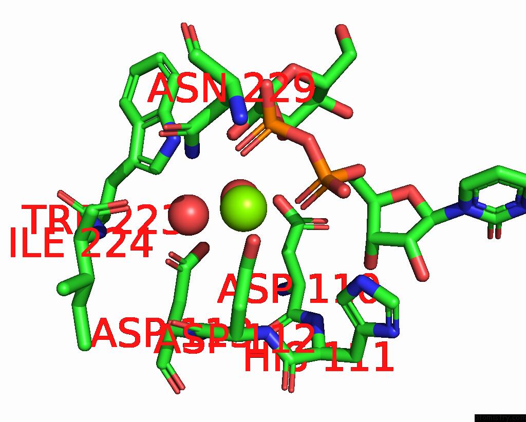
Mono view
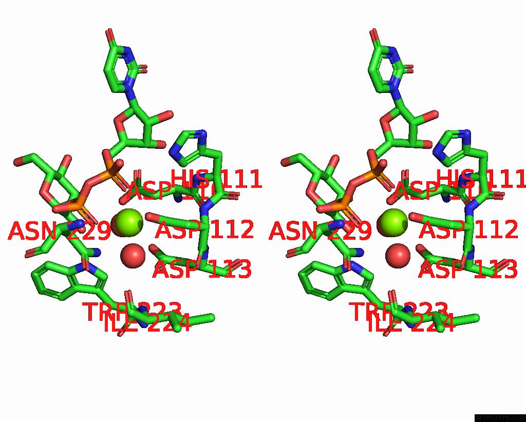
Stereo pair view
|
A full contact list of Magnesium with other atoms in the Mg binding
site number 5 of Crystal Structure of the L. Monocytogenes Rmlt in Complex with Udp- Glucose within 5.0Å range:
probe
|
atom
|
residue
|
distance (Å)
|
B
|
Occ
|
E:Mg703
b:56.9
occ:1.00
|
O
|
E:HOH867
|
2.1
|
53.5
|
1.0
|
OD1
|
E:ASP112
|
2.1
|
54.3
|
1.0
|
OD2
|
E:ASP112
|
2.1
|
59.3
|
1.0
|
O
|
E:HOH806
|
2.1
|
49.0
|
1.0
|
O2B
|
E:UPG701
|
2.2
|
60.4
|
1.0
|
O2A
|
E:UPG701
|
2.2
|
56.8
|
1.0
|
CG
|
E:ASP112
|
2.4
|
55.0
|
1.0
|
PB
|
E:UPG701
|
3.3
|
64.5
|
1.0
|
PA
|
E:UPG701
|
3.3
|
62.6
|
1.0
|
O3A
|
E:UPG701
|
3.5
|
60.2
|
1.0
|
ND2
|
E:ASN229
|
3.7
|
68.1
|
1.0
|
O3B
|
E:UPG701
|
3.8
|
61.3
|
1.0
|
CB
|
E:ASP112
|
3.9
|
54.6
|
1.0
|
OD1
|
E:ASP113
|
4.0
|
51.1
|
1.0
|
O1A
|
E:UPG701
|
4.1
|
60.9
|
1.0
|
NE1
|
E:TRP223
|
4.3
|
59.5
|
1.0
|
OD2
|
E:ASP113
|
4.4
|
54.9
|
1.0
|
CB
|
E:ASP110
|
4.4
|
54.9
|
1.0
|
CD1
|
E:TRP223
|
4.5
|
53.5
|
1.0
|
O
|
E:ILE224
|
4.5
|
52.4
|
1.0
|
CG
|
E:ASP113
|
4.5
|
53.5
|
1.0
|
O5C
|
E:UPG701
|
4.6
|
62.6
|
1.0
|
O1B
|
E:UPG701
|
4.6
|
65.9
|
1.0
|
O2'
|
E:UPG701
|
4.7
|
64.1
|
1.0
|
CA
|
E:ASP112
|
4.7
|
56.0
|
1.0
|
N
|
E:ASP112
|
4.7
|
53.1
|
1.0
|
C5C
|
E:UPG701
|
4.8
|
63.5
|
1.0
|
C
|
E:ASP112
|
4.8
|
50.0
|
1.0
|
CG
|
E:ASN229
|
4.9
|
76.8
|
1.0
|
C1'
|
E:UPG701
|
5.0
|
66.4
|
1.0
|
OD2
|
E:ASP110
|
5.0
|
57.2
|
1.0
|
O
|
E:ASP112
|
5.0
|
51.4
|
1.0
|
CD2
|
E:HIS111
|
5.0
|
60.3
|
1.0
|
|
Magnesium binding site 6 out
of 6 in 8bz6
Go back to
Magnesium Binding Sites List in 8bz6
Magnesium binding site 6 out
of 6 in the Crystal Structure of the L. Monocytogenes Rmlt in Complex with Udp- Glucose
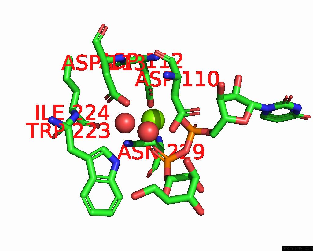
Mono view
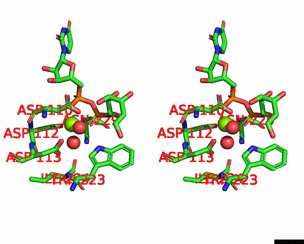
Stereo pair view
|
A full contact list of Magnesium with other atoms in the Mg binding
site number 6 of Crystal Structure of the L. Monocytogenes Rmlt in Complex with Udp- Glucose within 5.0Å range:
probe
|
atom
|
residue
|
distance (Å)
|
B
|
Occ
|
F:Mg703
b:74.0
occ:1.00
|
OD2
|
F:ASP112
|
2.1
|
74.9
|
1.0
|
O
|
F:HOH809
|
2.1
|
70.0
|
1.0
|
O2B
|
F:UPG701
|
2.1
|
83.0
|
1.0
|
O
|
F:HOH829
|
2.1
|
70.7
|
1.0
|
O2A
|
F:UPG701
|
2.1
|
78.8
|
1.0
|
OD1
|
F:ASP112
|
2.1
|
71.0
|
1.0
|
CG
|
F:ASP112
|
2.4
|
74.6
|
1.0
|
PA
|
F:UPG701
|
3.1
|
75.0
|
1.0
|
PB
|
F:UPG701
|
3.3
|
81.1
|
1.0
|
O3A
|
F:UPG701
|
3.4
|
83.0
|
1.0
|
O1A
|
F:UPG701
|
3.8
|
74.8
|
1.0
|
CB
|
F:ASP112
|
3.9
|
76.0
|
1.0
|
ND2
|
F:ASN229
|
3.9
|
113.9
|
1.0
|
OD1
|
F:ASP113
|
4.0
|
67.2
|
1.0
|
OD2
|
F:ASP113
|
4.1
|
68.3
|
1.0
|
O1B
|
F:UPG701
|
4.2
|
95.0
|
1.0
|
CB
|
F:ASP110
|
4.3
|
72.1
|
1.0
|
NE1
|
F:TRP223
|
4.4
|
72.1
|
1.0
|
CG
|
F:ASP113
|
4.4
|
70.1
|
1.0
|
CB
|
F:ASN229
|
4.4
|
122.3
|
1.0
|
O3B
|
F:UPG701
|
4.4
|
80.8
|
1.0
|
O5C
|
F:UPG701
|
4.5
|
80.6
|
1.0
|
O
|
F:ILE224
|
4.5
|
75.0
|
1.0
|
CD1
|
F:TRP223
|
4.6
|
65.9
|
1.0
|
CG
|
F:ASN229
|
4.7
|
118.8
|
1.0
|
OD2
|
F:ASP110
|
4.7
|
74.6
|
1.0
|
CA
|
F:ASP112
|
4.7
|
72.0
|
1.0
|
N
|
F:ASP112
|
4.7
|
78.1
|
1.0
|
C1'
|
F:UPG701
|
4.8
|
82.5
|
1.0
|
C5C
|
F:UPG701
|
4.8
|
74.7
|
1.0
|
C
|
F:ASP112
|
4.9
|
71.7
|
1.0
|
CG
|
F:ASP110
|
4.9
|
72.0
|
1.0
|
|
Reference:
R.Monteiro,
T.B.Cereija,
J.H.Morais-Cabral,
D.Cabanes.
Crystal Structure of the L. Monocytogenes Rmlt in Complex with Udp-Glucose To Be Published.
Page generated: Thu Oct 3 19:56:04 2024
|