Magnesium »
PDB 8c0k-8c6e »
8c3f »
Magnesium in PDB 8c3f: Double Mutant I(L177)H/F(M197)H Structure of Photosynthetic Reaction Center From Cereibacter Sphaeroides Strain Rv
Protein crystallography data
The structure of Double Mutant I(L177)H/F(M197)H Structure of Photosynthetic Reaction Center From Cereibacter Sphaeroides Strain Rv, PDB code: 8c3f
was solved by
A.G.Gabdulkhakov,
G.K.Selikhanov,
T.Y.Fufina,
L.G.Vasilieva,
with X-Ray Crystallography technique. A brief refinement statistics is given in the table below:
| Resolution Low / High (Å) | 46.12 / 2.60 |
| Space group | P 31 2 1 |
| Cell size a, b, c (Å), α, β, γ (°) | 139.346, 139.346, 184.574, 90, 90, 120 |
| R / Rfree (%) | 19.4 / 22 |
Other elements in 8c3f:
The structure of Double Mutant I(L177)H/F(M197)H Structure of Photosynthetic Reaction Center From Cereibacter Sphaeroides Strain Rv also contains other interesting chemical elements:
| Potassium | (K) | 2 atoms |
| Chlorine | (Cl) | 1 atom |
| Iron | (Fe) | 1 atom |
Magnesium Binding Sites:
The binding sites of Magnesium atom in the Double Mutant I(L177)H/F(M197)H Structure of Photosynthetic Reaction Center From Cereibacter Sphaeroides Strain Rv
(pdb code 8c3f). This binding sites where shown within
5.0 Angstroms radius around Magnesium atom.
In total 4 binding sites of Magnesium where determined in the Double Mutant I(L177)H/F(M197)H Structure of Photosynthetic Reaction Center From Cereibacter Sphaeroides Strain Rv, PDB code: 8c3f:
Jump to Magnesium binding site number: 1; 2; 3; 4;
In total 4 binding sites of Magnesium where determined in the Double Mutant I(L177)H/F(M197)H Structure of Photosynthetic Reaction Center From Cereibacter Sphaeroides Strain Rv, PDB code: 8c3f:
Jump to Magnesium binding site number: 1; 2; 3; 4;
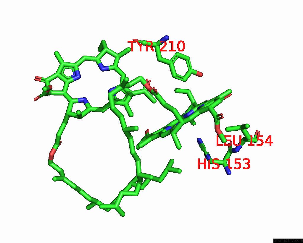
Magnesium binding site 1 out of 4 in 8c3f
Go back to
Magnesium binding site 1 out
of 4 in the Double Mutant I(L177)H/F(M197)H Structure of Photosynthetic Reaction Center From Cereibacter Sphaeroides Strain Rv
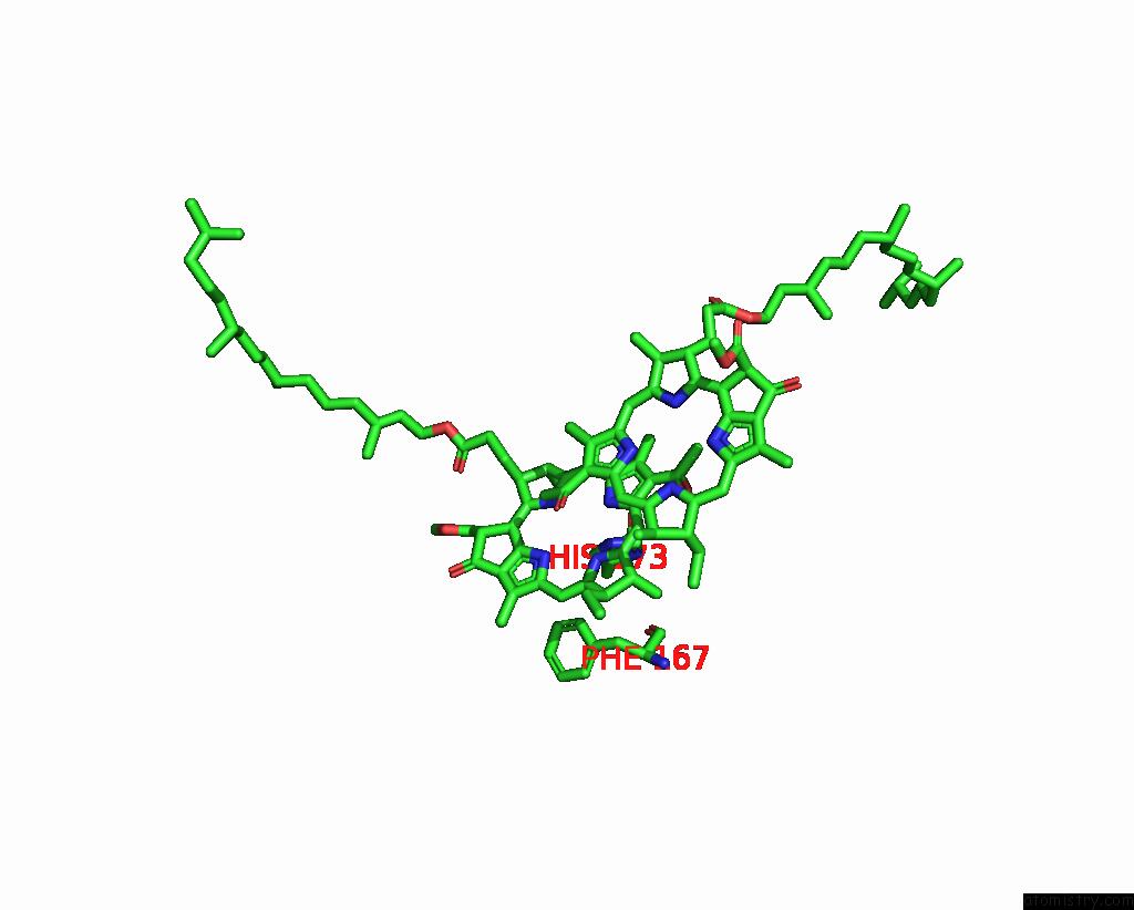
Mono view
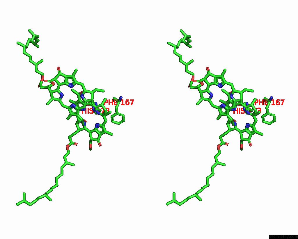
Stereo pair view
Mono view
Stereo pair view
A full contact list of Magnesium with other atoms in the Mg binding
site number 1 of Double Mutant I(L177)H/F(M197)H Structure of Photosynthetic Reaction Center From Cereibacter Sphaeroides Strain Rv within 5.0Å range:
|
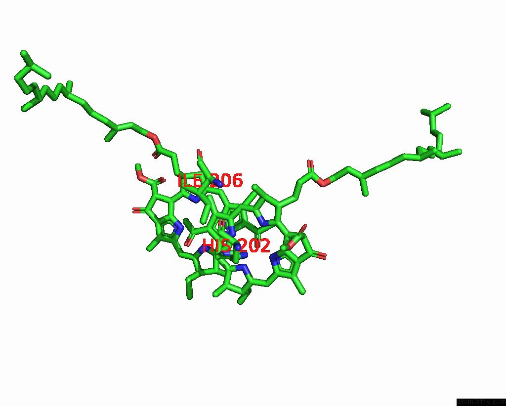
Magnesium binding site 2 out of 4 in 8c3f
Go back to
Magnesium binding site 2 out
of 4 in the Double Mutant I(L177)H/F(M197)H Structure of Photosynthetic Reaction Center From Cereibacter Sphaeroides Strain Rv
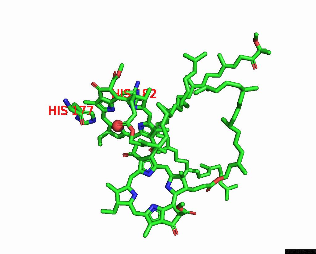
Mono view
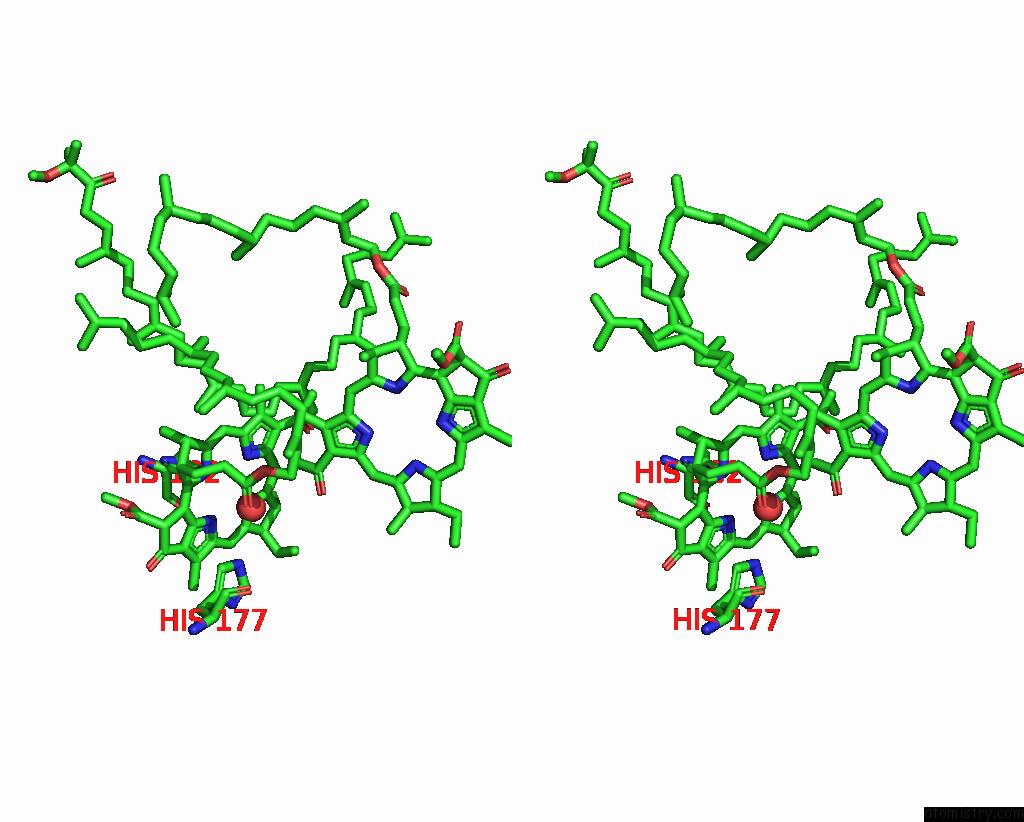
Stereo pair view
Mono view
Stereo pair view
A full contact list of Magnesium with other atoms in the Mg binding
site number 2 of Double Mutant I(L177)H/F(M197)H Structure of Photosynthetic Reaction Center From Cereibacter Sphaeroides Strain Rv within 5.0Å range:
|
Magnesium binding site 3 out of 4 in 8c3f
Go back to
Magnesium binding site 3 out
of 4 in the Double Mutant I(L177)H/F(M197)H Structure of Photosynthetic Reaction Center From Cereibacter Sphaeroides Strain Rv
Mono view

Stereo pair view
Mono view
Stereo pair view
A full contact list of Magnesium with other atoms in the Mg binding
site number 3 of Double Mutant I(L177)H/F(M197)H Structure of Photosynthetic Reaction Center From Cereibacter Sphaeroides Strain Rv within 5.0Å range:
|
Magnesium binding site 4 out of 4 in 8c3f
Go back to
Magnesium binding site 4 out
of 4 in the Double Mutant I(L177)H/F(M197)H Structure of Photosynthetic Reaction Center From Cereibacter Sphaeroides Strain Rv
Mono view

Stereo pair view
Mono view
Stereo pair view
A full contact list of Magnesium with other atoms in the Mg binding
site number 4 of Double Mutant I(L177)H/F(M197)H Structure of Photosynthetic Reaction Center From Cereibacter Sphaeroides Strain Rv within 5.0Å range:
|
Reference:
T.Y.Fufina,
G.K.Selikhanov,
A.G.Gabdulkhakov,
L.G.Vasilieva.
Properties and Crystal Structure of the Cereibacter Sphaeroides Photosynthetic Reaction Center with Double Amino Acid Substitution I(L177)H + F(M197)H. Membranes (Basel) V. 13 2023.
ISSN: ESSN 2077-0375
PubMed: 36837660
DOI: 10.3390/MEMBRANES13020157
Page generated: Thu Oct 3 20:05:07 2024
ISSN: ESSN 2077-0375
PubMed: 36837660
DOI: 10.3390/MEMBRANES13020157
Last articles
Zn in 9J0NZn in 9J0O
Zn in 9J0P
Zn in 9FJX
Zn in 9EKB
Zn in 9C0F
Zn in 9CAH
Zn in 9CH0
Zn in 9CH3
Zn in 9CH1