Magnesium »
PDB 8dh6-8dr5 »
8dq5 »
Magnesium in PDB 8dq5: X-Ray Crystal Structure of Flavobacterium Johnsoniae Dimanganese(II) Class Id Ribonucleotide Reductase T191I Variant
Enzymatic activity of X-Ray Crystal Structure of Flavobacterium Johnsoniae Dimanganese(II) Class Id Ribonucleotide Reductase T191I Variant
All present enzymatic activity of X-Ray Crystal Structure of Flavobacterium Johnsoniae Dimanganese(II) Class Id Ribonucleotide Reductase T191I Variant:
1.17.4.1;
1.17.4.1;
Protein crystallography data
The structure of X-Ray Crystal Structure of Flavobacterium Johnsoniae Dimanganese(II) Class Id Ribonucleotide Reductase T191I Variant, PDB code: 8dq5
was solved by
H.R.Rose,
A.K.Boal,
with X-Ray Crystallography technique. A brief refinement statistics is given in the table below:
| Resolution Low / High (Å) | 46.57 / 2.10 |
| Space group | P 31 |
| Cell size a, b, c (Å), α, β, γ (°) | 53.771, 53.771, 221.288, 90, 90, 120 |
| R / Rfree (%) | 24.7 / 28.2 |
Other elements in 8dq5:
The structure of X-Ray Crystal Structure of Flavobacterium Johnsoniae Dimanganese(II) Class Id Ribonucleotide Reductase T191I Variant also contains other interesting chemical elements:
| Manganese | (Mn) | 4 atoms |
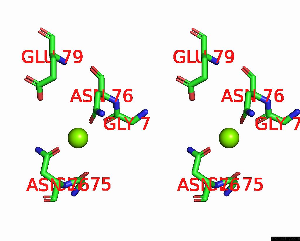
Magnesium Binding Sites:
The binding sites of Magnesium atom in the X-Ray Crystal Structure of Flavobacterium Johnsoniae Dimanganese(II) Class Id Ribonucleotide Reductase T191I Variant
(pdb code 8dq5). This binding sites where shown within
5.0 Angstroms radius around Magnesium atom.
In total only one binding site of Magnesium was determined in the X-Ray Crystal Structure of Flavobacterium Johnsoniae Dimanganese(II) Class Id Ribonucleotide Reductase T191I Variant, PDB code: 8dq5:
In total only one binding site of Magnesium was determined in the X-Ray Crystal Structure of Flavobacterium Johnsoniae Dimanganese(II) Class Id Ribonucleotide Reductase T191I Variant, PDB code: 8dq5:
Magnesium binding site 1 out of 1 in 8dq5
Go back to
Magnesium binding site 1 out
of 1 in the X-Ray Crystal Structure of Flavobacterium Johnsoniae Dimanganese(II) Class Id Ribonucleotide Reductase T191I Variant

Mono view
Stereo pair view
Mono view
Stereo pair view
A full contact list of Magnesium with other atoms in the Mg binding
site number 1 of X-Ray Crystal Structure of Flavobacterium Johnsoniae Dimanganese(II) Class Id Ribonucleotide Reductase T191I Variant within 5.0Å range:
|
Reference:
H.R.Rose,
A.K.Boal.
X-Ray Crystal Structure of Flavobacterium Johnsoniae Dimanganese(II) Class Id Ribonucleotide Reductase T191I Variant To Be Published.
Page generated: Fri Oct 4 00:48:02 2024
Last articles
Cl in 6BMUCl in 6BMX
Cl in 6BMR
Cl in 6BMV
Cl in 6BMT
Cl in 6BLA
Cl in 6BMC
Cl in 6BLG
Cl in 6BMA
Cl in 6BL4