Magnesium »
PDB 8els-8ez2 »
8etd »
Magnesium in PDB 8etd: Crystal Structure of Schizosaccharomyces Pombe RHO1
Protein crystallography data
The structure of Crystal Structure of Schizosaccharomyces Pombe RHO1, PDB code: 8etd
was solved by
Q.Huang,
J.Xie,
J.Seetharaman,
with X-Ray Crystallography technique. A brief refinement statistics is given in the table below:
| Resolution Low / High (Å) | 39.14 / 2.78 |
| Space group | C 1 2 1 |
| Cell size a, b, c (Å), α, β, γ (°) | 105.691, 66.349, 75.551, 90, 112.78, 90 |
| R / Rfree (%) | 19.1 / 23.2 |
Magnesium Binding Sites:
The binding sites of Magnesium atom in the Crystal Structure of Schizosaccharomyces Pombe RHO1
(pdb code 8etd). This binding sites where shown within
5.0 Angstroms radius around Magnesium atom.
In total 2 binding sites of Magnesium where determined in the Crystal Structure of Schizosaccharomyces Pombe RHO1, PDB code: 8etd:
Jump to Magnesium binding site number: 1; 2;
In total 2 binding sites of Magnesium where determined in the Crystal Structure of Schizosaccharomyces Pombe RHO1, PDB code: 8etd:
Jump to Magnesium binding site number: 1; 2;
Magnesium binding site 1 out of 2 in 8etd
Go back to
Magnesium binding site 1 out
of 2 in the Crystal Structure of Schizosaccharomyces Pombe RHO1
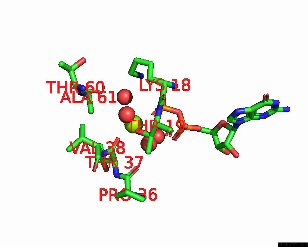
Mono view

Stereo pair view
Mono view
Stereo pair view
A full contact list of Magnesium with other atoms in the Mg binding
site number 1 of Crystal Structure of Schizosaccharomyces Pombe RHO1 within 5.0Å range:
|
Magnesium binding site 2 out of 2 in 8etd
Go back to
Magnesium binding site 2 out
of 2 in the Crystal Structure of Schizosaccharomyces Pombe RHO1
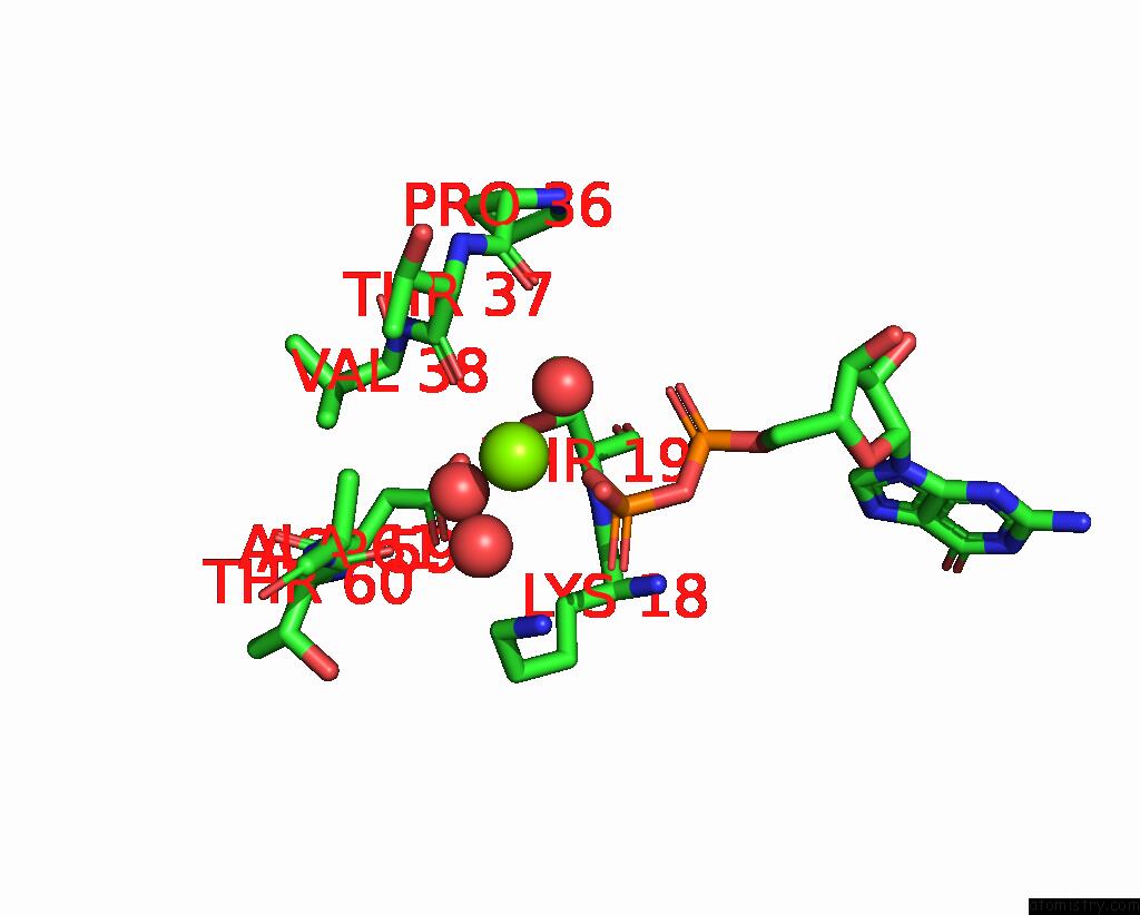
Mono view
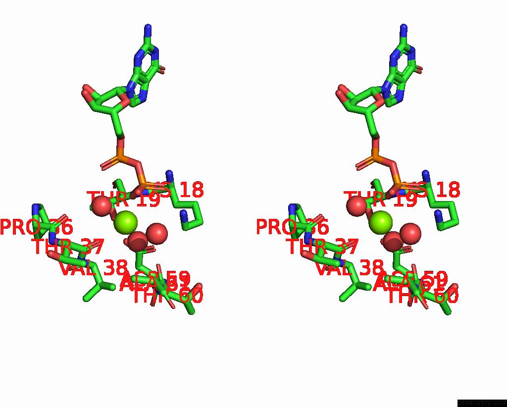
Stereo pair view
Mono view
Stereo pair view
A full contact list of Magnesium with other atoms in the Mg binding
site number 2 of Crystal Structure of Schizosaccharomyces Pombe RHO1 within 5.0Å range:
|
Reference:
Q.Huang,
J.Xie,
J.Seetharaman.
Crystal Structure of Schizosaccharomyces Pombe RHO1 Reveals Its Evolutionary Relationship with Other Rho Gtpases. Biology (Basel) V. 11 2022.
ISSN: ESSN 2079-7737
PubMed: 36358328
DOI: 10.3390/BIOLOGY11111627
Page generated: Fri Oct 4 01:20:16 2024
ISSN: ESSN 2079-7737
PubMed: 36358328
DOI: 10.3390/BIOLOGY11111627
Last articles
Zn in 9MJ5Zn in 9HNW
Zn in 9G0L
Zn in 9FNE
Zn in 9DZN
Zn in 9E0I
Zn in 9D32
Zn in 9DAK
Zn in 8ZXC
Zn in 8ZUF