Magnesium »
PDB 8fs8-8g47 »
8g1n »
Magnesium in PDB 8g1n: Structure of Campylobacter Concisus Pglc I57M/Q175M Variant with Modeled C-Terminus
Enzymatic activity of Structure of Campylobacter Concisus Pglc I57M/Q175M Variant with Modeled C-Terminus
All present enzymatic activity of Structure of Campylobacter Concisus Pglc I57M/Q175M Variant with Modeled C-Terminus:
2.7.8.36;
2.7.8.36;
Protein crystallography data
The structure of Structure of Campylobacter Concisus Pglc I57M/Q175M Variant with Modeled C-Terminus, PDB code: 8g1n
was solved by
G.J.Dodge,
L.C.Ray,
D.Das,
B.Imperiali,
K.N.Allen,
with X-Ray Crystallography technique. A brief refinement statistics is given in the table below:
| Resolution Low / High (Å) | 62.81 / 2.74 |
| Space group | P 32 2 1 |
| Cell size a, b, c (Å), α, β, γ (°) | 70.802, 70.802, 188.442, 90, 90, 120 |
| R / Rfree (%) | 25.9 / 29.7 |
Magnesium Binding Sites:
The binding sites of Magnesium atom in the Structure of Campylobacter Concisus Pglc I57M/Q175M Variant with Modeled C-Terminus
(pdb code 8g1n). This binding sites where shown within
5.0 Angstroms radius around Magnesium atom.
In total 2 binding sites of Magnesium where determined in the Structure of Campylobacter Concisus Pglc I57M/Q175M Variant with Modeled C-Terminus, PDB code: 8g1n:
Jump to Magnesium binding site number: 1; 2;
In total 2 binding sites of Magnesium where determined in the Structure of Campylobacter Concisus Pglc I57M/Q175M Variant with Modeled C-Terminus, PDB code: 8g1n:
Jump to Magnesium binding site number: 1; 2;
Magnesium binding site 1 out of 2 in 8g1n
Go back to
Magnesium binding site 1 out
of 2 in the Structure of Campylobacter Concisus Pglc I57M/Q175M Variant with Modeled C-Terminus
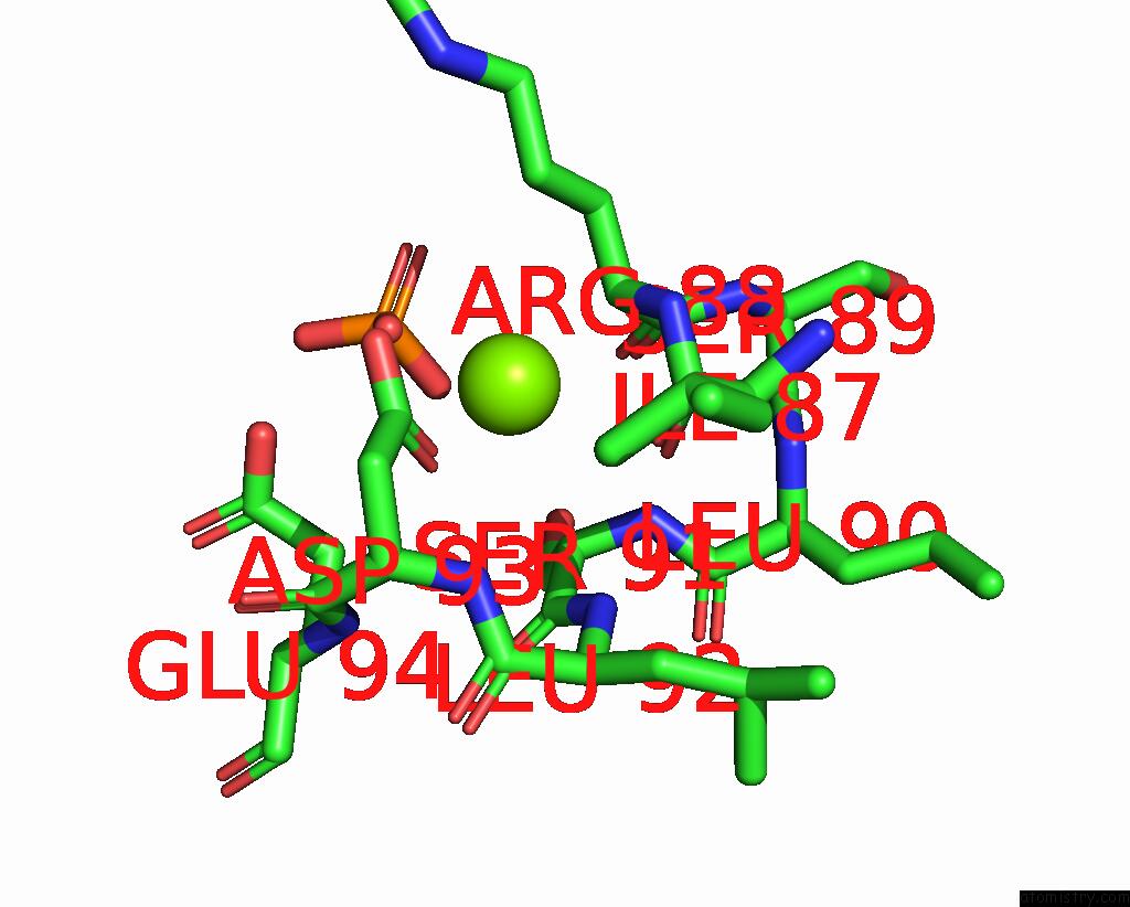
Mono view
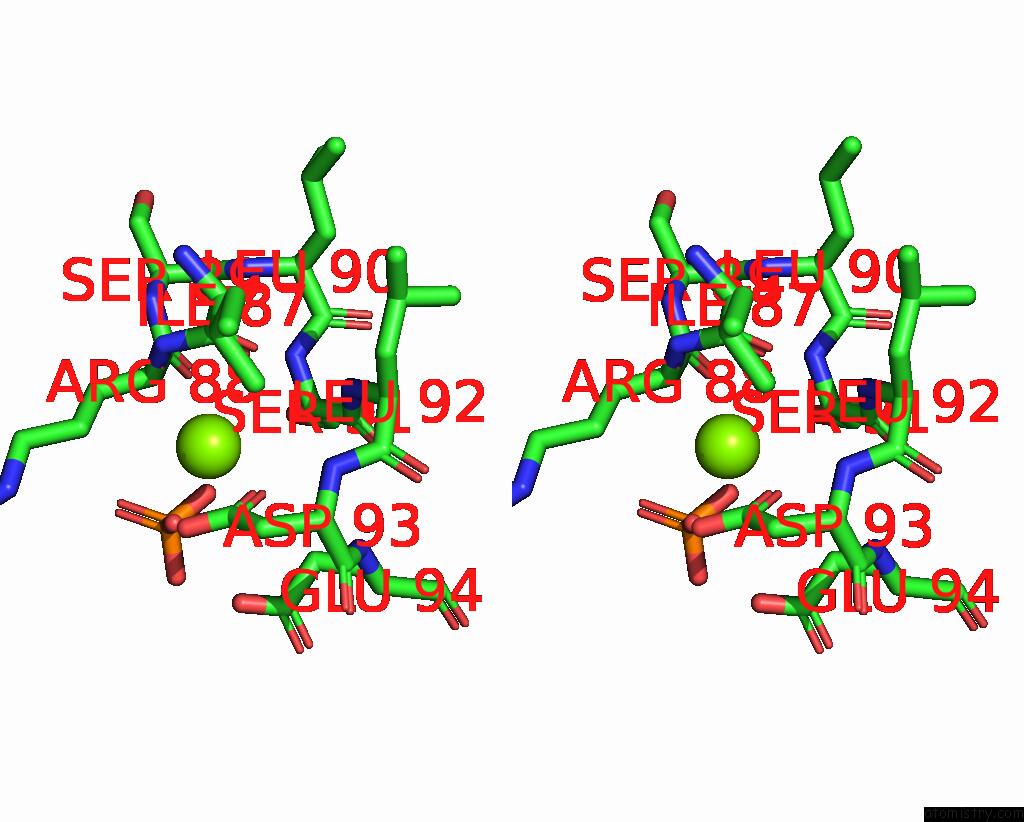
Stereo pair view
Mono view
Stereo pair view
A full contact list of Magnesium with other atoms in the Mg binding
site number 1 of Structure of Campylobacter Concisus Pglc I57M/Q175M Variant with Modeled C-Terminus within 5.0Å range:
|
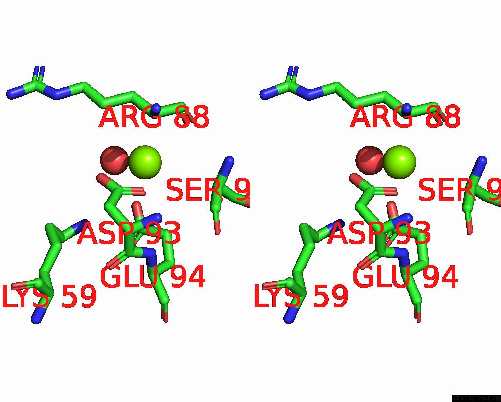
Magnesium binding site 2 out of 2 in 8g1n
Go back to
Magnesium binding site 2 out
of 2 in the Structure of Campylobacter Concisus Pglc I57M/Q175M Variant with Modeled C-Terminus
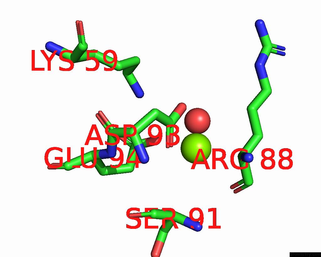
Mono view
Stereo pair view
Mono view
Stereo pair view
A full contact list of Magnesium with other atoms in the Mg binding
site number 2 of Structure of Campylobacter Concisus Pglc I57M/Q175M Variant with Modeled C-Terminus within 5.0Å range:
|
Reference:
A.J.Anderson,
G.J.Dodge,
K.N.Allen,
B.Imperiali.
Co-Conserved Sequence Motifs Are Predictive of Substrate Specificity in A Family of Monotopic Phosphoglycosyl Transferases. Protein Sci. E4646 2023.
ISSN: ESSN 1469-896X
PubMed: 37096962
DOI: 10.1002/PRO.4646
Page generated: Fri Oct 4 02:57:16 2024
ISSN: ESSN 1469-896X
PubMed: 37096962
DOI: 10.1002/PRO.4646
Last articles
Fe in 2YXOFe in 2YRS
Fe in 2YXC
Fe in 2YNM
Fe in 2YVJ
Fe in 2YP1
Fe in 2YU2
Fe in 2YU1
Fe in 2YQB
Fe in 2YOO