Magnesium »
PDB 8fs8-8g47 »
8g1s »
Magnesium in PDB 8g1s: Cryo-Em Structure of 3DVA Component 1 of Escherichia Coli Que-Pec (Paused Elongation Complex) Rna Polymerase Minus PREQ1 Ligand
Enzymatic activity of Cryo-Em Structure of 3DVA Component 1 of Escherichia Coli Que-Pec (Paused Elongation Complex) Rna Polymerase Minus PREQ1 Ligand
All present enzymatic activity of Cryo-Em Structure of 3DVA Component 1 of Escherichia Coli Que-Pec (Paused Elongation Complex) Rna Polymerase Minus PREQ1 Ligand:
2.7.7.6;
2.7.7.6;
Magnesium Binding Sites:
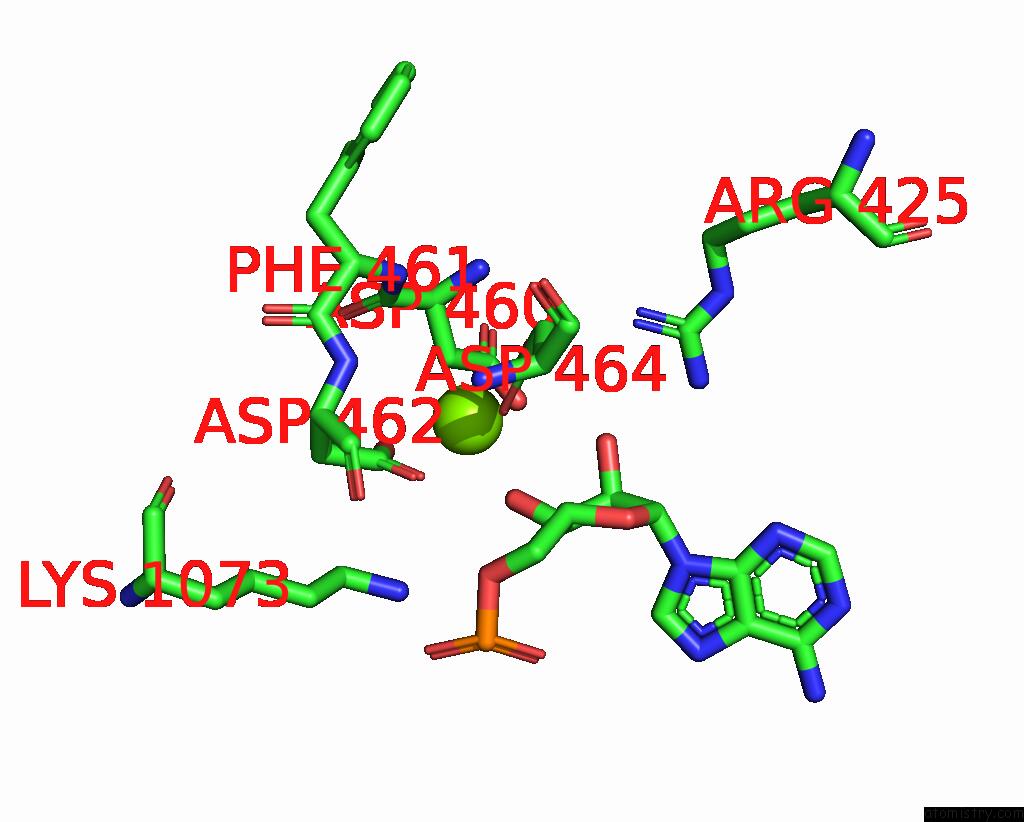
The binding sites of Magnesium atom in the Cryo-Em Structure of 3DVA Component 1 of Escherichia Coli Que-Pec (Paused Elongation Complex) Rna Polymerase Minus PREQ1 Ligand
(pdb code 8g1s). This binding sites where shown within
5.0 Angstroms radius around Magnesium atom.
In total only one binding site of Magnesium was determined in the Cryo-Em Structure of 3DVA Component 1 of Escherichia Coli Que-Pec (Paused Elongation Complex) Rna Polymerase Minus PREQ1 Ligand, PDB code: 8g1s:
In total only one binding site of Magnesium was determined in the Cryo-Em Structure of 3DVA Component 1 of Escherichia Coli Que-Pec (Paused Elongation Complex) Rna Polymerase Minus PREQ1 Ligand, PDB code: 8g1s:
Magnesium binding site 1 out of 1 in 8g1s
Go back to
Magnesium binding site 1 out
of 1 in the Cryo-Em Structure of 3DVA Component 1 of Escherichia Coli Que-Pec (Paused Elongation Complex) Rna Polymerase Minus PREQ1 Ligand

Mono view

Stereo pair view

Mono view

Stereo pair view
A full contact list of Magnesium with other atoms in the Mg binding
site number 1 of Cryo-Em Structure of 3DVA Component 1 of Escherichia Coli Que-Pec (Paused Elongation Complex) Rna Polymerase Minus PREQ1 Ligand within 5.0Å range:
|
Reference:
A.Chauvier,
J.C.Porta,
I.Deb,
E.Ellinger,
K.Meze,
A.T.Frank,
M.D.Ohi,
N.G.Walter.
Structural Basis For Control of Bacterial Rna Polymerase Pausing By A Riboswitch and Its Ligand. Nat.Struct.Mol.Biol. 2023.
ISSN: ESSN 1545-9985
PubMed: 37264140
DOI: 10.1038/S41594-023-01002-X
Page generated: Fri Oct 4 02:57:43 2024
ISSN: ESSN 1545-9985
PubMed: 37264140
DOI: 10.1038/S41594-023-01002-X
Last articles
Zn in 9MJ5Zn in 9HNW
Zn in 9G0L
Zn in 9FNE
Zn in 9DZN
Zn in 9E0I
Zn in 9D32
Zn in 9DAK
Zn in 8ZXC
Zn in 8ZUF