Magnesium »
PDB 8gi3-8gwe »
8gsr »
Magnesium in PDB 8gsr: Crystal Structure of L-2,4-Diketo-3-Deoxyrhamnonate Hydrolase From Sphingomonas Sp. (Apo-Form)
Protein crystallography data
The structure of Crystal Structure of L-2,4-Diketo-3-Deoxyrhamnonate Hydrolase From Sphingomonas Sp. (Apo-Form), PDB code: 8gsr
was solved by
S.Fukuhara,
Y.Watanabe,
S.Watanabe,
H.Nishiwaki,
with X-Ray Crystallography technique. A brief refinement statistics is given in the table below:
| Resolution Low / High (Å) | 49.07 / 1.73 |
| Space group | P 1 |
| Cell size a, b, c (Å), α, β, γ (°) | 64.217, 66.412, 75.202, 90.15, 91.8, 104.94 |
| R / Rfree (%) | 16.8 / 20.5 |
Magnesium Binding Sites:
The binding sites of Magnesium atom in the Crystal Structure of L-2,4-Diketo-3-Deoxyrhamnonate Hydrolase From Sphingomonas Sp. (Apo-Form)
(pdb code 8gsr). This binding sites where shown within
5.0 Angstroms radius around Magnesium atom.
In total 8 binding sites of Magnesium where determined in the Crystal Structure of L-2,4-Diketo-3-Deoxyrhamnonate Hydrolase From Sphingomonas Sp. (Apo-Form), PDB code: 8gsr:
Jump to Magnesium binding site number: 1; 2; 3; 4; 5; 6; 7; 8;
In total 8 binding sites of Magnesium where determined in the Crystal Structure of L-2,4-Diketo-3-Deoxyrhamnonate Hydrolase From Sphingomonas Sp. (Apo-Form), PDB code: 8gsr:
Jump to Magnesium binding site number: 1; 2; 3; 4; 5; 6; 7; 8;
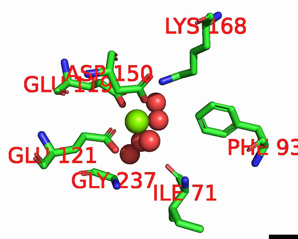
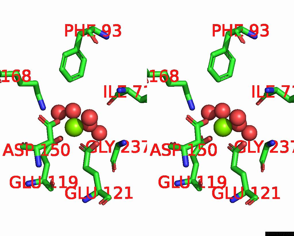
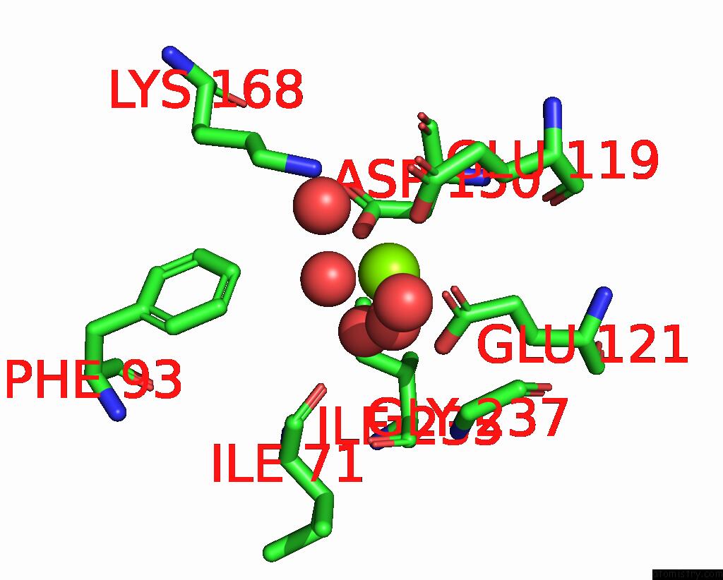
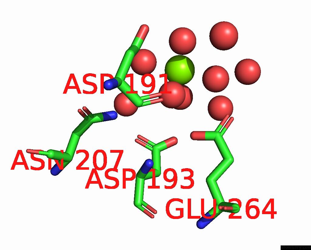
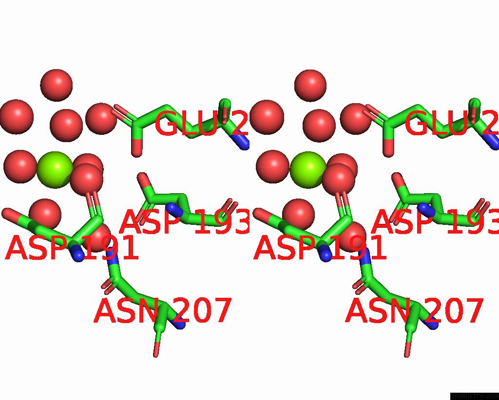
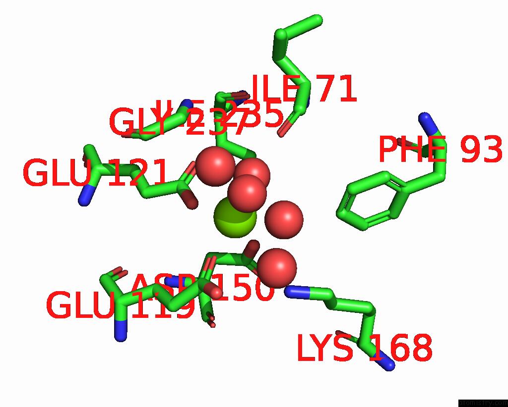
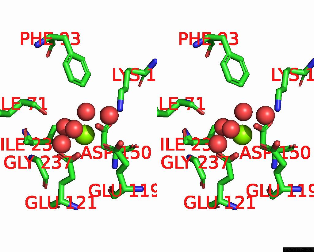
Magnesium binding site 1 out of 8 in 8gsr
Go back to
Magnesium binding site 1 out
of 8 in the Crystal Structure of L-2,4-Diketo-3-Deoxyrhamnonate Hydrolase From Sphingomonas Sp. (Apo-Form)

Mono view

Stereo pair view
Mono view
Stereo pair view
A full contact list of Magnesium with other atoms in the Mg binding
site number 1 of Crystal Structure of L-2,4-Diketo-3-Deoxyrhamnonate Hydrolase From Sphingomonas Sp. (Apo-Form) within 5.0Å range:
|
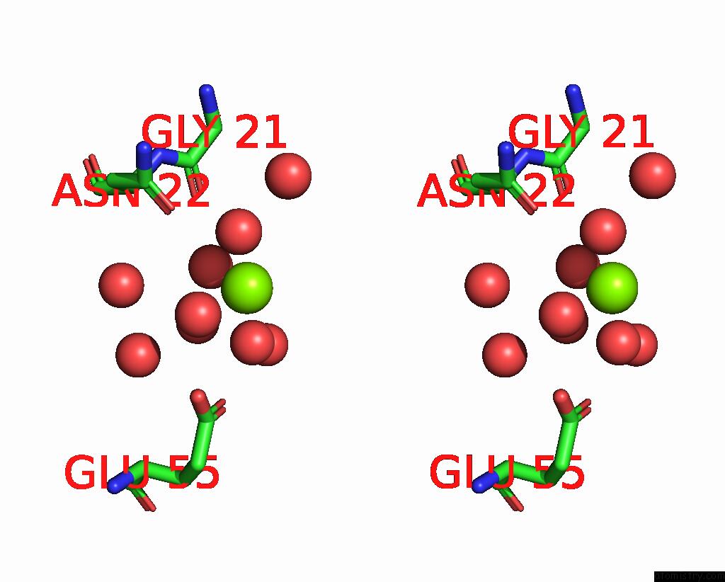
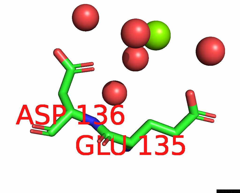
Magnesium binding site 2 out of 8 in 8gsr
Go back to
Magnesium binding site 2 out
of 8 in the Crystal Structure of L-2,4-Diketo-3-Deoxyrhamnonate Hydrolase From Sphingomonas Sp. (Apo-Form)
Mono view
Stereo pair view
Mono view
Stereo pair view
A full contact list of Magnesium with other atoms in the Mg binding
site number 2 of Crystal Structure of L-2,4-Diketo-3-Deoxyrhamnonate Hydrolase From Sphingomonas Sp. (Apo-Form) within 5.0Å range:
|
Magnesium binding site 3 out of 8 in 8gsr
Go back to
Magnesium binding site 3 out
of 8 in the Crystal Structure of L-2,4-Diketo-3-Deoxyrhamnonate Hydrolase From Sphingomonas Sp. (Apo-Form)

Mono view
Stereo pair view
Mono view
Stereo pair view
A full contact list of Magnesium with other atoms in the Mg binding
site number 3 of Crystal Structure of L-2,4-Diketo-3-Deoxyrhamnonate Hydrolase From Sphingomonas Sp. (Apo-Form) within 5.0Å range:
|
Magnesium binding site 4 out of 8 in 8gsr
Go back to
Magnesium binding site 4 out
of 8 in the Crystal Structure of L-2,4-Diketo-3-Deoxyrhamnonate Hydrolase From Sphingomonas Sp. (Apo-Form)
Mono view

Stereo pair view
Mono view
Stereo pair view
A full contact list of Magnesium with other atoms in the Mg binding
site number 4 of Crystal Structure of L-2,4-Diketo-3-Deoxyrhamnonate Hydrolase From Sphingomonas Sp. (Apo-Form) within 5.0Å range:
|
Magnesium binding site 5 out of 8 in 8gsr
Go back to
Magnesium binding site 5 out
of 8 in the Crystal Structure of L-2,4-Diketo-3-Deoxyrhamnonate Hydrolase From Sphingomonas Sp. (Apo-Form)

Mono view
Stereo pair view
Mono view
Stereo pair view
A full contact list of Magnesium with other atoms in the Mg binding
site number 5 of Crystal Structure of L-2,4-Diketo-3-Deoxyrhamnonate Hydrolase From Sphingomonas Sp. (Apo-Form) within 5.0Å range:
|
Magnesium binding site 6 out of 8 in 8gsr
Go back to
Magnesium binding site 6 out
of 8 in the Crystal Structure of L-2,4-Diketo-3-Deoxyrhamnonate Hydrolase From Sphingomonas Sp. (Apo-Form)
Mono view
Stereo pair view
Mono view
Stereo pair view
A full contact list of Magnesium with other atoms in the Mg binding
site number 6 of Crystal Structure of L-2,4-Diketo-3-Deoxyrhamnonate Hydrolase From Sphingomonas Sp. (Apo-Form) within 5.0Å range:
|
Magnesium binding site 7 out of 8 in 8gsr
Go back to
Magnesium binding site 7 out
of 8 in the Crystal Structure of L-2,4-Diketo-3-Deoxyrhamnonate Hydrolase From Sphingomonas Sp. (Apo-Form)
Mono view

Stereo pair view
Mono view
Stereo pair view
A full contact list of Magnesium with other atoms in the Mg binding
site number 7 of Crystal Structure of L-2,4-Diketo-3-Deoxyrhamnonate Hydrolase From Sphingomonas Sp. (Apo-Form) within 5.0Å range:
|
Magnesium binding site 8 out of 8 in 8gsr
Go back to
Magnesium binding site 8 out
of 8 in the Crystal Structure of L-2,4-Diketo-3-Deoxyrhamnonate Hydrolase From Sphingomonas Sp. (Apo-Form)
Mono view
Stereo pair view
Mono view
Stereo pair view
A full contact list of Magnesium with other atoms in the Mg binding
site number 8 of Crystal Structure of L-2,4-Diketo-3-Deoxyrhamnonate Hydrolase From Sphingomonas Sp. (Apo-Form) within 5.0Å range:
|
Reference:
S.Fukuhara,
S.Watanabe,
Y.Watanabe,
H.Nishiwaki.
Crystal Structure of L-2,4-Diketo-3-Deoxyrhamnonate Hydrolase Involved in the Nonphosphorylated L-Rhamnose Pathway From Bacteria. Biochemistry V. 62 524 2023.
ISSN: ISSN 0006-2960
PubMed: 36563174
DOI: 10.1021/ACS.BIOCHEM.2C00596
Page generated: Fri Oct 4 03:55:33 2024
ISSN: ISSN 0006-2960
PubMed: 36563174
DOI: 10.1021/ACS.BIOCHEM.2C00596
Last articles
Zn in 9MJ5Zn in 9HNW
Zn in 9G0L
Zn in 9FNE
Zn in 9DZN
Zn in 9E0I
Zn in 9D32
Zn in 9DAK
Zn in 8ZXC
Zn in 8ZUF