Magnesium »
PDB 8gwk-8h5m »
8h0c »
Magnesium in PDB 8h0c: Structure of the Thermolabile Hemolysin From Vibrio Alginolyticus (in Complex with Arachidonic Acid)
Protein crystallography data
The structure of Structure of the Thermolabile Hemolysin From Vibrio Alginolyticus (in Complex with Arachidonic Acid), PDB code: 8h0c
was solved by
Q.Ma,
C.Wang,
with X-Ray Crystallography technique. A brief refinement statistics is given in the table below:
| Resolution Low / High (Å) | 64.75 / 1.98 |
| Space group | P 1 21 1 |
| Cell size a, b, c (Å), α, β, γ (°) | 66.104, 72.888, 83.899, 90, 101.6, 90 |
| R / Rfree (%) | 19.3 / 22.7 |
Magnesium Binding Sites:
The binding sites of Magnesium atom in the Structure of the Thermolabile Hemolysin From Vibrio Alginolyticus (in Complex with Arachidonic Acid)
(pdb code 8h0c). This binding sites where shown within
5.0 Angstroms radius around Magnesium atom.
In total 2 binding sites of Magnesium where determined in the Structure of the Thermolabile Hemolysin From Vibrio Alginolyticus (in Complex with Arachidonic Acid), PDB code: 8h0c:
Jump to Magnesium binding site number: 1; 2;
In total 2 binding sites of Magnesium where determined in the Structure of the Thermolabile Hemolysin From Vibrio Alginolyticus (in Complex with Arachidonic Acid), PDB code: 8h0c:
Jump to Magnesium binding site number: 1; 2;
Magnesium binding site 1 out of 2 in 8h0c
Go back to
Magnesium binding site 1 out
of 2 in the Structure of the Thermolabile Hemolysin From Vibrio Alginolyticus (in Complex with Arachidonic Acid)

Mono view
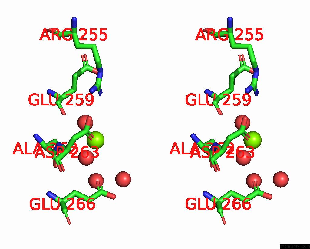
Stereo pair view
Mono view
Stereo pair view
A full contact list of Magnesium with other atoms in the Mg binding
site number 1 of Structure of the Thermolabile Hemolysin From Vibrio Alginolyticus (in Complex with Arachidonic Acid) within 5.0Å range:
|
Magnesium binding site 2 out of 2 in 8h0c
Go back to
Magnesium binding site 2 out
of 2 in the Structure of the Thermolabile Hemolysin From Vibrio Alginolyticus (in Complex with Arachidonic Acid)
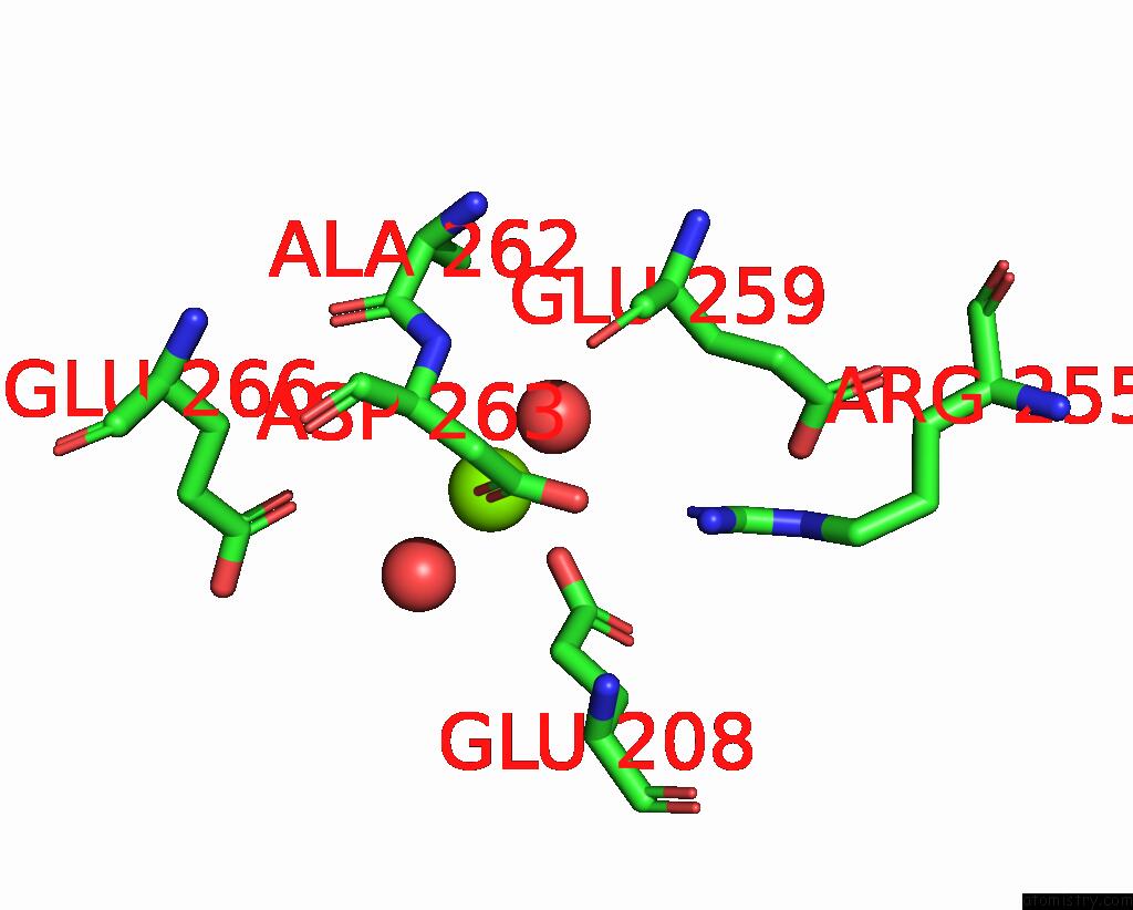
Mono view
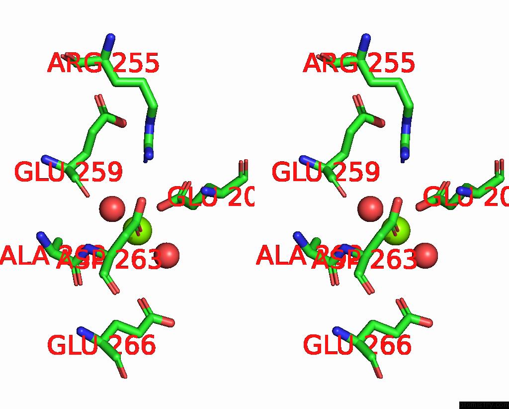
Stereo pair view
Mono view
Stereo pair view
A full contact list of Magnesium with other atoms in the Mg binding
site number 2 of Structure of the Thermolabile Hemolysin From Vibrio Alginolyticus (in Complex with Arachidonic Acid) within 5.0Å range:
|
Reference:
C.Wang,
C.Liu,
X.Zhu,
Q.Peng,
Q.Ma.
Catalytic Site Flexibility Facilitates the Substrate and Catalytic Promiscuity of Vibrio Dual Lipase/Transferase. Nat Commun V. 14 4795 2023.
ISSN: ESSN 2041-1723
PubMed: 37558668
DOI: 10.1038/S41467-023-40455-Y
Page generated: Fri Oct 4 04:16:50 2024
ISSN: ESSN 2041-1723
PubMed: 37558668
DOI: 10.1038/S41467-023-40455-Y
Last articles
Zn in 9MJ5Zn in 9HNW
Zn in 9G0L
Zn in 9FNE
Zn in 9DZN
Zn in 9E0I
Zn in 9D32
Zn in 9DAK
Zn in 8ZXC
Zn in 8ZUF