Magnesium »
PDB 8gwk-8h5m »
8h0l »
Magnesium in PDB 8h0l: Sulfur Binding Domain of Hga Complexed with Phosphorothioated Dna
Protein crystallography data
The structure of Sulfur Binding Domain of Hga Complexed with Phosphorothioated Dna, PDB code: 8h0l
was solved by
G.Liu,
X.He,
W.Hu,
B.Yang,
Q.Xiao,
with X-Ray Crystallography technique. A brief refinement statistics is given in the table below:
| Resolution Low / High (Å) | 24.13 / 1.80 |
| Space group | P 21 21 21 |
| Cell size a, b, c (Å), α, β, γ (°) | 39.865, 57.53, 177.362, 90, 90, 90 |
| R / Rfree (%) | 19.8 / 22.4 |
Magnesium Binding Sites:
The binding sites of Magnesium atom in the Sulfur Binding Domain of Hga Complexed with Phosphorothioated Dna
(pdb code 8h0l). This binding sites where shown within
5.0 Angstroms radius around Magnesium atom.
In total 2 binding sites of Magnesium where determined in the Sulfur Binding Domain of Hga Complexed with Phosphorothioated Dna, PDB code: 8h0l:
Jump to Magnesium binding site number: 1; 2;
In total 2 binding sites of Magnesium where determined in the Sulfur Binding Domain of Hga Complexed with Phosphorothioated Dna, PDB code: 8h0l:
Jump to Magnesium binding site number: 1; 2;
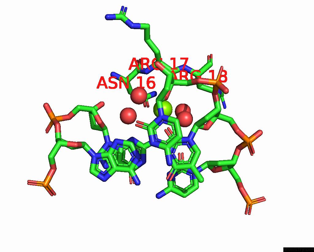
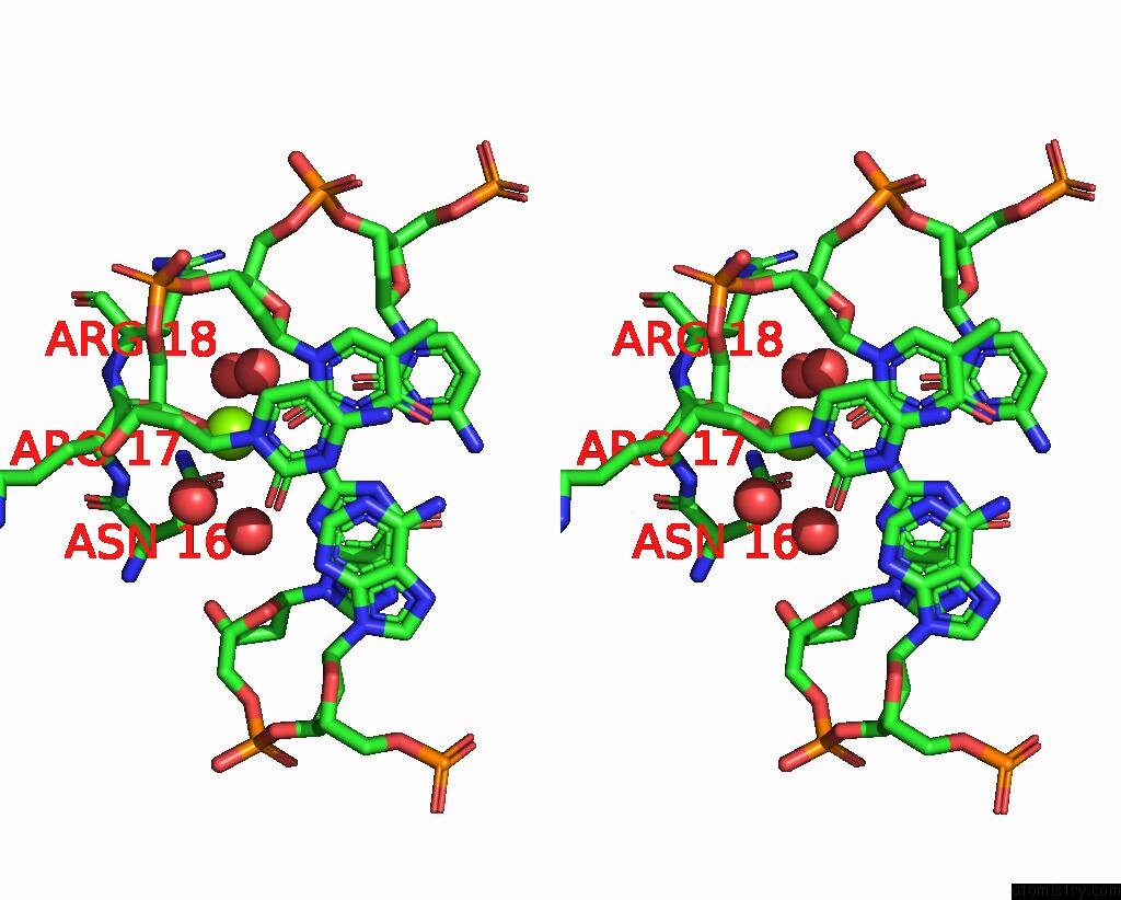
Magnesium binding site 1 out of 2 in 8h0l
Go back to
Magnesium binding site 1 out
of 2 in the Sulfur Binding Domain of Hga Complexed with Phosphorothioated Dna
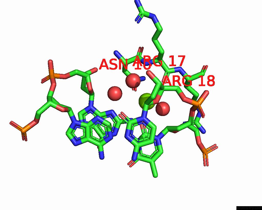
Mono view

Stereo pair view
Mono view
Stereo pair view
A full contact list of Magnesium with other atoms in the Mg binding
site number 1 of Sulfur Binding Domain of Hga Complexed with Phosphorothioated Dna within 5.0Å range:
|
Magnesium binding site 2 out of 2 in 8h0l
Go back to
Magnesium binding site 2 out
of 2 in the Sulfur Binding Domain of Hga Complexed with Phosphorothioated Dna
Mono view
Stereo pair view
Mono view
Stereo pair view
A full contact list of Magnesium with other atoms in the Mg binding
site number 2 of Sulfur Binding Domain of Hga Complexed with Phosphorothioated Dna within 5.0Å range:
|
Reference:
W.Hu,
B.Yang,
Q.Xiao,
Y.Wang,
Y.Shuai,
G.Zhao,
L.Zhang,
Z.Deng,
X.He,
G.Liu.
Characterization of A Promiscuous Dna Sulfur Binding Domain and Application in Site-Directed Rna Base Editing. Nucleic Acids Res. 2023.
ISSN: ESSN 1362-4962
PubMed: 37702119
DOI: 10.1093/NAR/GKAD743
Page generated: Fri Oct 4 04:16:50 2024
ISSN: ESSN 1362-4962
PubMed: 37702119
DOI: 10.1093/NAR/GKAD743
Last articles
Zn in 9MJ5Zn in 9HNW
Zn in 9G0L
Zn in 9FNE
Zn in 9DZN
Zn in 9E0I
Zn in 9D32
Zn in 9DAK
Zn in 8ZXC
Zn in 8ZUF