Magnesium in PDB 8hmq: Crystal Structure of PKM2 Mutant P403A

Enzymatic activity of Crystal Structure of PKM2 Mutant P403A

All present enzymatic activity of Crystal Structure of PKM2 Mutant P403A:
2.7.1.40; 2.7.10.2; 2.7.11.1;

Protein crystallography data

The structure of Crystal Structure of PKM2 Mutant P403A, PDB code: 8hmq was solved by S.Upadhyay, A.Kumar, A.K.Patel, with X-Ray Crystallography technique. A brief refinement statistics is given in the table below:

Resolution Low / High (Å) 46.73 / 2.50
Space group P 21 21 21
Cell size a, b, c (Å), α, β, γ (°) 99.829, 136.839, 149.16, 90, 90, 90
R / Rfree (%) 21.4 / 26.3

Magnesium Binding Sites:

The binding sites of Magnesium atom in the Crystal Structure of PKM2 Mutant P403A (pdb code 8hmq). This binding sites where shown within 5.0 Angstroms radius around Magnesium atom.
In total 9 binding sites of Magnesium where determined in the Crystal Structure of PKM2 Mutant P403A, PDB code: 8hmq:
Jump to Magnesium binding site number: 1; 2; 3; 4; 5; 6; 7; 8; 9;

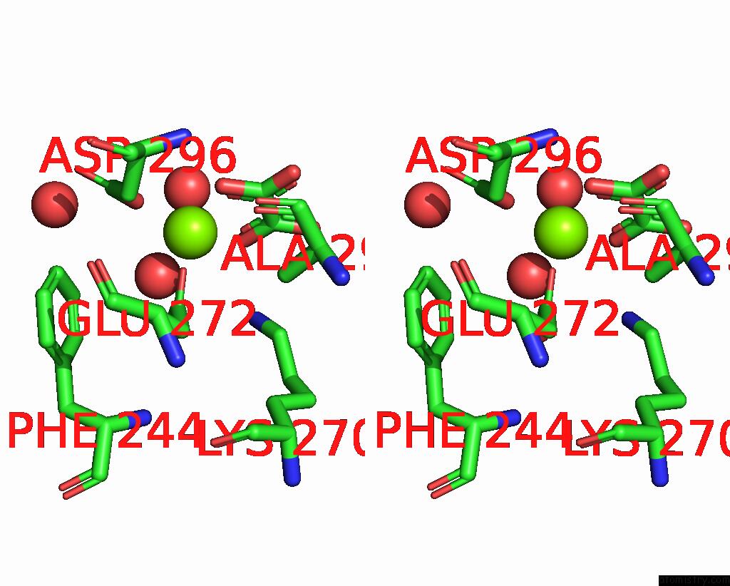
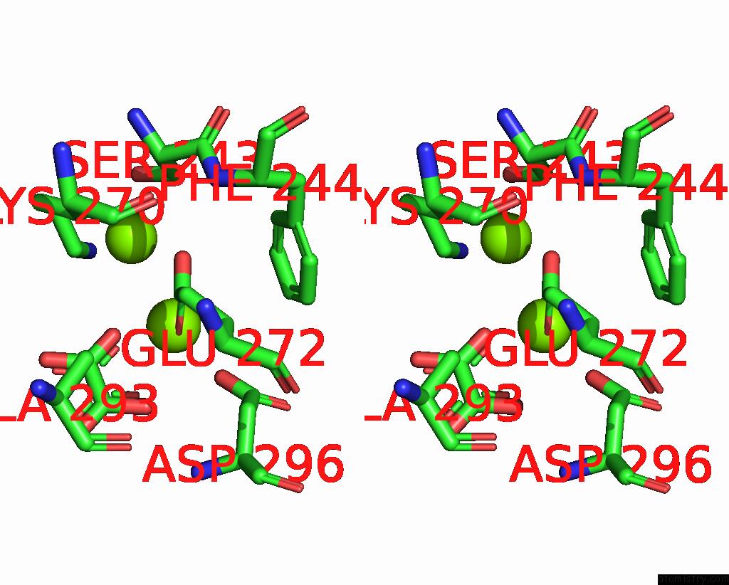
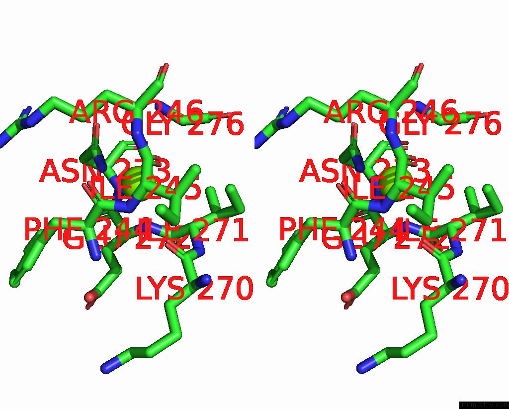
Magnesium binding site 1 out of 9 in 8hmq

Go back to Magnesium Binding Sites List in 8hmq
Magnesium binding site 1 out of 9 in the Crystal Structure of PKM2 Mutant P403A


Mono view


Stereo pair view

A full contact list of Magnesium with other atoms in the Mg binding site number 1 of Crystal Structure of PKM2 Mutant P403A within 5.0Å range:
probe atom residue distance (Å) B Occ
A:Mg607

b:40.7
occ:1.00
OE1 A:GLU272 1.9 44.9 1.0
O2 A:OXL601 2.0 45.3 1.0
OD2 A:ASP296 2.1 48.5 1.0
O A:HOH710 2.2 38.5 1.0
O A:HOH714 2.2 51.5 1.0
O1 A:OXL601 2.2 43.3 1.0
C2 A:OXL601 2.7 49.4 1.0
C1 A:OXL601 2.9 47.7 1.0
CD A:GLU272 3.0 46.7 1.0
CG A:ASP296 3.1 50.4 1.0
OE2 A:GLU272 3.4 40.4 1.0
CB A:ASP296 3.6 47.7 1.0
O4 A:OXL601 4.0 47.7 1.0
NZ A:LYS270 4.0 40.0 1.0
O3 A:OXL601 4.1 43.8 1.0
OD1 A:ASP296 4.2 46.1 1.0
CG A:GLU272 4.3 41.4 1.0
N A:ASP296 4.3 42.0 1.0
CE2 A:PHE244 4.3 52.7 1.0
CE A:LYS270 4.5 45.4 1.0
CB A:ALA293 4.5 39.6 1.0
CA A:ASP296 4.6 41.6 1.0
CZ A:PHE244 4.6 54.7 1.0
O A:HOH735 4.8 48.4 1.0
CB A:GLU272 4.8 43.6 1.0

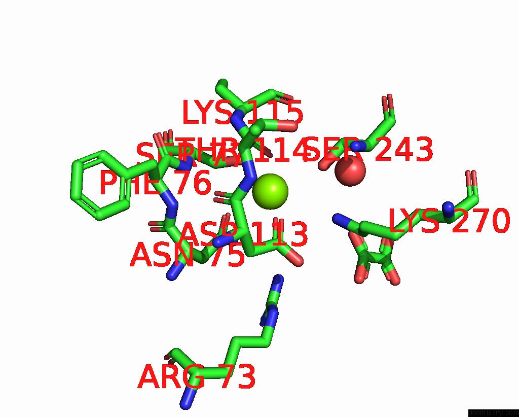
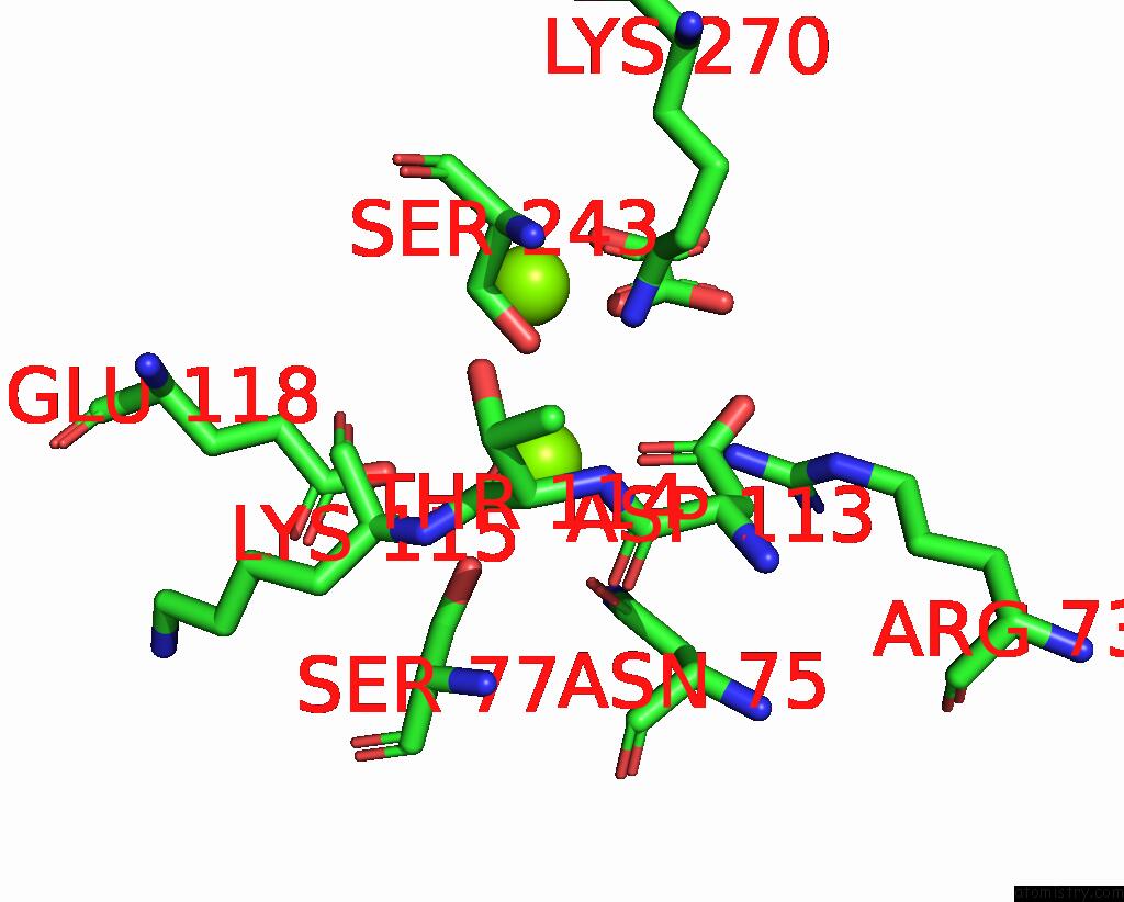
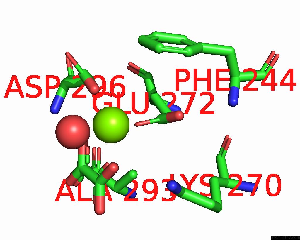
Magnesium binding site 2 out of 9 in 8hmq

Go back to Magnesium Binding Sites List in 8hmq
Magnesium binding site 2 out of 9 in the Crystal Structure of PKM2 Mutant P403A


Mono view


Stereo pair view

A full contact list of Magnesium with other atoms in the Mg binding site number 2 of Crystal Structure of PKM2 Mutant P403A within 5.0Å range:
probe atom residue distance (Å) B Occ
A:Mg608

b:54.9
occ:1.00
OD1 A:ASP113 2.4 47.1 1.0
OD1 A:ASN75 2.5 54.2 1.0
O A:THR114 2.7 50.1 1.0
OG A:SER77 2.9 56.4 1.0
CG A:ASN75 3.4 51.4 1.0
OG A:SER243 3.5 42.2 1.0
CG A:ASP113 3.6 45.2 1.0
NZ A:LYS270 3.7 40.0 1.0
C A:THR114 3.7 50.6 1.0
ND2 A:ASN75 3.8 51.8 1.0
CB A:SER77 4.0 58.8 1.0
O A:ASP113 4.0 46.9 1.0
CA A:LYS115 4.3 55.3 1.0
NH2 A:ARG73 4.3 39.3 1.0
C A:ASP113 4.3 51.5 1.0
N A:SER77 4.3 50.5 1.0
N A:LYS115 4.4 52.8 1.0
OD2 A:ASP113 4.4 45.4 1.0
O A:HOH714 4.4 51.5 1.0
CB A:ASP113 4.5 43.4 1.0
O A:LYS115 4.5 53.5 1.0
N A:THR114 4.6 40.8 1.0
CB A:SER243 4.6 46.5 1.0
CA A:SER77 4.7 62.5 1.0
CB A:ASN75 4.7 45.1 1.0
C A:LYS115 4.8 51.3 1.0
CA A:THR114 4.8 44.1 1.0
O2 A:OXL601 4.9 45.3 1.0
N A:PHE76 4.9 49.3 1.0
CA A:ASN75 5.0 44.8 1.0

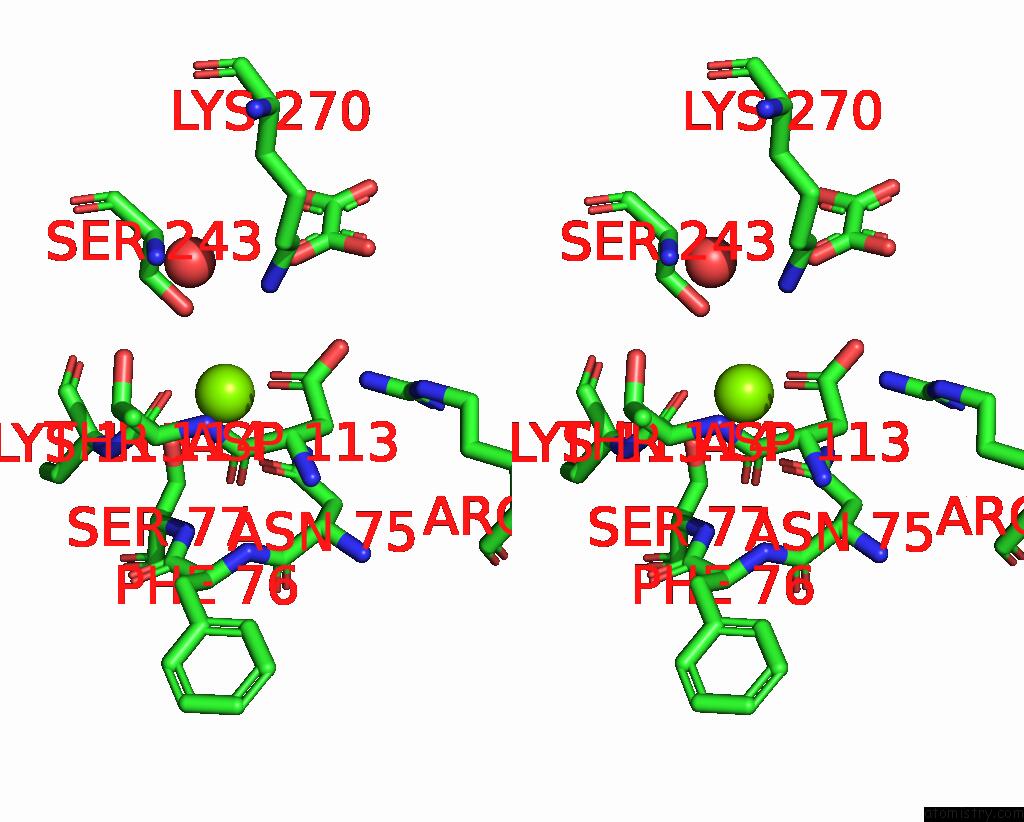
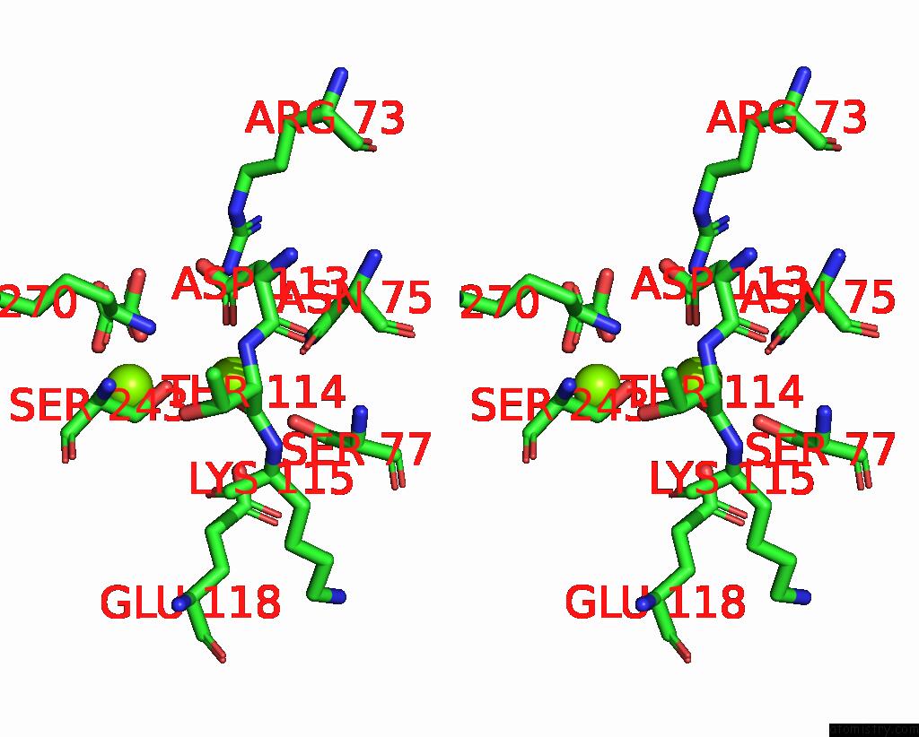
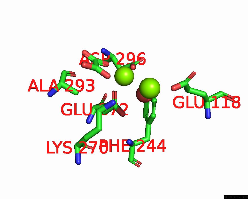
Magnesium binding site 3 out of 9 in 8hmq

Go back to Magnesium Binding Sites List in 8hmq
Magnesium binding site 3 out of 9 in the Crystal Structure of PKM2 Mutant P403A


Mono view


Stereo pair view

A full contact list of Magnesium with other atoms in the Mg binding site number 3 of Crystal Structure of PKM2 Mutant P403A within 5.0Å range:
probe atom residue distance (Å) B Occ
A:Mg609

b:63.7
occ:1.00
NE2 A:GLN458 2.2 54.7 1.0
OD1 A:ASP238 2.7 36.8 1.0
O A:ASP236 2.8 41.4 1.0
OD1 A:ASP236 3.1 64.7 1.0
NH2 A:ARG461 3.1 48.4 1.0
CD A:GLN458 3.4 54.1 1.0
CG A:ASP236 3.5 59.1 1.0
CG A:ASP238 3.5 42.5 1.0
O A:VAL237 3.6 48.1 1.0
O A:HOH720 3.6 44.7 1.0
C A:ASP236 3.7 53.4 1.0
OD2 A:ASP236 3.7 57.9 1.0
C A:VAL237 3.7 45.4 1.0
N A:ASP238 4.0 47.6 1.0
NE A:ARG461 4.0 40.8 1.0
CA A:ASP238 4.0 48.0 1.0
CZ A:ARG461 4.0 42.9 1.0
CG A:GLN458 4.1 47.4 1.0
OD2 A:ASP238 4.3 40.0 1.0
CB A:ASP238 4.3 42.7 1.0
O2 A:EDO604 4.3 67.1 1.0
CB A:GLN458 4.4 49.2 1.0
OE1 A:GLN458 4.4 59.2 1.0
CB A:ASP236 4.4 51.2 1.0
N A:VAL237 4.4 42.7 1.0
CA A:VAL237 4.5 43.3 1.0
CA A:ASP236 4.5 52.4 1.0
C2 A:EDO604 4.7 73.0 1.0
C1 A:EDO604 4.7 64.5 1.0
CA A:GLN458 4.9 43.4 1.0

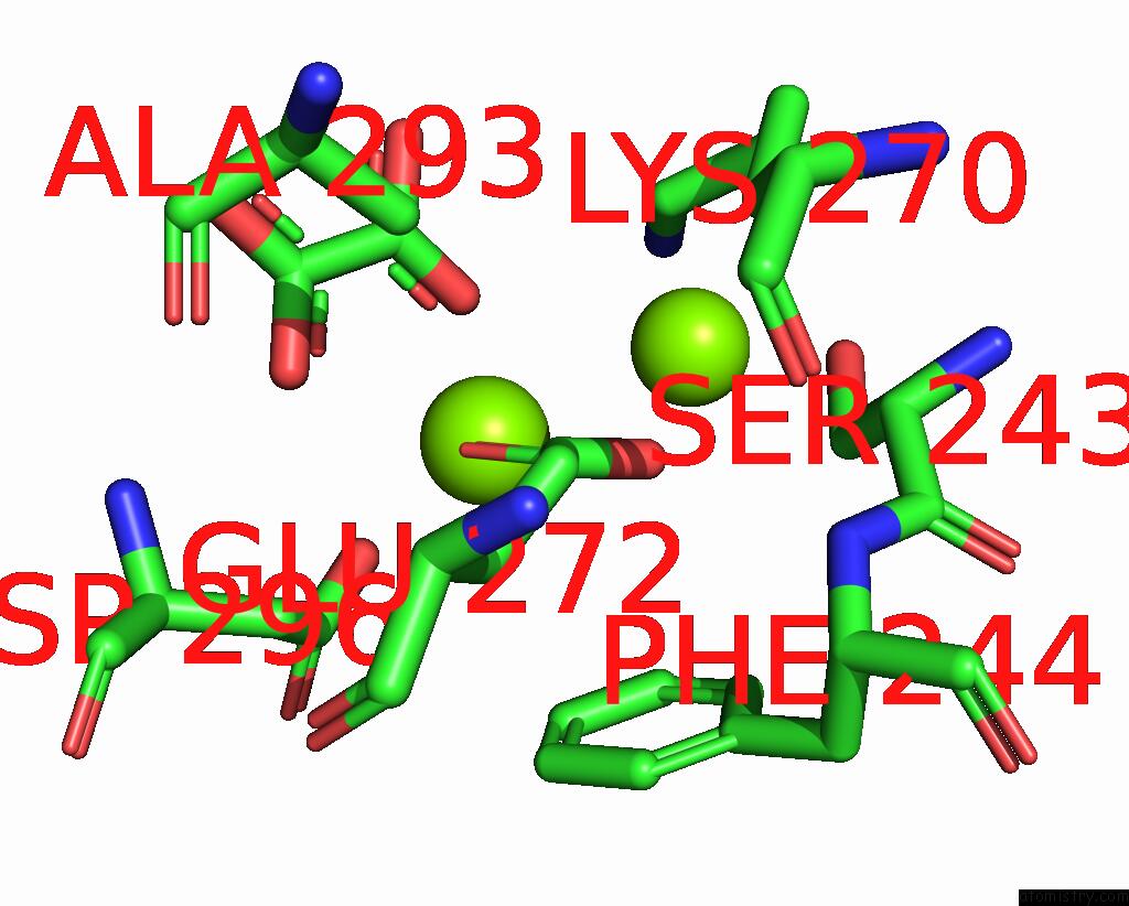
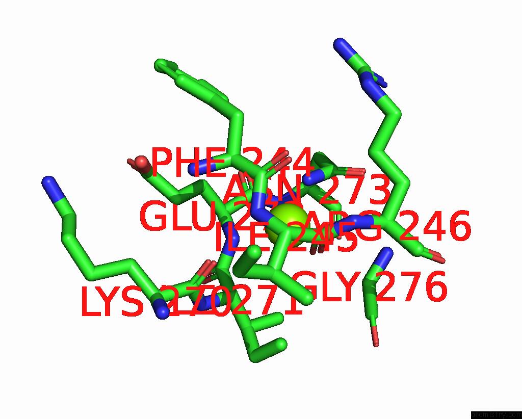
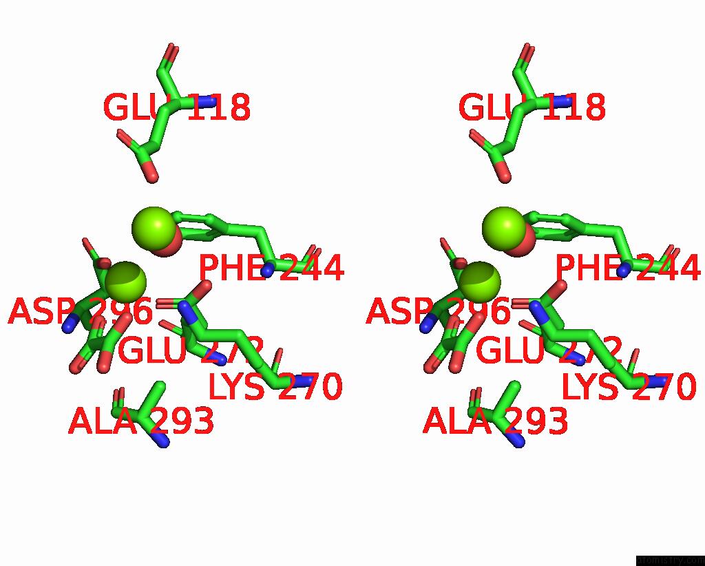
Magnesium binding site 4 out of 9 in 8hmq

Go back to Magnesium Binding Sites List in 8hmq
Magnesium binding site 4 out of 9 in the Crystal Structure of PKM2 Mutant P403A


Mono view


Stereo pair view

A full contact list of Magnesium with other atoms in the Mg binding site number 4 of Crystal Structure of PKM2 Mutant P403A within 5.0Å range:
probe atom residue distance (Å) B Occ
B:Mg605

b:50.2
occ:1.00
O4 B:OXL601 2.0 53.7 1.0
OE1 B:GLU272 2.0 50.0 1.0
OD2 B:ASP296 2.3 52.8 1.0
CD B:GLU272 2.8 51.5 1.0
O3 B:OXL601 2.9 51.7 1.0
OE2 B:GLU272 2.9 51.6 1.0
C2 B:OXL601 3.0 55.1 1.0
CG B:ASP296 3.4 53.4 1.0
C1 B:OXL601 3.5 53.1 1.0
NZ B:LYS270 3.6 48.5 1.0
CZ B:PHE244 3.9 63.2 1.0
CE2 B:PHE244 4.1 66.9 1.0
CB B:ASP296 4.1 52.2 1.0
O2 B:OXL601 4.2 53.0 1.0
CE B:LYS270 4.2 50.7 1.0
CG B:GLU272 4.3 51.0 1.0
OD1 B:ASP296 4.4 54.4 1.0
MG B:MG606 4.6 54.7 1.0
CE1 B:PHE244 4.6 62.0 1.0
O1 B:OXL601 4.7 50.0 1.0
CB B:GLU272 4.8 53.7 1.0
CD2 B:PHE244 4.9 57.0 1.0
CB B:SER243 4.9 51.5 1.0
CB B:ALA293 4.9 41.1 1.0

Magnesium binding site 5 out of 9 in 8hmq

Go back to Magnesium Binding Sites List in 8hmq
Magnesium binding site 5 out of 9 in the Crystal Structure of PKM2 Mutant P403A


Mono view


Stereo pair view

A full contact list of Magnesium with other atoms in the Mg binding site number 5 of Crystal Structure of PKM2 Mutant P403A within 5.0Å range:
probe atom residue distance (Å) B Occ
B:Mg606

b:54.7
occ:1.00
OG B:SER77 2.7 71.4 1.0
OD1 B:ASP113 2.7 56.7 1.0
OD1 B:ASN75 2.8 61.5 1.0
O B:THR114 3.0 55.0 1.0
OG B:SER243 3.3 53.5 1.0
OE2 B:GLU118 3.5 72.3 1.0
NZ B:LYS270 3.6 48.5 1.0
CG B:ASN75 3.7 63.9 1.0
CG B:ASP113 3.8 51.7 1.0
ND2 B:ASN75 3.9 57.7 1.0
CB B:SER77 4.1 73.4 1.0
C B:THR114 4.1 57.8 1.0
CB B:SER243 4.3 51.5 1.0
NH2 B:ARG73 4.3 49.4 1.0
O B:LYS115 4.4 63.9 1.0
OD2 B:ASP113 4.4 52.5 1.0
CA B:LYS115 4.5 64.1 1.0
O4 B:OXL601 4.5 53.7 1.0
MG B:MG605 4.6 50.2 1.0
CD B:GLU118 4.7 71.7 1.0
N B:SER77 4.8 66.4 1.0
N B:LYS115 4.8 63.1 1.0
C B:LYS115 4.8 61.5 1.0
O B:ASP113 4.8 57.0 1.0
CB B:ASP113 4.9 50.0 1.0
C B:ASP113 4.9 53.7 1.0
CA B:SER77 5.0 68.0 1.0

Magnesium binding site 6 out of 9 in 8hmq

Go back to Magnesium Binding Sites List in 8hmq
Magnesium binding site 6 out of 9 in the Crystal Structure of PKM2 Mutant P403A


Mono view


Stereo pair view

A full contact list of Magnesium with other atoms in the Mg binding site number 6 of Crystal Structure of PKM2 Mutant P403A within 5.0Å range:
probe atom residue distance (Å) B Occ
B:Mg607

b:63.4
occ:1.00
ND2 B:ASN273 2.1 58.3 1.0
O B:ILE245 2.5 47.7 1.0
N B:GLU272 2.9 51.2 1.0
CG B:ASN273 3.2 54.6 1.0
N B:ASN273 3.4 53.9 1.0
C B:ILE245 3.4 51.9 1.0
O B:LYS270 3.5 46.4 1.0
CA B:ILE271 3.6 49.7 1.0
N B:ILE245 3.7 54.4 1.0
C B:ILE271 3.7 46.8 1.0
CA B:GLU272 3.8 52.6 1.0
C B:PHE244 3.8 50.0 1.0
OD1 B:ASN273 3.8 67.0 1.0
CA B:GLY276 3.9 49.2 1.0
CA B:PHE244 3.9 49.3 1.0
C B:GLU272 3.9 49.6 1.0
CB B:ASN273 4.1 50.4 1.0
CA B:ILE245 4.2 53.5 1.0
CB B:GLU272 4.2 53.7 1.0
CA B:ASN273 4.2 53.7 1.0
N B:GLY276 4.2 54.6 1.0
N B:ARG246 4.3 49.5 1.0
O B:ASN273 4.4 49.3 1.0
C B:LYS270 4.4 48.4 1.0
CG2 B:ILE271 4.4 46.5 1.0
O B:PHE244 4.4 51.9 1.0
N B:ILE271 4.5 49.1 1.0
CG B:GLU272 4.5 51.0 1.0
CA B:ARG246 4.5 55.1 1.0
CD1 B:ILE271 4.5 39.9 1.0
CB B:ILE271 4.6 47.4 1.0
C B:ASN273 4.7 47.7 1.0
N B:PHE244 4.8 45.6 1.0
CG B:ARG246 4.9 61.8 1.0
O B:ILE271 4.9 44.6 1.0
CB B:ILE245 4.9 53.4 1.0
CB B:PHE244 5.0 59.1 1.0

Magnesium binding site 7 out of 9 in 8hmq

Go back to Magnesium Binding Sites List in 8hmq
Magnesium binding site 7 out of 9 in the Crystal Structure of PKM2 Mutant P403A


Mono view


Stereo pair view

A full contact list of Magnesium with other atoms in the Mg binding site number 7 of Crystal Structure of PKM2 Mutant P403A within 5.0Å range:
probe atom residue distance (Å) B Occ
C:Mg605

b:62.9
occ:1.00
OD2 C:ASP296 2.0 60.1 1.0
O2 C:OXL601 2.0 63.7 1.0
OE1 C:GLU272 2.0 71.9 1.0
O1 C:OXL601 2.3 63.0 1.0
O C:HOH709 2.4 61.8 1.0
C2 C:OXL601 2.8 57.0 1.0
C1 C:OXL601 3.0 60.0 1.0
CD C:GLU272 3.1 64.9 1.0
CG C:ASP296 3.1 68.2 1.0
OE2 C:GLU272 3.4 67.1 1.0
CB C:ASP296 3.8 65.7 1.0
O4 C:OXL601 4.1 49.5 1.0
NZ C:LYS270 4.1 64.9 1.0
OD1 C:ASP296 4.1 58.2 1.0
O3 C:OXL601 4.2 57.8 1.0
CE2 C:PHE244 4.3 79.5 1.0
CG C:GLU272 4.4 58.3 1.0
CZ C:PHE244 4.5 75.0 1.0
N C:ASP296 4.6 62.1 1.0
CE C:LYS270 4.6 61.1 1.0
CB C:ALA293 4.7 56.2 1.0
CB C:GLU272 4.8 60.3 1.0
CA C:ASP296 4.8 63.5 1.0

Magnesium binding site 8 out of 9 in 8hmq

Go back to Magnesium Binding Sites List in 8hmq
Magnesium binding site 8 out of 9 in the Crystal Structure of PKM2 Mutant P403A


Mono view


Stereo pair view

A full contact list of Magnesium with other atoms in the Mg binding site number 8 of Crystal Structure of PKM2 Mutant P403A within 5.0Å range:
probe atom residue distance (Å) B Occ
D:Mg606

b:56.6
occ:1.00
O3 D:OXL601 2.0 52.4 1.0
OD2 D:ASP296 2.1 56.5 1.0
OE1 D:GLU272 2.1 53.1 1.0
O4 D:OXL601 2.4 50.5 1.0
O D:HOH708 2.6 56.3 1.0
C1 D:OXL601 2.9 50.9 1.0
C2 D:OXL601 3.0 51.1 1.0
CD D:GLU272 3.1 56.1 1.0
CG D:ASP296 3.2 56.9 1.0
OE2 D:GLU272 3.5 49.0 1.0
NZ D:LYS270 3.8 48.0 1.0
CB D:ASP296 3.9 49.8 1.0
O1 D:OXL601 4.1 51.2 1.0
CE2 D:PHE244 4.2 59.6 1.0
OD1 D:ASP296 4.3 53.4 1.0
O2 D:OXL601 4.3 57.3 1.0
CE D:LYS270 4.4 51.5 1.0
CG D:GLU272 4.5 49.1 1.0
CZ D:PHE244 4.6 54.8 1.0
N D:ASP296 4.6 54.0 1.0
MG D:MG607 4.6 66.7 1.0
CB D:ALA293 4.6 47.0 1.0
OE2 D:GLU118 4.7 71.7 1.0
CA D:ASP296 4.9 56.1 1.0
CB D:GLU272 5.0 54.7 1.0
CD2 D:PHE244 5.0 58.7 1.0

Magnesium binding site 9 out of 9 in 8hmq

Go back to Magnesium Binding Sites List in 8hmq
Magnesium binding site 9 out of 9 in the Crystal Structure of PKM2 Mutant P403A


Mono view


Stereo pair view

A full contact list of Magnesium with other atoms in the Mg binding site number 9 of Crystal Structure of PKM2 Mutant P403A within 5.0Å range:
probe atom residue distance (Å) B Occ
D:Mg607

b:66.7
occ:1.00
OD1 D:ASP113 2.6 59.4 1.0
OD1 D:ASN75 2.8 67.9 1.0
O D:HOH708 3.0 56.3 1.0
OE2 D:GLU118 3.2 71.7 1.0
O D:THR114 3.4 59.4 1.0
OG D:SER243 3.5 56.5 1.0
NZ D:LYS270 3.5 48.0 1.0
CG D:ASN75 3.5 66.4 1.0
ND2 D:ASN75 3.6 60.1 1.0
CG D:ASP113 3.7 58.0 1.0
OG D:SER77 3.7 74.6 1.0
CB D:SER77 4.1 69.7 1.0
NH2 D:ARG73 4.1 56.6 1.0
O4 D:OXL601 4.2 50.5 1.0
OD2 D:ASP113 4.3 62.0 1.0
CD D:GLU118 4.4 79.7 1.0
C D:THR114 4.5 61.9 1.0
CB D:SER243 4.6 53.0 1.0
MG D:MG606 4.6 56.6 1.0
CB D:ASP113 4.6 56.4 1.0
O D:LYS115 4.8 71.8 1.0
OE1 D:GLU118 4.8 59.2 1.0
O D:ASP113 4.9 61.5 1.0
CB D:ASN75 4.9 54.3 1.0
CE D:LYS270 4.9 51.5 1.0
C D:ASP113 5.0 57.9 1.0

Reference:

S.Upadhyay, A.Kumar, A.K.Patel. Structural and Mechanistic Insights Into Cancer Patient-Derived Mutations in Pyruvate Kinase Muscle Isoform 2 To Be Published.
Page generated: Fri Apr 7 11:26:08 2023

Last articles

Zn in 8WB0
Zn in 8WAX
Zn in 8WAU
Zn in 8WAZ
Zn in 8WAY
Zn in 8WAV
Zn in 8WAW
Zn in 8WAT
Zn in 8W7M
Zn in 8WD3
© Copyright 2008-2020 by atomistry.com
Home   |    Site Map   |    Copyright   |    Contact us   |    Privacy