Magnesium »
PDB 8hs0-8i24 »
8i07 »
Magnesium in PDB 8i07: Crystal Structure of Escherichia Coli Glyoxylate Carboligase Double Mutant in Complex with Glycolaldehyde
Enzymatic activity of Crystal Structure of Escherichia Coli Glyoxylate Carboligase Double Mutant in Complex with Glycolaldehyde
All present enzymatic activity of Crystal Structure of Escherichia Coli Glyoxylate Carboligase Double Mutant in Complex with Glycolaldehyde:
4.1.1.47;
4.1.1.47;
Protein crystallography data
The structure of Crystal Structure of Escherichia Coli Glyoxylate Carboligase Double Mutant in Complex with Glycolaldehyde, PDB code: 8i07
was solved by
J.H.Kim,
J.S.Kim,
with X-Ray Crystallography technique. A brief refinement statistics is given in the table below:
| Resolution Low / High (Å) | 49.89 / 1.99 |
| Space group | P 41 21 2 |
| Cell size a, b, c (Å), α, β, γ (°) | 189.299, 189.299, 246.982, 90, 90, 90 |
| R / Rfree (%) | 20.8 / 24.2 |
Magnesium Binding Sites:
The binding sites of Magnesium atom in the Crystal Structure of Escherichia Coli Glyoxylate Carboligase Double Mutant in Complex with Glycolaldehyde
(pdb code 8i07). This binding sites where shown within
5.0 Angstroms radius around Magnesium atom.
In total 10 binding sites of Magnesium where determined in the Crystal Structure of Escherichia Coli Glyoxylate Carboligase Double Mutant in Complex with Glycolaldehyde, PDB code: 8i07:
Jump to Magnesium binding site number: 1; 2; 3; 4; 5; 6; 7; 8; 9; 10;
In total 10 binding sites of Magnesium where determined in the Crystal Structure of Escherichia Coli Glyoxylate Carboligase Double Mutant in Complex with Glycolaldehyde, PDB code: 8i07:
Jump to Magnesium binding site number: 1; 2; 3; 4; 5; 6; 7; 8; 9; 10;
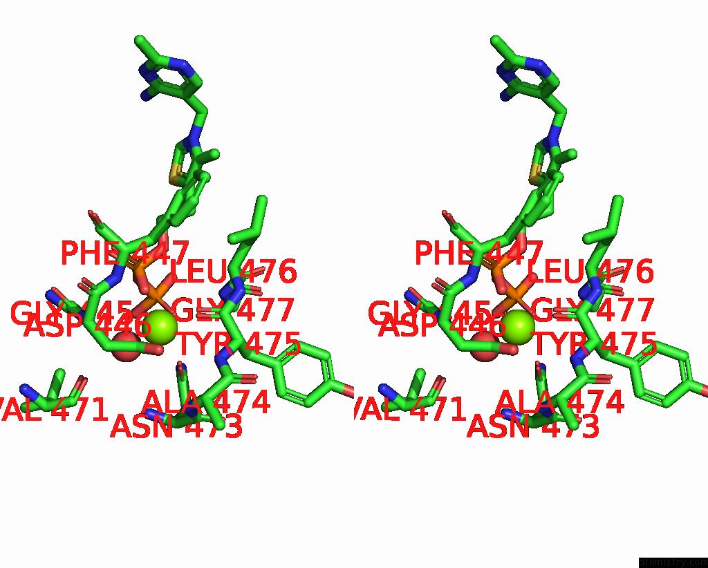
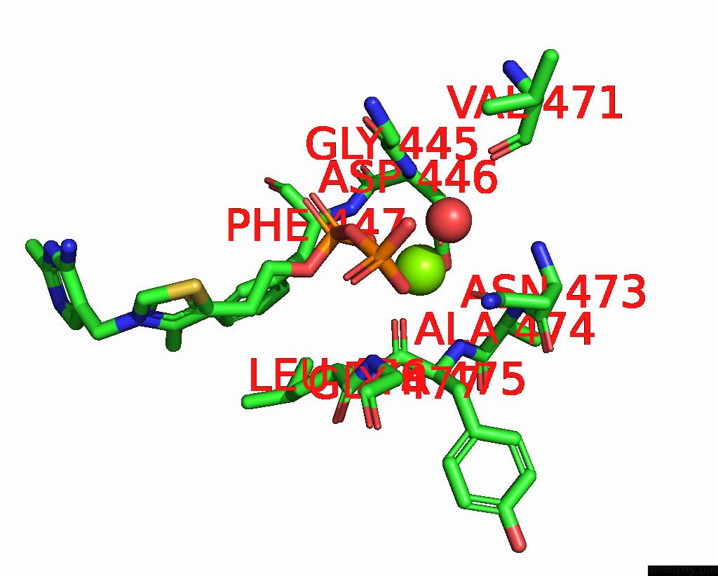
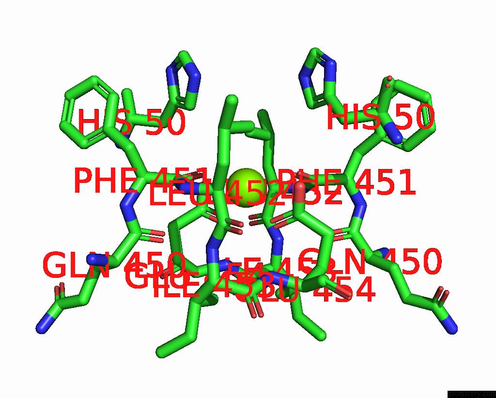
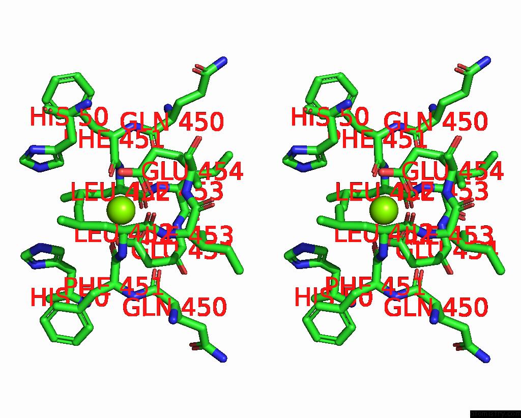
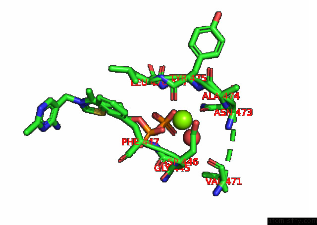
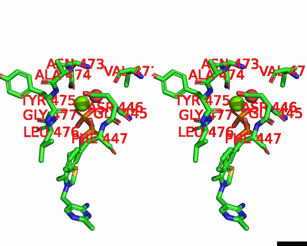
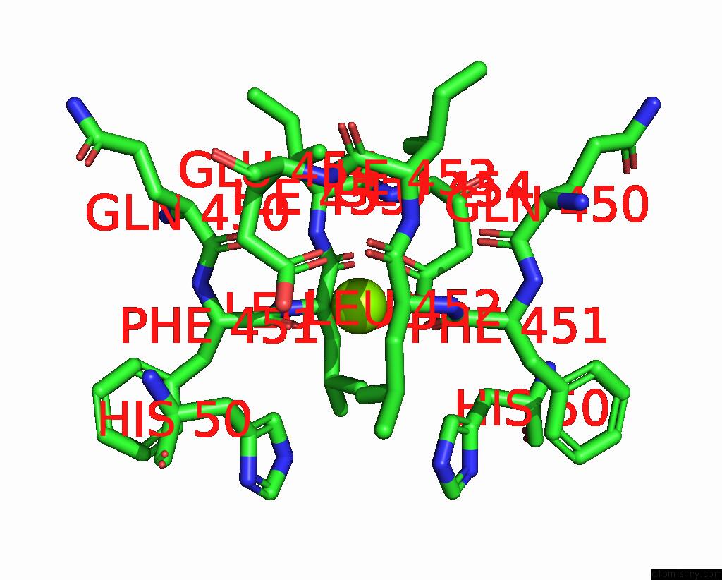
Magnesium binding site 1 out of 10 in 8i07
Go back to
Magnesium binding site 1 out
of 10 in the Crystal Structure of Escherichia Coli Glyoxylate Carboligase Double Mutant in Complex with Glycolaldehyde
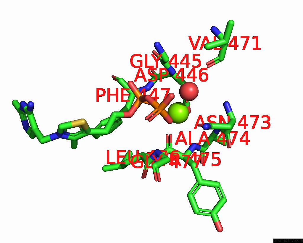
Mono view
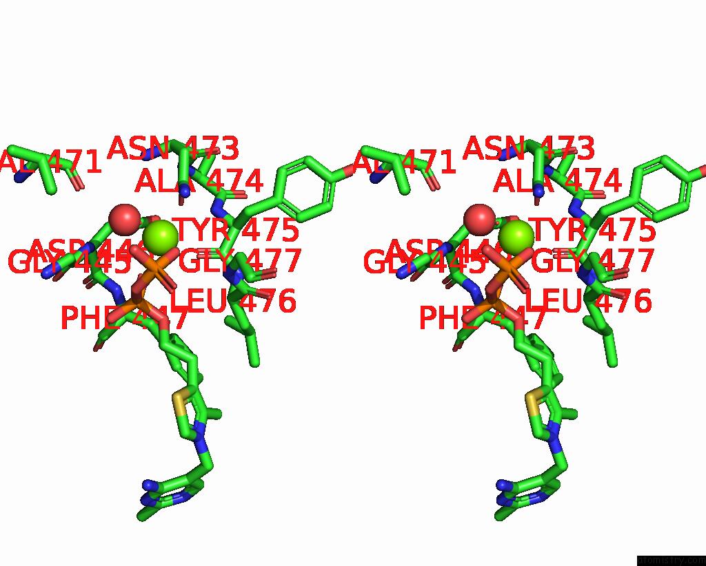
Stereo pair view
Mono view
Stereo pair view
A full contact list of Magnesium with other atoms in the Mg binding
site number 1 of Crystal Structure of Escherichia Coli Glyoxylate Carboligase Double Mutant in Complex with Glycolaldehyde within 5.0Å range:
|
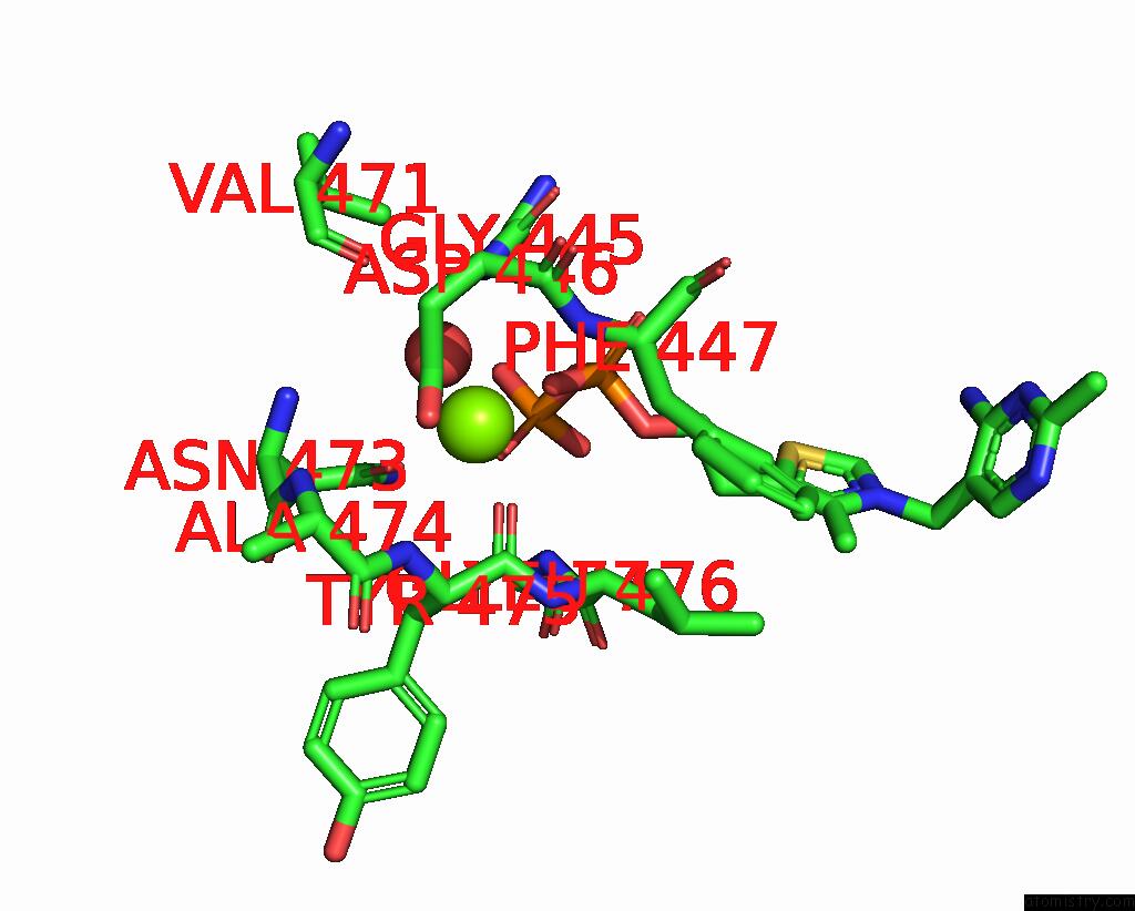
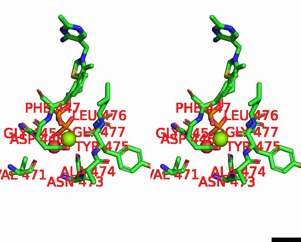
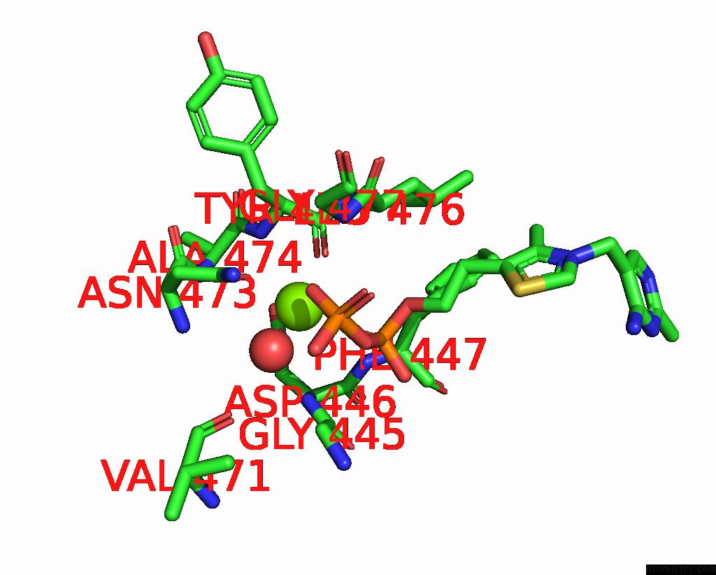
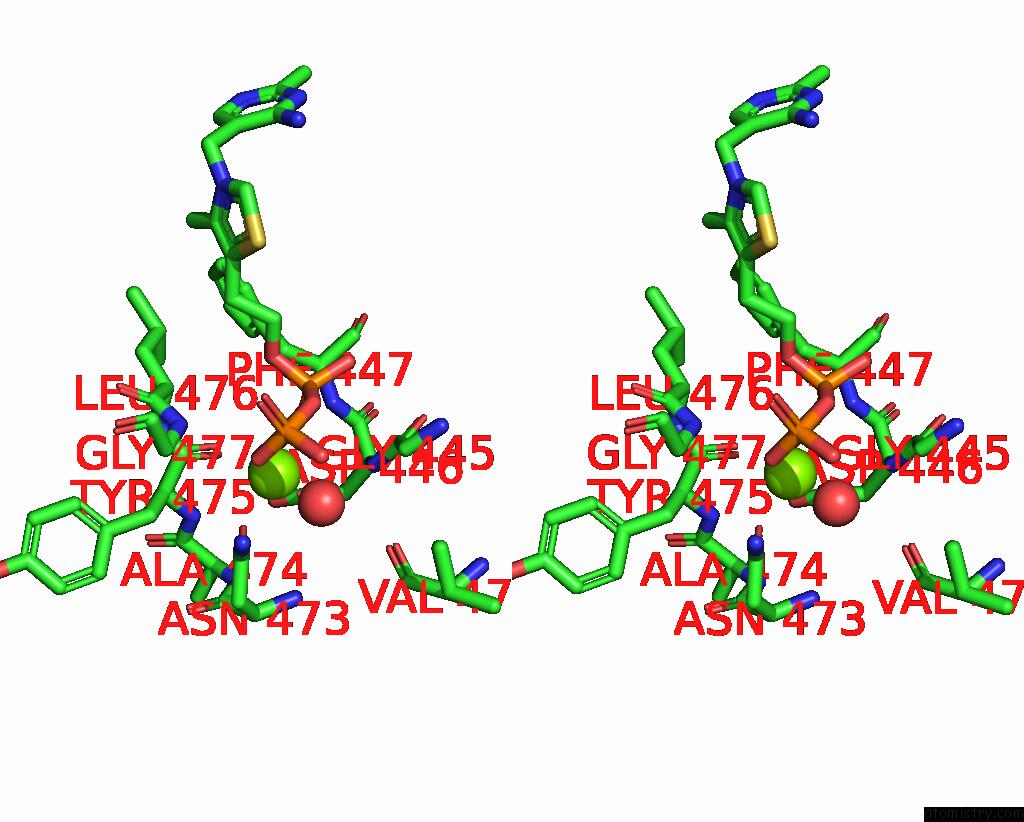
Magnesium binding site 2 out of 10 in 8i07
Go back to
Magnesium binding site 2 out
of 10 in the Crystal Structure of Escherichia Coli Glyoxylate Carboligase Double Mutant in Complex with Glycolaldehyde

Mono view

Stereo pair view
Mono view
Stereo pair view
A full contact list of Magnesium with other atoms in the Mg binding
site number 2 of Crystal Structure of Escherichia Coli Glyoxylate Carboligase Double Mutant in Complex with Glycolaldehyde within 5.0Å range:
|
Magnesium binding site 3 out of 10 in 8i07
Go back to
Magnesium binding site 3 out
of 10 in the Crystal Structure of Escherichia Coli Glyoxylate Carboligase Double Mutant in Complex with Glycolaldehyde

Mono view
Stereo pair view
Mono view
Stereo pair view
A full contact list of Magnesium with other atoms in the Mg binding
site number 3 of Crystal Structure of Escherichia Coli Glyoxylate Carboligase Double Mutant in Complex with Glycolaldehyde within 5.0Å range:
|
Magnesium binding site 4 out of 10 in 8i07
Go back to
Magnesium binding site 4 out
of 10 in the Crystal Structure of Escherichia Coli Glyoxylate Carboligase Double Mutant in Complex with Glycolaldehyde

Mono view

Stereo pair view
Mono view
Stereo pair view
A full contact list of Magnesium with other atoms in the Mg binding
site number 4 of Crystal Structure of Escherichia Coli Glyoxylate Carboligase Double Mutant in Complex with Glycolaldehyde within 5.0Å range:
|
Magnesium binding site 5 out of 10 in 8i07
Go back to
Magnesium binding site 5 out
of 10 in the Crystal Structure of Escherichia Coli Glyoxylate Carboligase Double Mutant in Complex with Glycolaldehyde
Mono view

Stereo pair view
Mono view
Stereo pair view
A full contact list of Magnesium with other atoms in the Mg binding
site number 5 of Crystal Structure of Escherichia Coli Glyoxylate Carboligase Double Mutant in Complex with Glycolaldehyde within 5.0Å range:
|
Magnesium binding site 6 out of 10 in 8i07
Go back to
Magnesium binding site 6 out
of 10 in the Crystal Structure of Escherichia Coli Glyoxylate Carboligase Double Mutant in Complex with Glycolaldehyde
Mono view
Stereo pair view
Mono view
Stereo pair view
A full contact list of Magnesium with other atoms in the Mg binding
site number 6 of Crystal Structure of Escherichia Coli Glyoxylate Carboligase Double Mutant in Complex with Glycolaldehyde within 5.0Å range:
|
Magnesium binding site 7 out of 10 in 8i07
Go back to
Magnesium binding site 7 out
of 10 in the Crystal Structure of Escherichia Coli Glyoxylate Carboligase Double Mutant in Complex with Glycolaldehyde
Mono view
Stereo pair view
Mono view
Stereo pair view
A full contact list of Magnesium with other atoms in the Mg binding
site number 7 of Crystal Structure of Escherichia Coli Glyoxylate Carboligase Double Mutant in Complex with Glycolaldehyde within 5.0Å range:
|
Magnesium binding site 8 out of 10 in 8i07
Go back to
Magnesium binding site 8 out
of 10 in the Crystal Structure of Escherichia Coli Glyoxylate Carboligase Double Mutant in Complex with Glycolaldehyde
Mono view
Stereo pair view
Mono view
Stereo pair view
A full contact list of Magnesium with other atoms in the Mg binding
site number 8 of Crystal Structure of Escherichia Coli Glyoxylate Carboligase Double Mutant in Complex with Glycolaldehyde within 5.0Å range:
|
Magnesium binding site 9 out of 10 in 8i07
Go back to
Magnesium binding site 9 out
of 10 in the Crystal Structure of Escherichia Coli Glyoxylate Carboligase Double Mutant in Complex with Glycolaldehyde
Mono view
Stereo pair view
Mono view
Stereo pair view
A full contact list of Magnesium with other atoms in the Mg binding
site number 9 of Crystal Structure of Escherichia Coli Glyoxylate Carboligase Double Mutant in Complex with Glycolaldehyde within 5.0Å range:
|
Magnesium binding site 10 out of 10 in 8i07
Go back to
Magnesium binding site 10 out
of 10 in the Crystal Structure of Escherichia Coli Glyoxylate Carboligase Double Mutant in Complex with Glycolaldehyde
Mono view

Stereo pair view
Mono view
Stereo pair view
A full contact list of Magnesium with other atoms in the Mg binding
site number 10 of Crystal Structure of Escherichia Coli Glyoxylate Carboligase Double Mutant in Complex with Glycolaldehyde within 5.0Å range:
|
Reference:
J.H.Kim,
H.Cheon,
H.J.Jo,
J.W.Kim,
G.Y.Kim,
H.R.Seo,
P.W.Seo,
J.S.Kim,
J.B.Park.
Engineering of Two Thiamine Diphosphate-Dependent Enzymes For the Regioselective Condensation of C1-Formaldehyde Into C4-Erythrulose Int.J.Biol.Macromol. V. 253 27674 2023.
ISSN: ISSN 0141-8130
DOI: 10.1016/J.IJBIOMAC.2023.127674
Page generated: Fri Oct 4 09:00:45 2024
ISSN: ISSN 0141-8130
DOI: 10.1016/J.IJBIOMAC.2023.127674
Last articles
Zn in 9JYWZn in 9IR4
Zn in 9IR3
Zn in 9GMX
Zn in 9GMW
Zn in 9JEJ
Zn in 9ERF
Zn in 9ERE
Zn in 9EGV
Zn in 9EGW