Magnesium »
PDB 8i51-8ij9 »
8iaw »
Magnesium in PDB 8iaw: Crystal Structure of Streptococcus Pneumoniae Pyruvate Kinase in Complex with Phosphoenolpyruvate
Protein crystallography data
The structure of Crystal Structure of Streptococcus Pneumoniae Pyruvate Kinase in Complex with Phosphoenolpyruvate, PDB code: 8iaw
was solved by
R.Nakashima,
A.Taguchi,
with X-Ray Crystallography technique. A brief refinement statistics is given in the table below:
| Resolution Low / High (Å) | 48.58 / 2.89 |
| Space group | P 21 21 2 |
| Cell size a, b, c (Å), α, β, γ (°) | 96.057, 110.372, 130.931, 90, 90, 90 |
| R / Rfree (%) | 24.1 / 27.3 |
Magnesium Binding Sites:
The binding sites of Magnesium atom in the Crystal Structure of Streptococcus Pneumoniae Pyruvate Kinase in Complex with Phosphoenolpyruvate
(pdb code 8iaw). This binding sites where shown within
5.0 Angstroms radius around Magnesium atom.
In total 2 binding sites of Magnesium where determined in the Crystal Structure of Streptococcus Pneumoniae Pyruvate Kinase in Complex with Phosphoenolpyruvate, PDB code: 8iaw:
Jump to Magnesium binding site number: 1; 2;
In total 2 binding sites of Magnesium where determined in the Crystal Structure of Streptococcus Pneumoniae Pyruvate Kinase in Complex with Phosphoenolpyruvate, PDB code: 8iaw:
Jump to Magnesium binding site number: 1; 2;
Magnesium binding site 1 out of 2 in 8iaw
Go back to
Magnesium binding site 1 out
of 2 in the Crystal Structure of Streptococcus Pneumoniae Pyruvate Kinase in Complex with Phosphoenolpyruvate
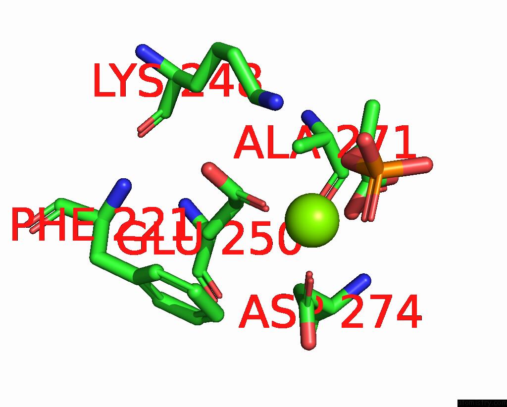
Mono view
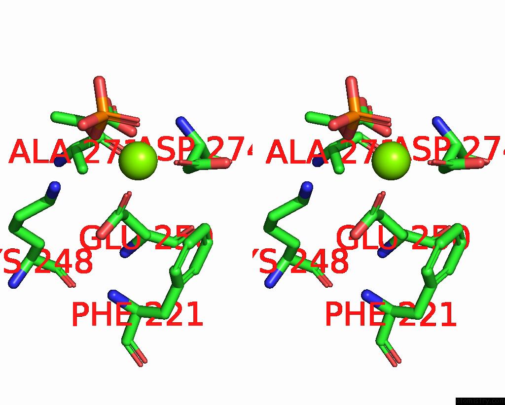
Stereo pair view
Mono view
Stereo pair view
A full contact list of Magnesium with other atoms in the Mg binding
site number 1 of Crystal Structure of Streptococcus Pneumoniae Pyruvate Kinase in Complex with Phosphoenolpyruvate within 5.0Å range:
|
Magnesium binding site 2 out of 2 in 8iaw
Go back to
Magnesium binding site 2 out
of 2 in the Crystal Structure of Streptococcus Pneumoniae Pyruvate Kinase in Complex with Phosphoenolpyruvate
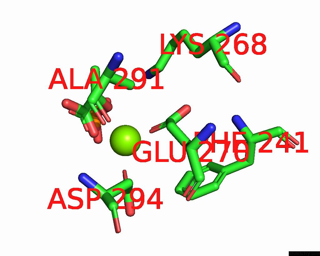
Mono view
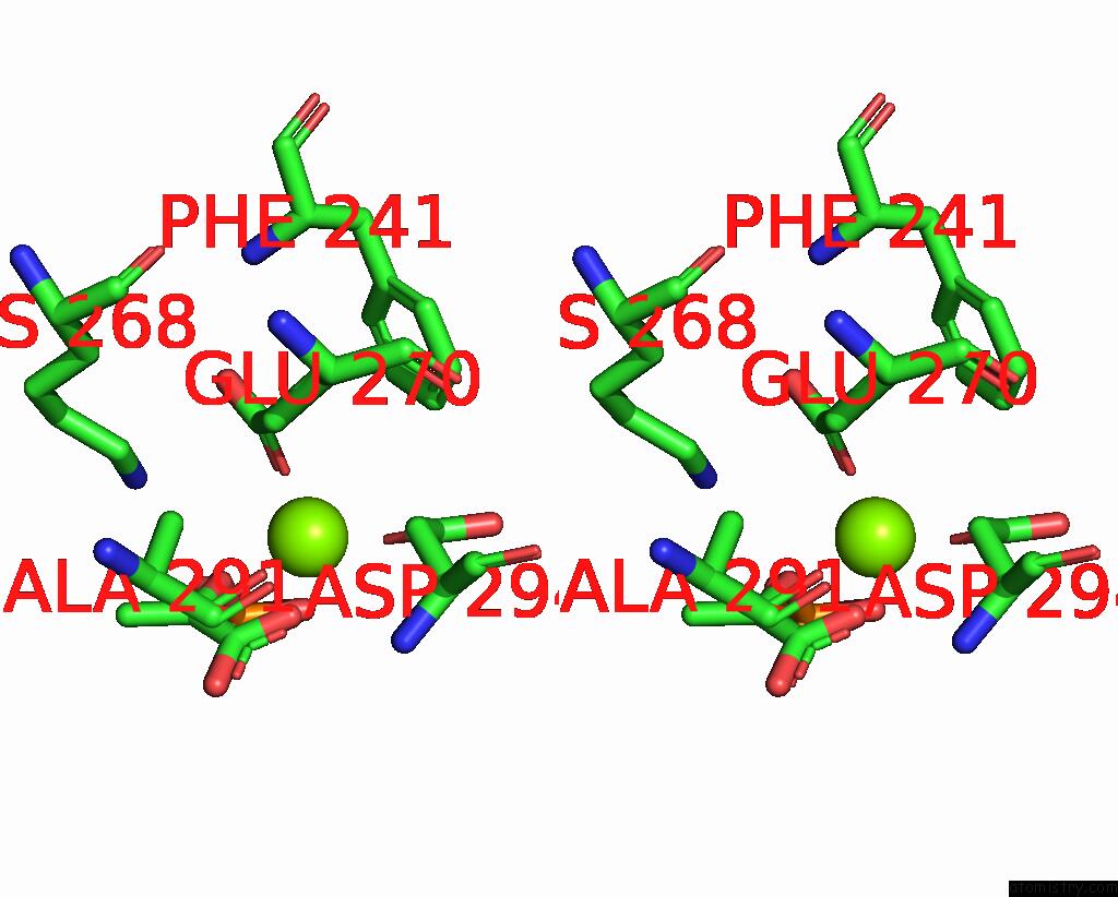
Stereo pair view
Mono view
Stereo pair view
A full contact list of Magnesium with other atoms in the Mg binding
site number 2 of Crystal Structure of Streptococcus Pneumoniae Pyruvate Kinase in Complex with Phosphoenolpyruvate within 5.0Å range:
|
Reference:
A.Taguchi,
R.Nakashima,
K.Nishino.
Functional and Structural Characterization of Streptococcus Pneumoniae Pyruvate Kinase Involved in Fosfomycin Resistance. J.Biol.Chem. 2023.
ISSN: ESSN 1083-351X
DOI: 10.1016/J.JBC.2023.104892
Page generated: Fri Oct 4 09:20:12 2024
ISSN: ESSN 1083-351X
DOI: 10.1016/J.JBC.2023.104892
Last articles
Zn in 9MJ5Zn in 9HNW
Zn in 9G0L
Zn in 9FNE
Zn in 9DZN
Zn in 9E0I
Zn in 9D32
Zn in 9DAK
Zn in 8ZXC
Zn in 8ZUF