Magnesium »
PDB 8j3y-8jgq »
8jbl »
Magnesium in PDB 8jbl: Crystal Structure of Na+,K+-Atpase in the E1.MG2+ State
Protein crystallography data
The structure of Crystal Structure of Na+,K+-Atpase in the E1.MG2+ State, PDB code: 8jbl
was solved by
R.Kanai,
B.Vilsen,
F.Cornelius,
C.Toyoshima,
with X-Ray Crystallography technique. A brief refinement statistics is given in the table below:
| Resolution Low / High (Å) | 11.99 / 3.00 |
| Space group | P 1 21 1 |
| Cell size a, b, c (Å), α, β, γ (°) | 196.797, 74.319, 163.553, 90, 116.29, 90 |
| R / Rfree (%) | 23.3 / 28.2 |
Magnesium Binding Sites:
The binding sites of Magnesium atom in the Crystal Structure of Na+,K+-Atpase in the E1.MG2+ State
(pdb code 8jbl). This binding sites where shown within
5.0 Angstroms radius around Magnesium atom.
In total 6 binding sites of Magnesium where determined in the Crystal Structure of Na+,K+-Atpase in the E1.MG2+ State, PDB code: 8jbl:
Jump to Magnesium binding site number: 1; 2; 3; 4; 5; 6;
In total 6 binding sites of Magnesium where determined in the Crystal Structure of Na+,K+-Atpase in the E1.MG2+ State, PDB code: 8jbl:
Jump to Magnesium binding site number: 1; 2; 3; 4; 5; 6;
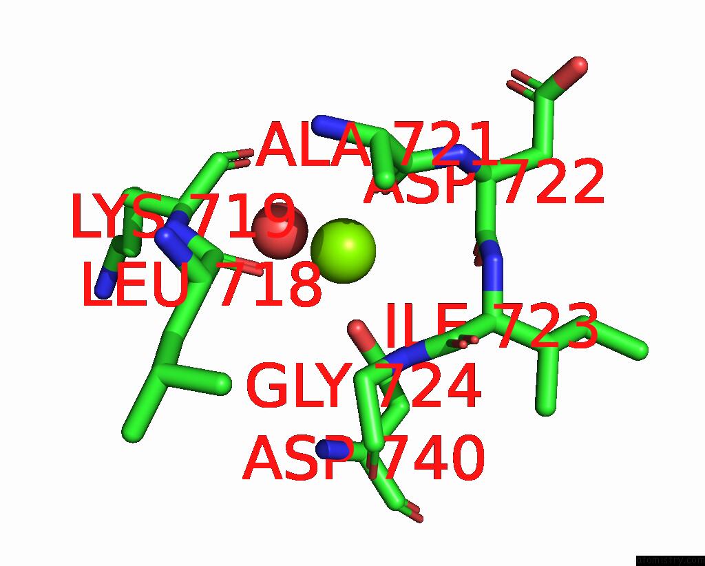
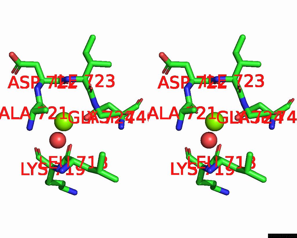
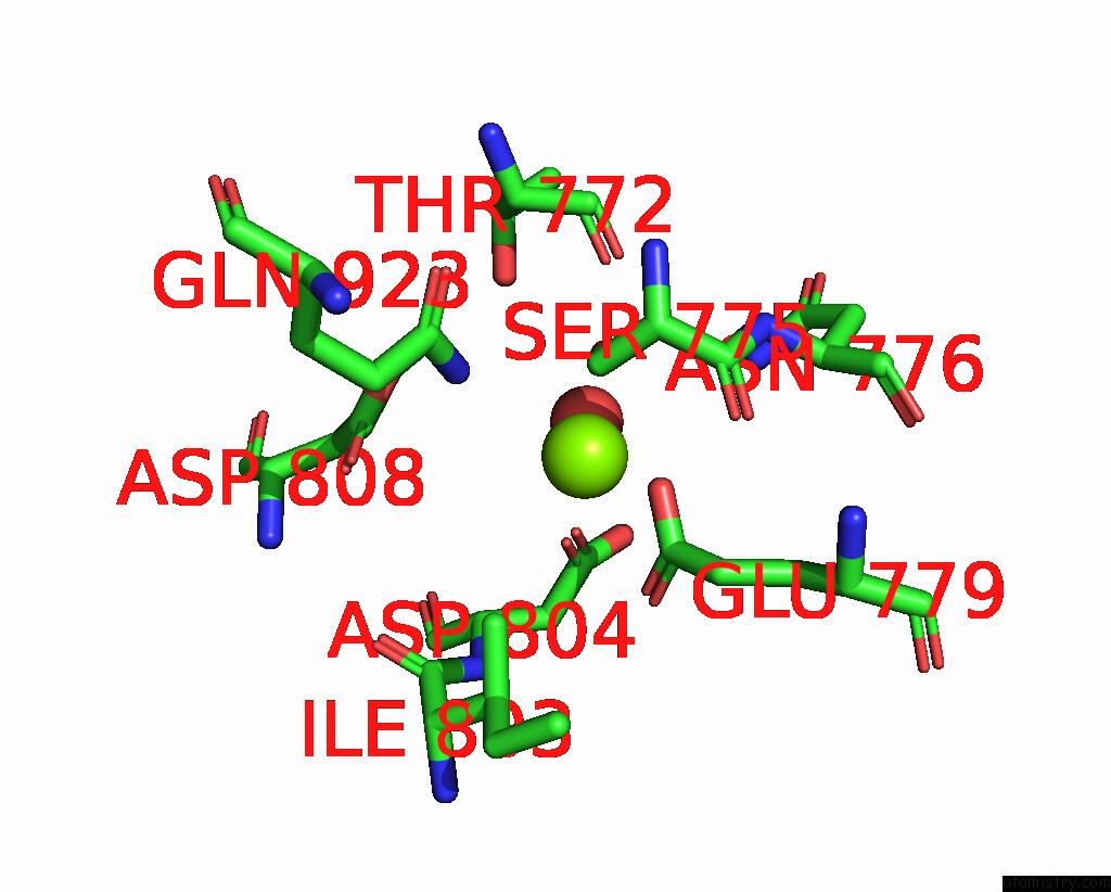
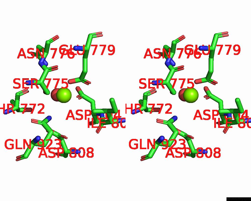
Magnesium binding site 1 out of 6 in 8jbl
Go back to
Magnesium binding site 1 out
of 6 in the Crystal Structure of Na+,K+-Atpase in the E1.MG2+ State
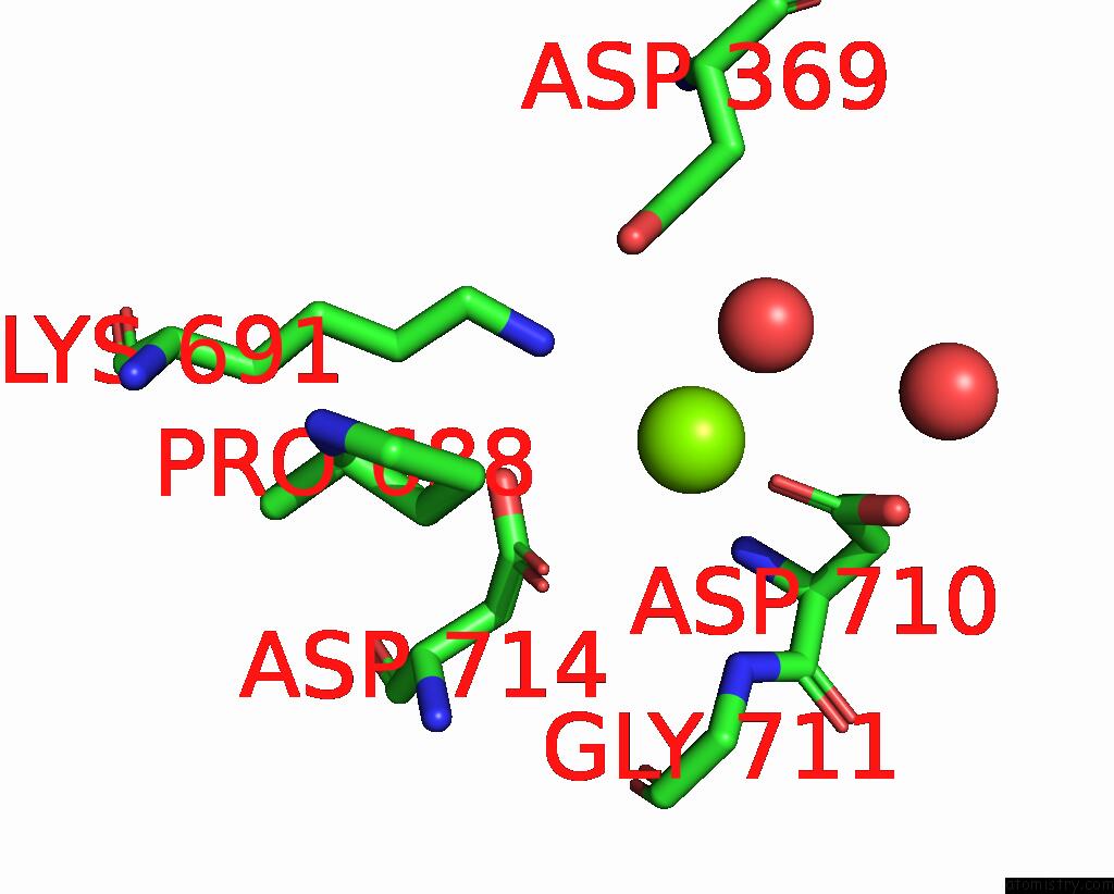
Mono view
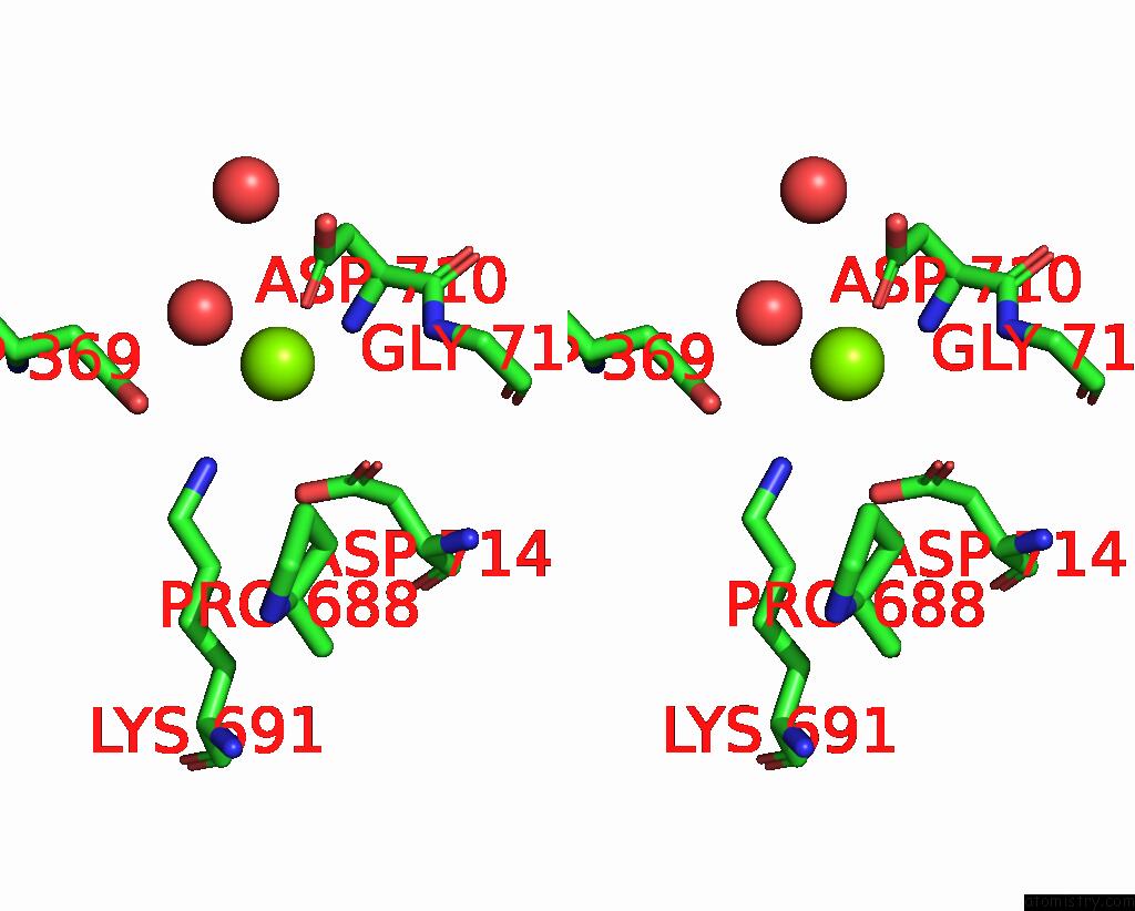
Stereo pair view
Mono view
Stereo pair view
A full contact list of Magnesium with other atoms in the Mg binding
site number 1 of Crystal Structure of Na+,K+-Atpase in the E1.MG2+ State within 5.0Å range:
|
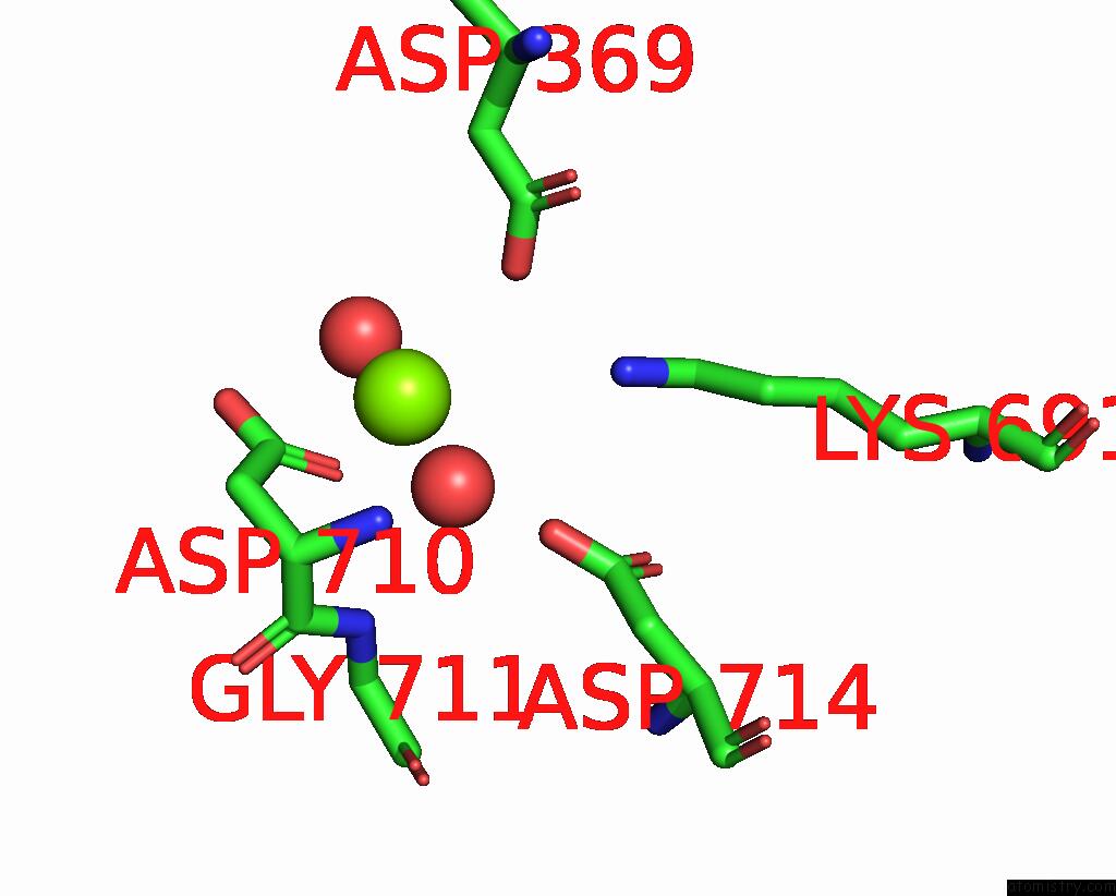
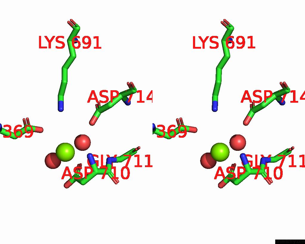
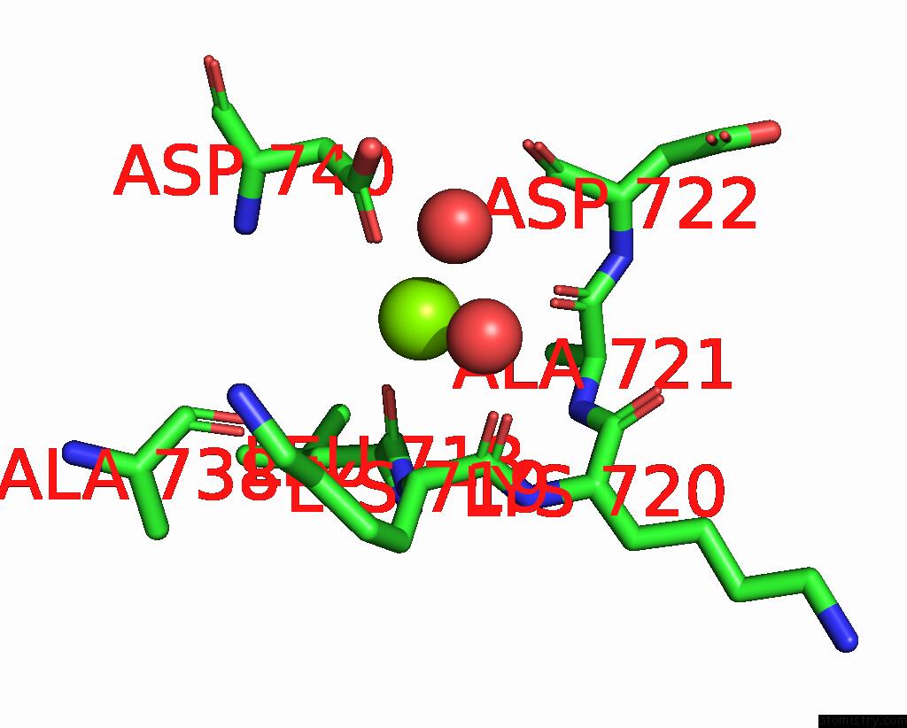
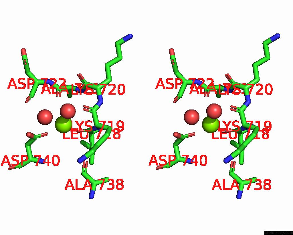
Magnesium binding site 2 out of 6 in 8jbl
Go back to
Magnesium binding site 2 out
of 6 in the Crystal Structure of Na+,K+-Atpase in the E1.MG2+ State
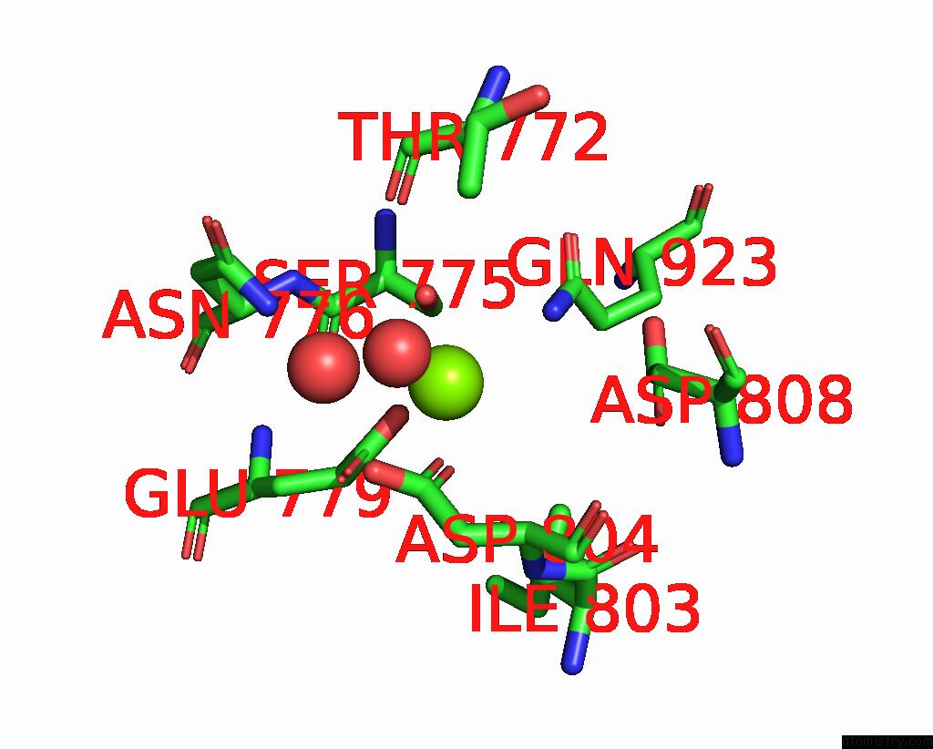
Mono view
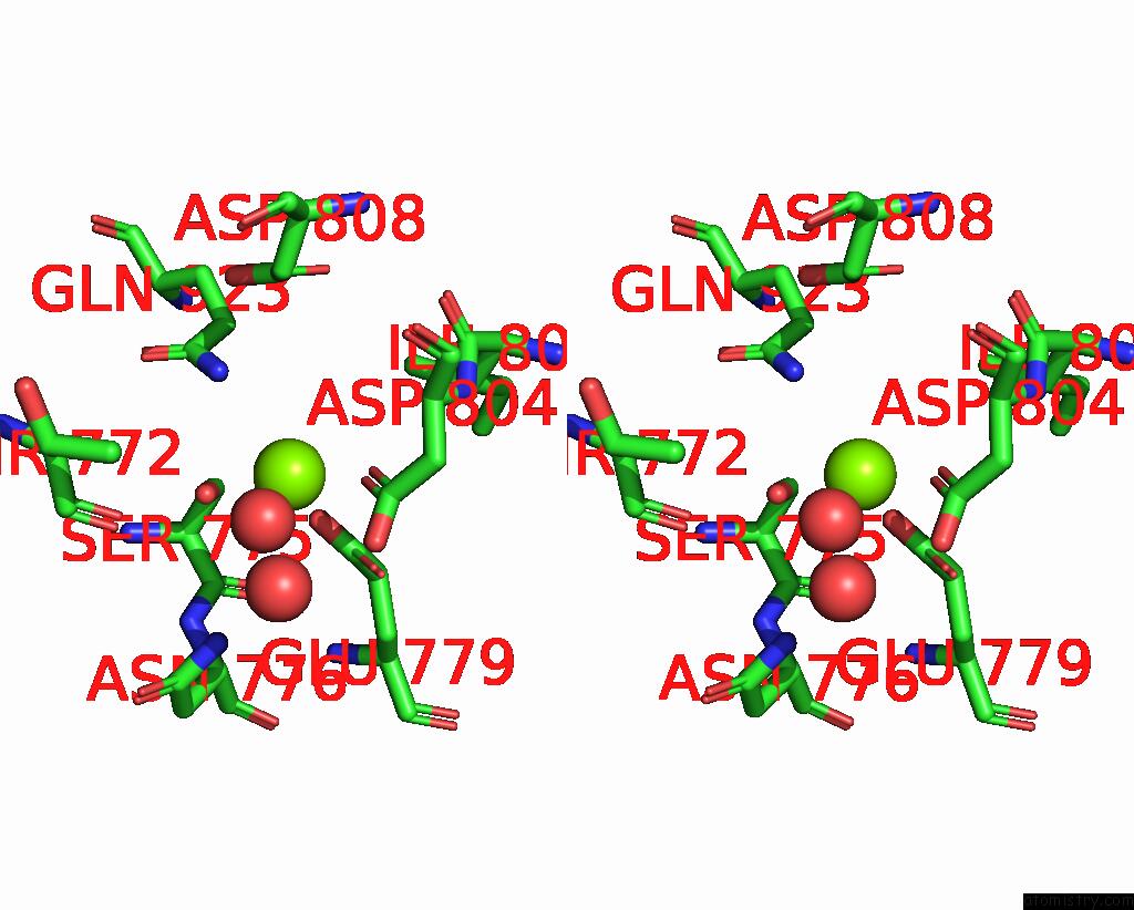
Stereo pair view
Mono view
Stereo pair view
A full contact list of Magnesium with other atoms in the Mg binding
site number 2 of Crystal Structure of Na+,K+-Atpase in the E1.MG2+ State within 5.0Å range:
|
Magnesium binding site 3 out of 6 in 8jbl
Go back to
Magnesium binding site 3 out
of 6 in the Crystal Structure of Na+,K+-Atpase in the E1.MG2+ State
Mono view
Stereo pair view
Mono view
Stereo pair view
A full contact list of Magnesium with other atoms in the Mg binding
site number 3 of Crystal Structure of Na+,K+-Atpase in the E1.MG2+ State within 5.0Å range:
|
Magnesium binding site 4 out of 6 in 8jbl
Go back to
Magnesium binding site 4 out
of 6 in the Crystal Structure of Na+,K+-Atpase in the E1.MG2+ State
Mono view
Stereo pair view
Mono view
Stereo pair view
A full contact list of Magnesium with other atoms in the Mg binding
site number 4 of Crystal Structure of Na+,K+-Atpase in the E1.MG2+ State within 5.0Å range:
|
Magnesium binding site 5 out of 6 in 8jbl
Go back to
Magnesium binding site 5 out
of 6 in the Crystal Structure of Na+,K+-Atpase in the E1.MG2+ State
Mono view
Stereo pair view
Mono view
Stereo pair view
A full contact list of Magnesium with other atoms in the Mg binding
site number 5 of Crystal Structure of Na+,K+-Atpase in the E1.MG2+ State within 5.0Å range:
|
Magnesium binding site 6 out of 6 in 8jbl
Go back to
Magnesium binding site 6 out
of 6 in the Crystal Structure of Na+,K+-Atpase in the E1.MG2+ State
Mono view
Stereo pair view
Mono view
Stereo pair view
A full contact list of Magnesium with other atoms in the Mg binding
site number 6 of Crystal Structure of Na+,K+-Atpase in the E1.MG2+ State within 5.0Å range:
|
Reference:
R.Kanai,
B.Vilsen,
F.Cornelius,
C.Toyoshima.
Crystal Structures of Na + ,K + -Atpase Reveal the Mechanism That Converts the K + -Bound Form to Na + -Bound Form and Opens and Closes the Cytoplasmic Gate. Febs Lett. 2023.
ISSN: ISSN 0014-5793
PubMed: 37357620
DOI: 10.1002/1873-3468.14689
Page generated: Fri Oct 4 12:21:43 2024
ISSN: ISSN 0014-5793
PubMed: 37357620
DOI: 10.1002/1873-3468.14689
Last articles
Zn in 9MJ5Zn in 9HNW
Zn in 9G0L
Zn in 9FNE
Zn in 9DZN
Zn in 9E0I
Zn in 9D32
Zn in 9DAK
Zn in 8ZXC
Zn in 8ZUF