Magnesium »
PDB 8psz-8q2n »
8q1c »
Magnesium in PDB 8q1c: Substrate-Free D10N,P146A Variant of Beta-Phosphoglucomutase From Lactococcus Lactis
Protein crystallography data
The structure of Substrate-Free D10N,P146A Variant of Beta-Phosphoglucomutase From Lactococcus Lactis, PDB code: 8q1c
was solved by
F.A.Cruz-Navarrete,
N.J.Baxter,
A.J.Flinders,
A.Buzoianu,
M.J.Cliff,
P.J.Baker,
J.P.Waltho,
with X-Ray Crystallography technique. A brief refinement statistics is given in the table below:
| Resolution Low / High (Å) | 52.70 / 1.68 |
| Space group | P 1 21 1 |
| Cell size a, b, c (Å), α, β, γ (°) | 38.65, 117.251, 53.25, 90, 98.64, 90 |
| R / Rfree (%) | 21.1 / 27 |
Magnesium Binding Sites:
The binding sites of Magnesium atom in the Substrate-Free D10N,P146A Variant of Beta-Phosphoglucomutase From Lactococcus Lactis
(pdb code 8q1c). This binding sites where shown within
5.0 Angstroms radius around Magnesium atom.
In total 2 binding sites of Magnesium where determined in the Substrate-Free D10N,P146A Variant of Beta-Phosphoglucomutase From Lactococcus Lactis, PDB code: 8q1c:
Jump to Magnesium binding site number: 1; 2;
In total 2 binding sites of Magnesium where determined in the Substrate-Free D10N,P146A Variant of Beta-Phosphoglucomutase From Lactococcus Lactis, PDB code: 8q1c:
Jump to Magnesium binding site number: 1; 2;
Magnesium binding site 1 out of 2 in 8q1c
Go back to
Magnesium binding site 1 out
of 2 in the Substrate-Free D10N,P146A Variant of Beta-Phosphoglucomutase From Lactococcus Lactis
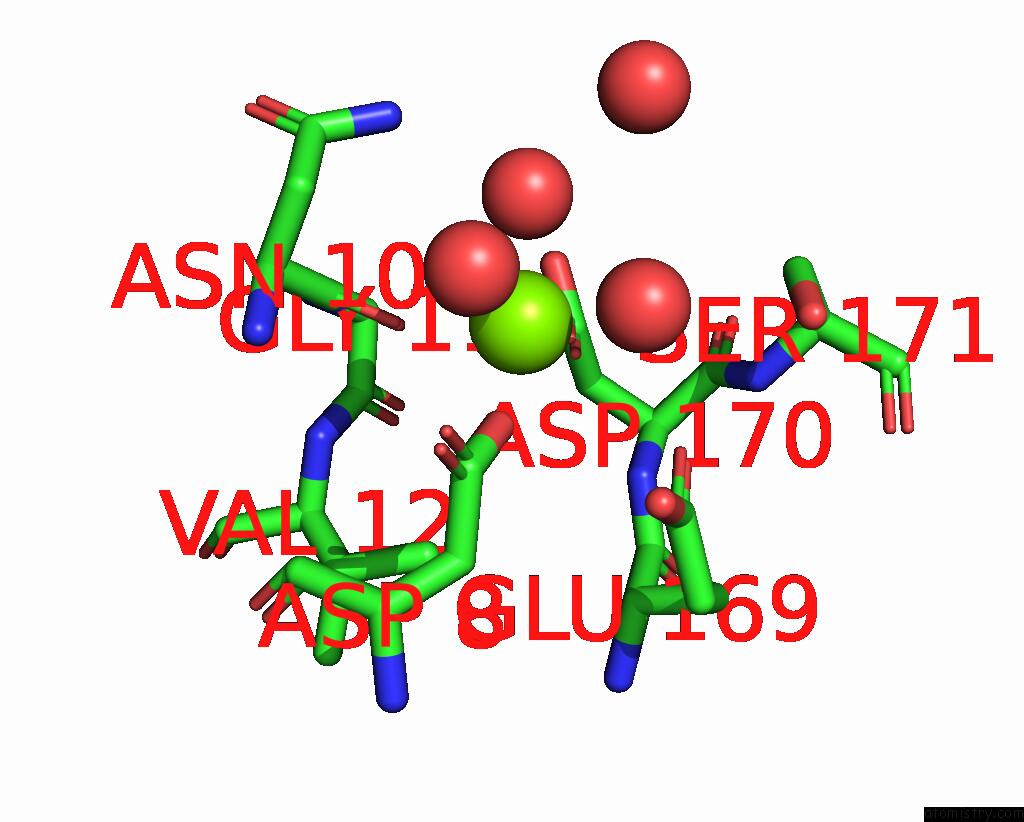
Mono view
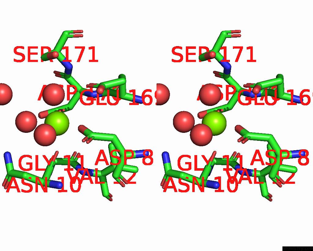
Stereo pair view
Mono view
Stereo pair view
A full contact list of Magnesium with other atoms in the Mg binding
site number 1 of Substrate-Free D10N,P146A Variant of Beta-Phosphoglucomutase From Lactococcus Lactis within 5.0Å range:
|
Magnesium binding site 2 out of 2 in 8q1c
Go back to
Magnesium binding site 2 out
of 2 in the Substrate-Free D10N,P146A Variant of Beta-Phosphoglucomutase From Lactococcus Lactis
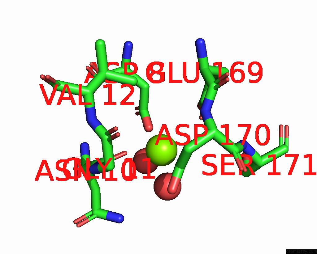
Mono view
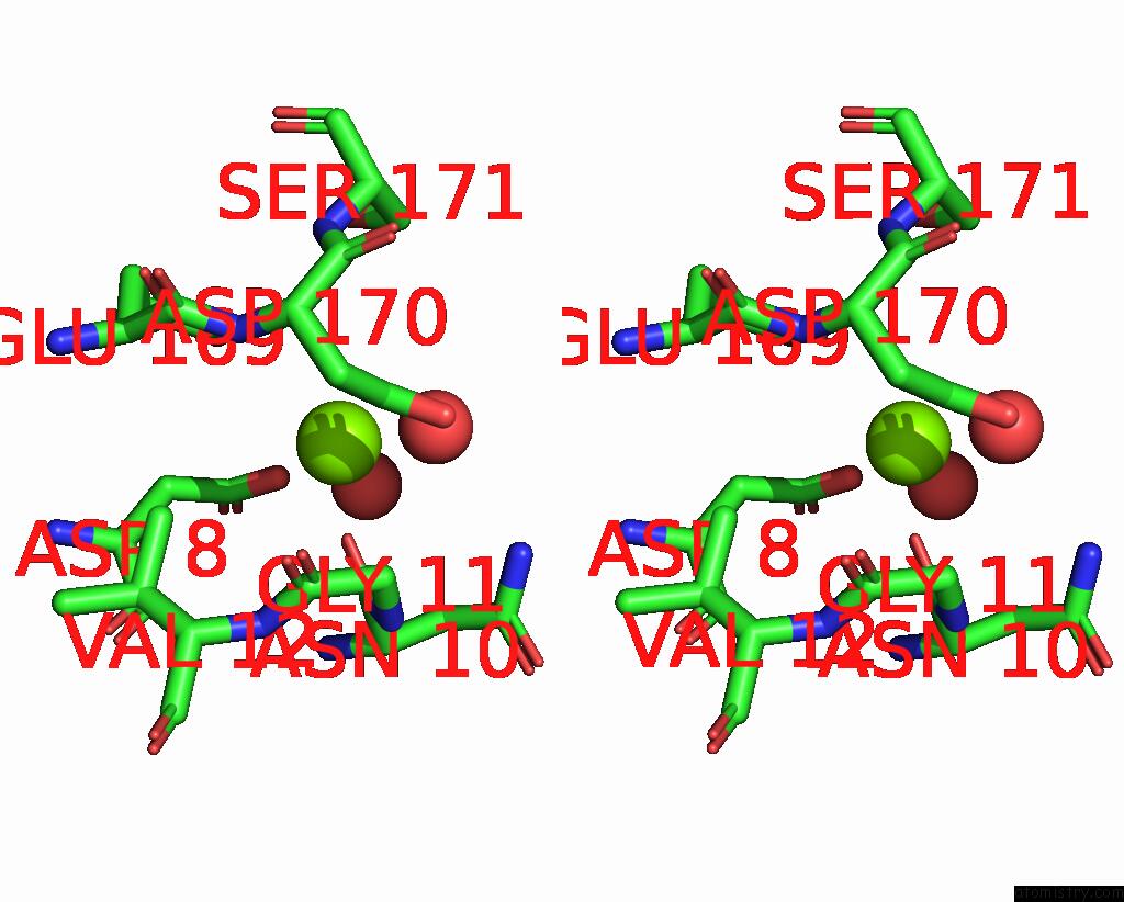
Stereo pair view
Mono view
Stereo pair view
A full contact list of Magnesium with other atoms in the Mg binding
site number 2 of Substrate-Free D10N,P146A Variant of Beta-Phosphoglucomutase From Lactococcus Lactis within 5.0Å range:
|
Reference:
F.A.Cruz-Navarrete,
N.J.Baxter,
A.J.Flinders,
A.Buzoianu,
M.J.Cliff,
P.J.Baker,
J.P.Waltho.
Peri Active Site Catalysis of Proline Isomerisation Is the Molecular Basis of Allomorphy in Beta-Phosphoglucomutase. Commun Biol V. 7 909 2024.
ISSN: ESSN 2399-3642
PubMed: 39068257
DOI: 10.1038/S42003-024-06577-9
Page generated: Fri Oct 4 16:29:10 2024
ISSN: ESSN 2399-3642
PubMed: 39068257
DOI: 10.1038/S42003-024-06577-9
Last articles
Zn in 9MJ5Zn in 9HNW
Zn in 9G0L
Zn in 9FNE
Zn in 9DZN
Zn in 9E0I
Zn in 9D32
Zn in 9DAK
Zn in 8ZXC
Zn in 8ZUF