Magnesium »
PDB 8qs8-8qyq »
8qwn »
Magnesium in PDB 8qwn: Structure of P53 Cancer Mutant Y220C (Hexagonal Crystal Form)
Protein crystallography data
The structure of Structure of P53 Cancer Mutant Y220C (Hexagonal Crystal Form), PDB code: 8qwn
was solved by
D.I.Balourdas,
S.Knapp,
A.C.Joerger,
Structural Genomics Consortium(Sgc),
with X-Ray Crystallography technique. A brief refinement statistics is given in the table below:
| Resolution Low / High (Å) | 39.30 / 1.44 |
| Space group | P 65 2 2 |
| Cell size a, b, c (Å), α, β, γ (°) | 45.33, 45.33, 332.64, 90, 90, 120 |
| R / Rfree (%) | 16.9 / 20.9 |
Other elements in 8qwn:
The structure of Structure of P53 Cancer Mutant Y220C (Hexagonal Crystal Form) also contains other interesting chemical elements:
| Chlorine | (Cl) | 1 atom |
| Zinc | (Zn) | 1 atom |
Magnesium Binding Sites:
The binding sites of Magnesium atom in the Structure of P53 Cancer Mutant Y220C (Hexagonal Crystal Form)
(pdb code 8qwn). This binding sites where shown within
5.0 Angstroms radius around Magnesium atom.
In total only one binding site of Magnesium was determined in the Structure of P53 Cancer Mutant Y220C (Hexagonal Crystal Form), PDB code: 8qwn:
In total only one binding site of Magnesium was determined in the Structure of P53 Cancer Mutant Y220C (Hexagonal Crystal Form), PDB code: 8qwn:
Magnesium binding site 1 out of 1 in 8qwn
Go back to
Magnesium binding site 1 out
of 1 in the Structure of P53 Cancer Mutant Y220C (Hexagonal Crystal Form)
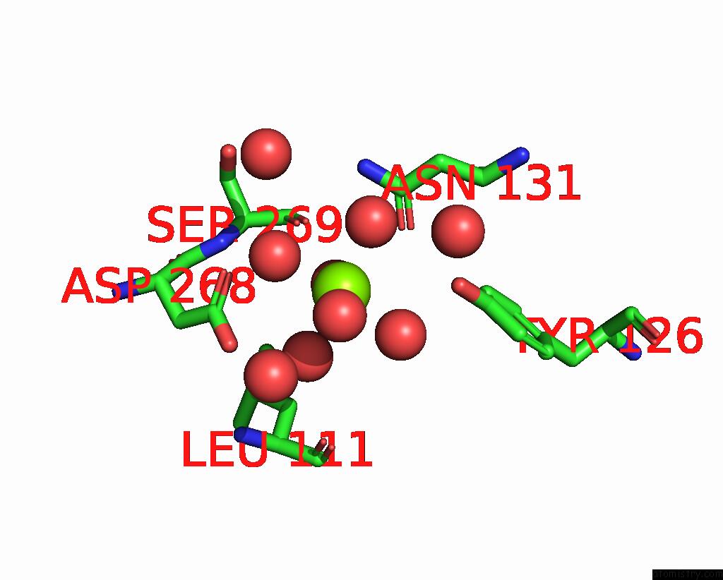
Mono view
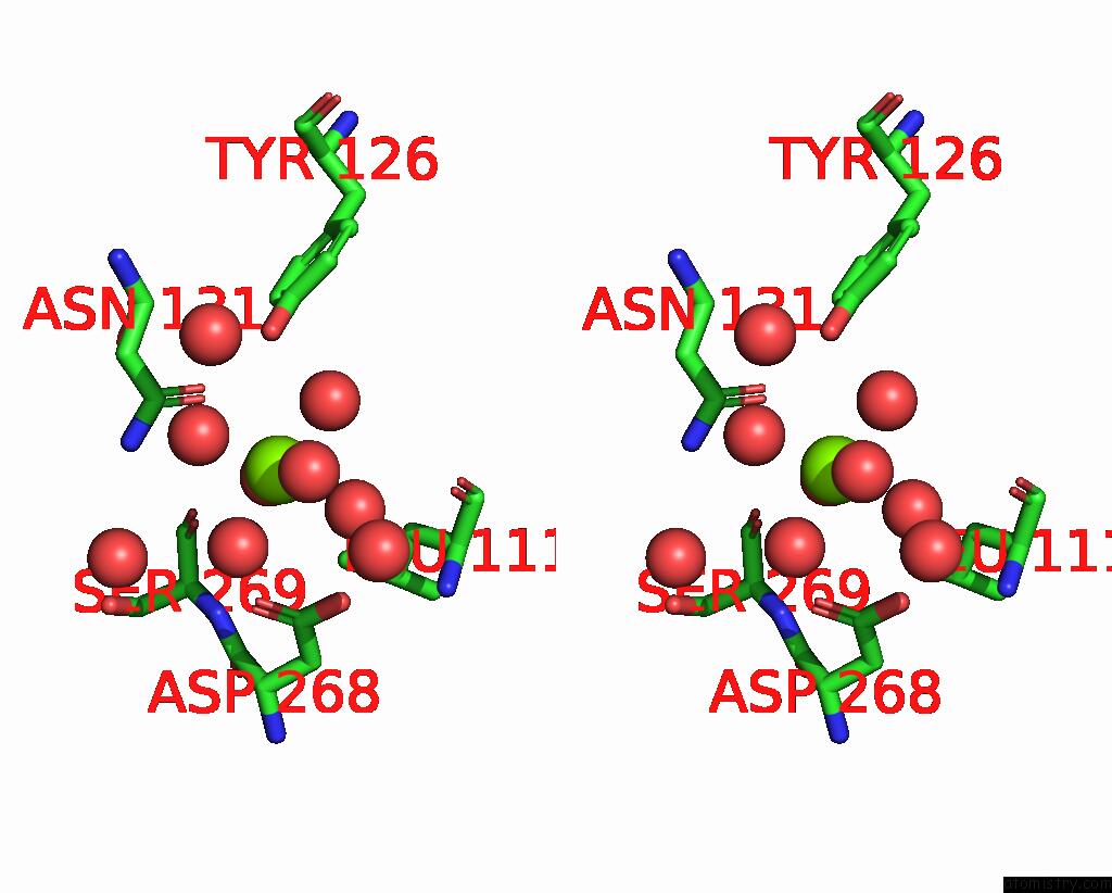
Stereo pair view
Mono view
Stereo pair view
A full contact list of Magnesium with other atoms in the Mg binding
site number 1 of Structure of P53 Cancer Mutant Y220C (Hexagonal Crystal Form) within 5.0Å range:
|
Reference:
D.I.Balourdas,
A.M.Markl,
A.Kramer,
G.Settanni,
A.C.Joerger.
Structural Basis of P53 Inactivation By Cavity-Creating Cancer Mutations and Its Implications For the Development of Mutant P53 Reactivators. Cell Death Dis V. 15 408 2024.
ISSN: ISSN 2041-4889
PubMed: 38862470
DOI: 10.1038/S41419-024-06739-X
Page generated: Fri Aug 15 13:36:58 2025
ISSN: ISSN 2041-4889
PubMed: 38862470
DOI: 10.1038/S41419-024-06739-X
Last articles
Mg in 8THYMg in 8THX
Mg in 8TF0
Mg in 8THD
Mg in 8THC
Mg in 8THB
Mg in 8TGS
Mg in 8TFW
Mg in 8TFA
Mg in 8TEF