Magnesium »
PDB 8uee-8un4 »
8uel »
Magnesium in PDB 8uel: Crystal Structure of Enolase From Litopenaeus Vannamei
Protein crystallography data
The structure of Crystal Structure of Enolase From Litopenaeus Vannamei, PDB code: 8uel
was solved by
X.Chang,
G.Zhao,
with X-Ray Crystallography technique. A brief refinement statistics is given in the table below:
| Resolution Low / High (Å) | 29.68 / 2.49 |
| Space group | P 21 21 21 |
| Cell size a, b, c (Å), α, β, γ (°) | 79.613, 86.571, 155.881, 90, 90, 90 |
| R / Rfree (%) | 16.8 / 22.4 |
Magnesium Binding Sites:
The binding sites of Magnesium atom in the Crystal Structure of Enolase From Litopenaeus Vannamei
(pdb code 8uel). This binding sites where shown within
5.0 Angstroms radius around Magnesium atom.
In total 4 binding sites of Magnesium where determined in the Crystal Structure of Enolase From Litopenaeus Vannamei, PDB code: 8uel:
Jump to Magnesium binding site number: 1; 2; 3; 4;
In total 4 binding sites of Magnesium where determined in the Crystal Structure of Enolase From Litopenaeus Vannamei, PDB code: 8uel:
Jump to Magnesium binding site number: 1; 2; 3; 4;
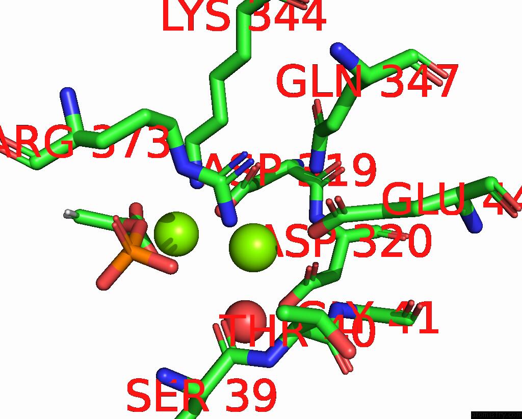
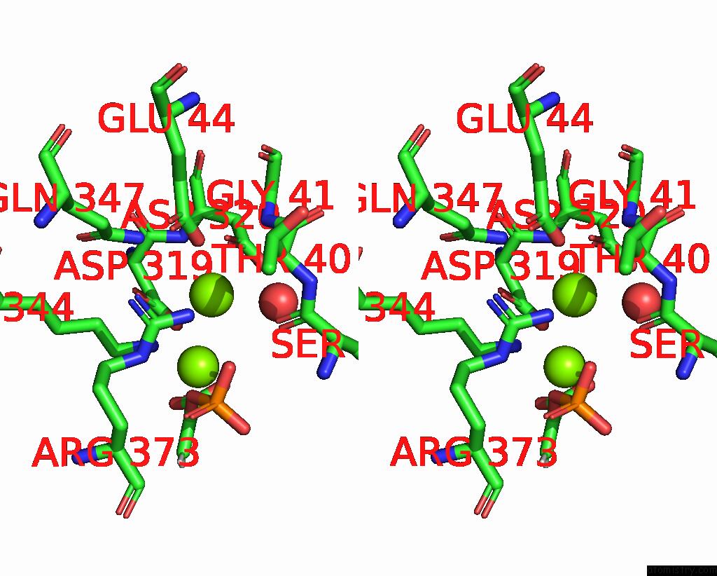
Magnesium binding site 1 out of 4 in 8uel
Go back to
Magnesium binding site 1 out
of 4 in the Crystal Structure of Enolase From Litopenaeus Vannamei

Mono view
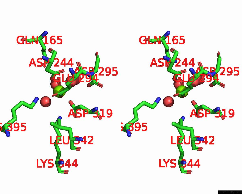
Stereo pair view
Mono view
Stereo pair view
A full contact list of Magnesium with other atoms in the Mg binding
site number 1 of Crystal Structure of Enolase From Litopenaeus Vannamei within 5.0Å range:
|
Magnesium binding site 2 out of 4 in 8uel
Go back to
Magnesium binding site 2 out
of 4 in the Crystal Structure of Enolase From Litopenaeus Vannamei

Mono view
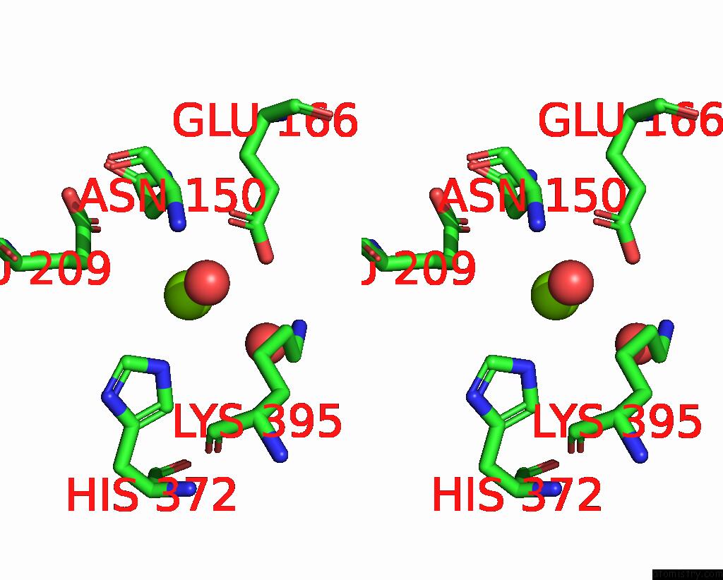
Stereo pair view
Mono view
Stereo pair view
A full contact list of Magnesium with other atoms in the Mg binding
site number 2 of Crystal Structure of Enolase From Litopenaeus Vannamei within 5.0Å range:
|
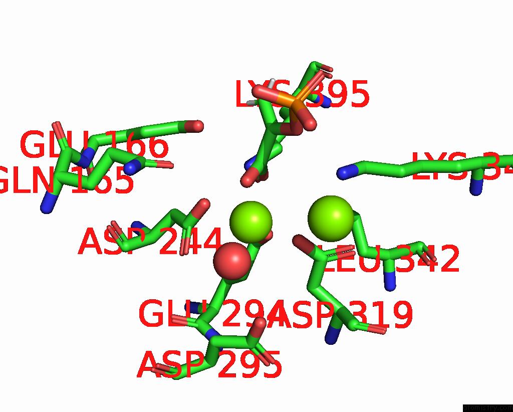
Magnesium binding site 3 out of 4 in 8uel
Go back to
Magnesium binding site 3 out
of 4 in the Crystal Structure of Enolase From Litopenaeus Vannamei
Mono view
Stereo pair view
Mono view
Stereo pair view
A full contact list of Magnesium with other atoms in the Mg binding
site number 3 of Crystal Structure of Enolase From Litopenaeus Vannamei within 5.0Å range:
|
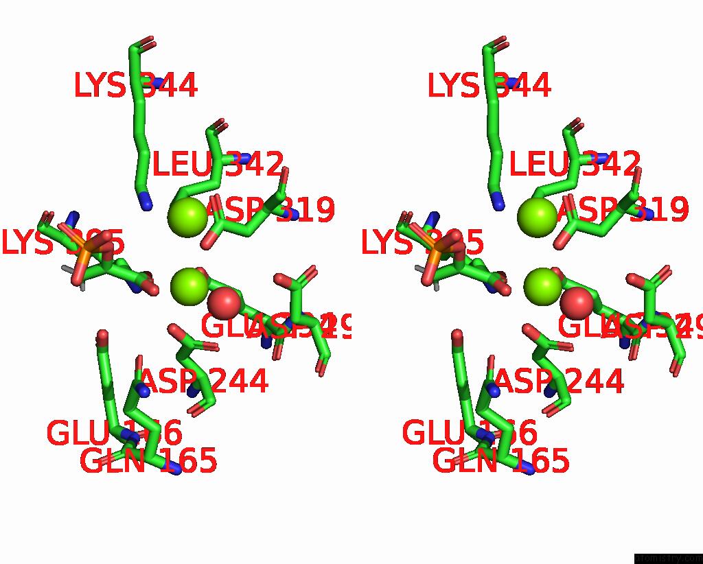
Magnesium binding site 4 out of 4 in 8uel
Go back to
Magnesium binding site 4 out
of 4 in the Crystal Structure of Enolase From Litopenaeus Vannamei
Mono view
Stereo pair view
Mono view
Stereo pair view
A full contact list of Magnesium with other atoms in the Mg binding
site number 4 of Crystal Structure of Enolase From Litopenaeus Vannamei within 5.0Å range:
|
Reference:
X.Chang,
T.Zhang,
J.Zang,
C.Lv,
G.Zhao.
Characterization and Structural Analyses of Enolase From Shrimp ( Litopenaeus Vannamei ). J.Agric.Food Chem. 2023.
ISSN: ESSN 1520-5118
PubMed: 38033172
DOI: 10.1021/ACS.JAFC.3C07135
Page generated: Fri Aug 15 16:54:55 2025
ISSN: ESSN 1520-5118
PubMed: 38033172
DOI: 10.1021/ACS.JAFC.3C07135
Last articles
Mg in 8WMOMg in 8WMN
Mg in 8WMM
Mg in 8WEY
Mg in 8WKG
Mg in 8WKF
Mg in 8WIM
Mg in 8WIL
Mg in 8WH2
Mg in 8WH0