Magnesium »
PDB 8uee-8un4 »
8uk4 »
Magnesium in PDB 8uk4: Crystal Structure of Human Polymerase Eta with Incoming Dtmpnpp Nucleotide Opposite Urea Lesion
Enzymatic activity of Crystal Structure of Human Polymerase Eta with Incoming Dtmpnpp Nucleotide Opposite Urea Lesion
All present enzymatic activity of Crystal Structure of Human Polymerase Eta with Incoming Dtmpnpp Nucleotide Opposite Urea Lesion:
2.7.7.7;
2.7.7.7;
Protein crystallography data
The structure of Crystal Structure of Human Polymerase Eta with Incoming Dtmpnpp Nucleotide Opposite Urea Lesion, PDB code: 8uk4
was solved by
R.Tomar,
M.Egli,
M.P.Stone,
with X-Ray Crystallography technique. A brief refinement statistics is given in the table below:
| Resolution Low / High (Å) | 42.78 / 3.02 |
| Space group | P 61 |
| Cell size a, b, c (Å), α, β, γ (°) | 98.795, 98.795, 80.29, 90, 90, 120 |
| R / Rfree (%) | 17.4 / 26.5 |
Magnesium Binding Sites:
The binding sites of Magnesium atom in the Crystal Structure of Human Polymerase Eta with Incoming Dtmpnpp Nucleotide Opposite Urea Lesion
(pdb code 8uk4). This binding sites where shown within
5.0 Angstroms radius around Magnesium atom.
In total only one binding site of Magnesium was determined in the Crystal Structure of Human Polymerase Eta with Incoming Dtmpnpp Nucleotide Opposite Urea Lesion, PDB code: 8uk4:
In total only one binding site of Magnesium was determined in the Crystal Structure of Human Polymerase Eta with Incoming Dtmpnpp Nucleotide Opposite Urea Lesion, PDB code: 8uk4:
Magnesium binding site 1 out of 1 in 8uk4
Go back to
Magnesium binding site 1 out
of 1 in the Crystal Structure of Human Polymerase Eta with Incoming Dtmpnpp Nucleotide Opposite Urea Lesion
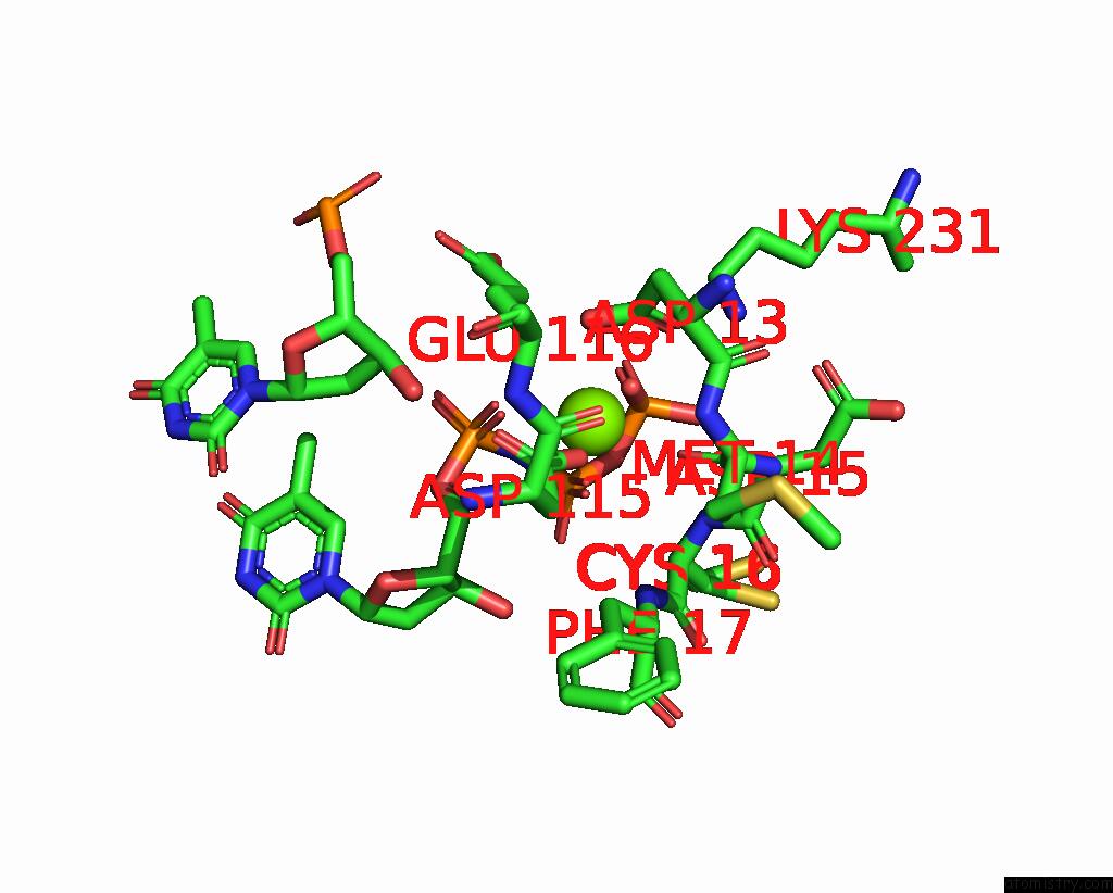
Mono view
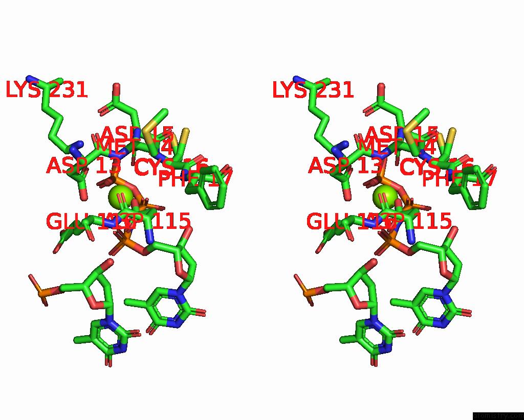
Stereo pair view
Mono view
Stereo pair view
A full contact list of Magnesium with other atoms in the Mg binding
site number 1 of Crystal Structure of Human Polymerase Eta with Incoming Dtmpnpp Nucleotide Opposite Urea Lesion within 5.0Å range:
|
Reference:
R.Tomar,
S.Li,
M.Egli,
M.P.Stone.
Replication Bypass of the N -(2-Deoxy-D-Erythro-Pentofuranosyl)-Urea Dna Lesion By Human Dna Polymerase Eta. Biochemistry 2024.
ISSN: ISSN 0006-2960
PubMed: 38413007
DOI: 10.1021/ACS.BIOCHEM.3C00569
Page generated: Fri Aug 15 16:58:19 2025
ISSN: ISSN 0006-2960
PubMed: 38413007
DOI: 10.1021/ACS.BIOCHEM.3C00569
Last articles
Mg in 8WMOMg in 8WMN
Mg in 8WMM
Mg in 8WEY
Mg in 8WKG
Mg in 8WKF
Mg in 8WIM
Mg in 8WIL
Mg in 8WH2
Mg in 8WH0