Magnesium »
PDB 8xw7-8y9l »
8xzr »
Magnesium in PDB 8xzr: Crystal Structure of Fole Riboswitch with 8-NH2 Guanine
Protein crystallography data
The structure of Crystal Structure of Fole Riboswitch with 8-NH2 Guanine, PDB code: 8xzr
was solved by
C.Y.Li,
A.M.Ren,
with X-Ray Crystallography technique. A brief refinement statistics is given in the table below:
| Resolution Low / High (Å) | 25.15 / 1.83 |
| Space group | P 21 21 21 |
| Cell size a, b, c (Å), α, β, γ (°) | 50.299, 58.59, 61.119, 90, 90, 90 |
| R / Rfree (%) | 21.6 / 25 |
Magnesium Binding Sites:
The binding sites of Magnesium atom in the Crystal Structure of Fole Riboswitch with 8-NH2 Guanine
(pdb code 8xzr). This binding sites where shown within
5.0 Angstroms radius around Magnesium atom.
In total 7 binding sites of Magnesium where determined in the Crystal Structure of Fole Riboswitch with 8-NH2 Guanine, PDB code: 8xzr:
Jump to Magnesium binding site number: 1; 2; 3; 4; 5; 6; 7;
In total 7 binding sites of Magnesium where determined in the Crystal Structure of Fole Riboswitch with 8-NH2 Guanine, PDB code: 8xzr:
Jump to Magnesium binding site number: 1; 2; 3; 4; 5; 6; 7;
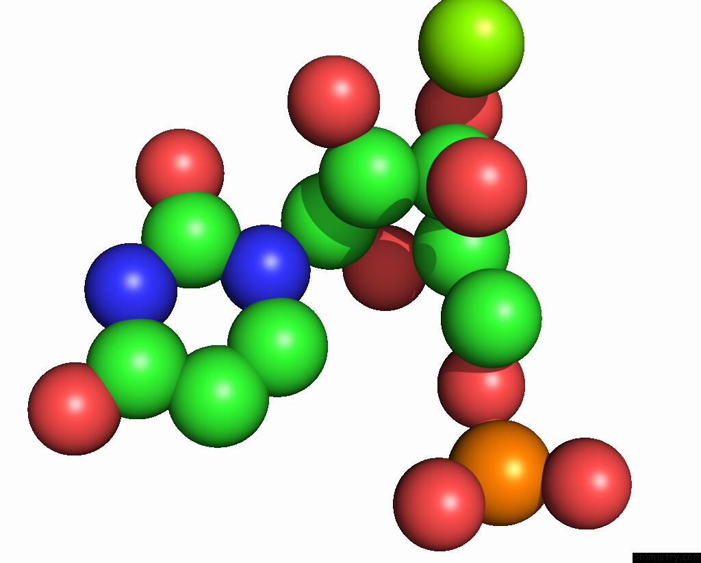
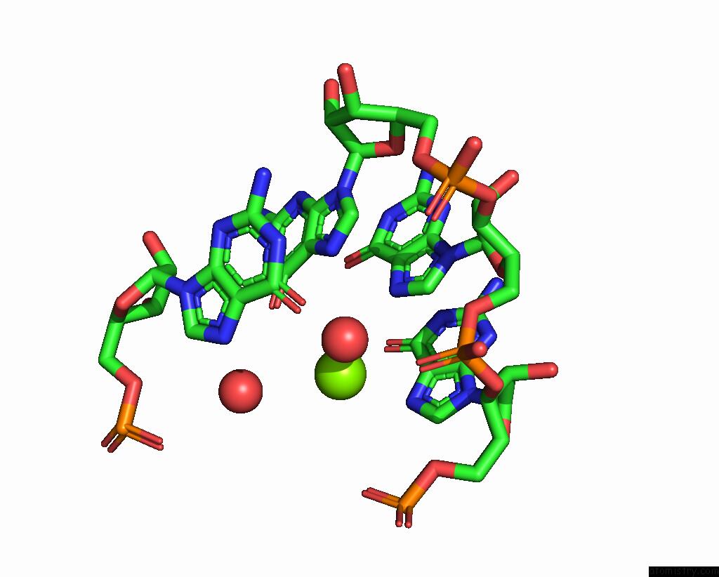
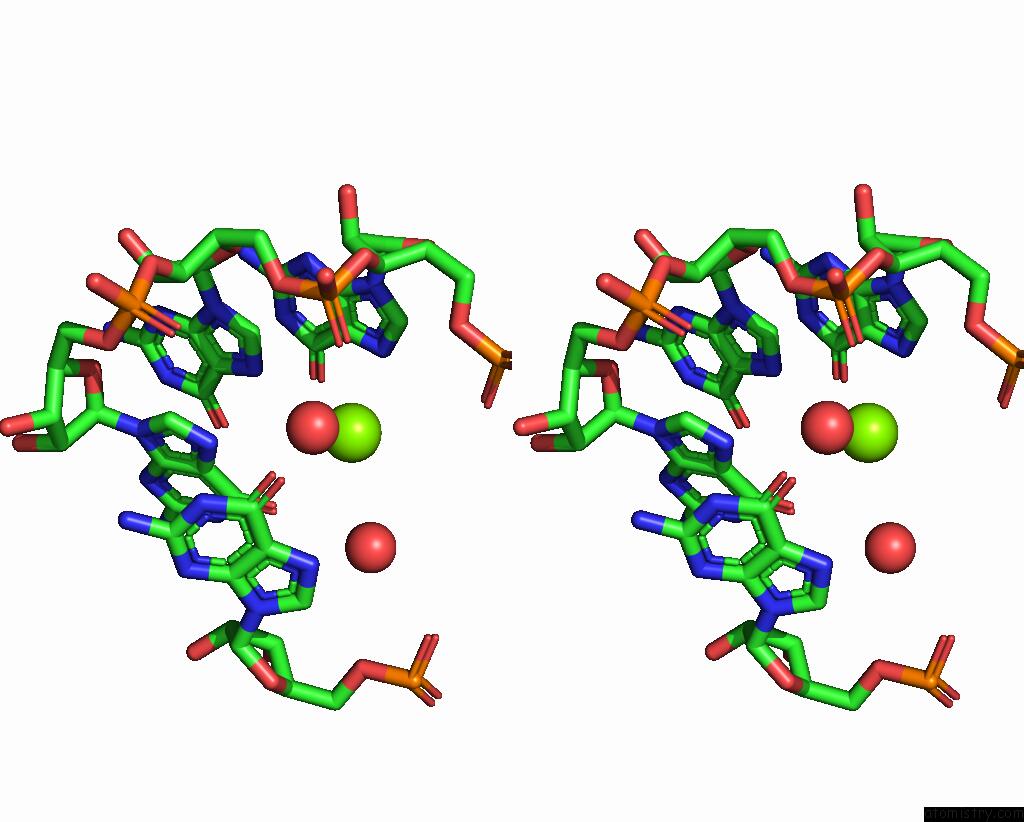
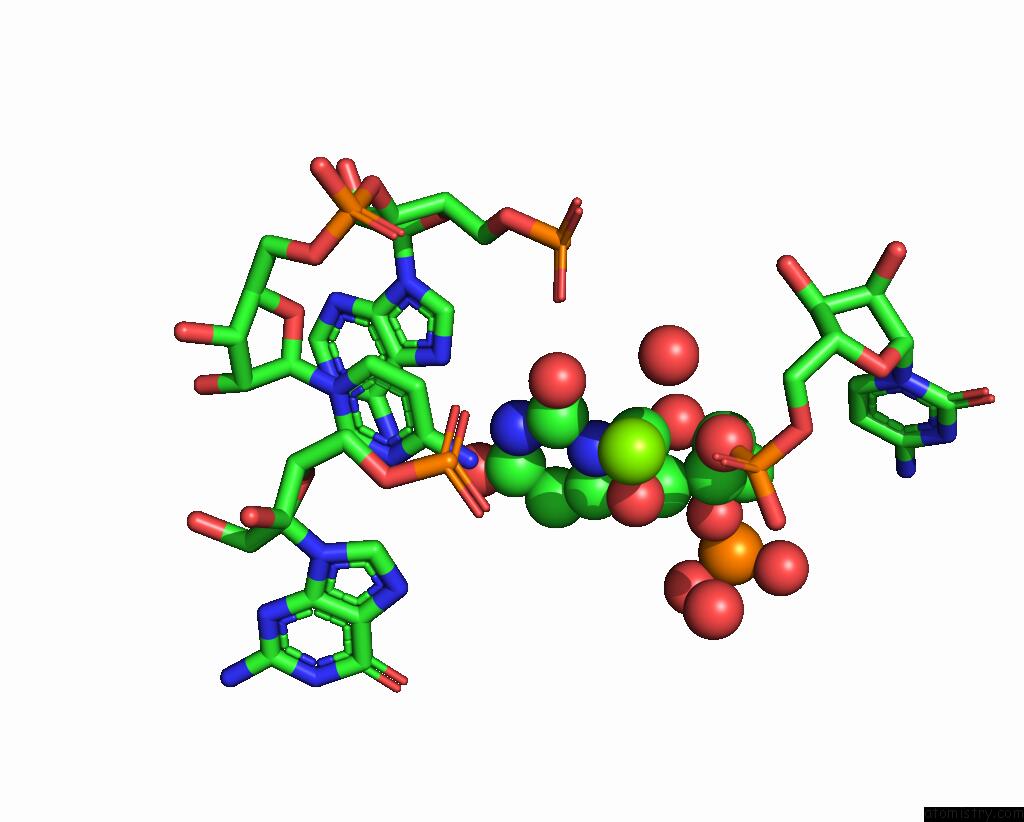
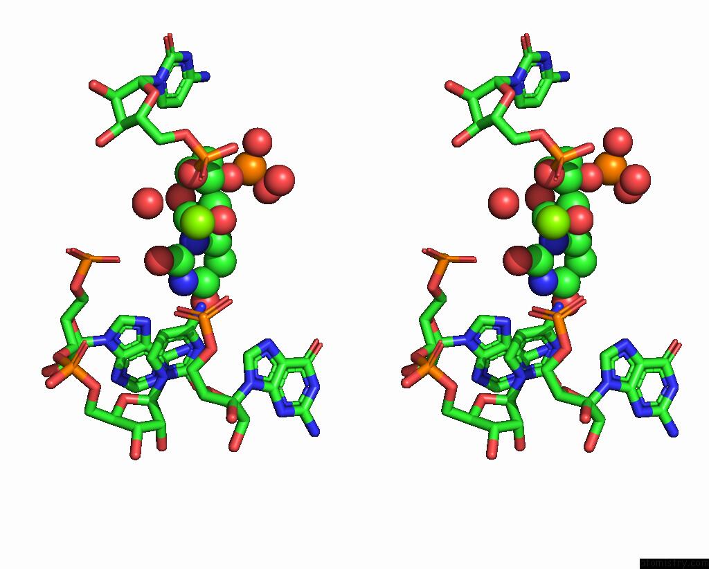
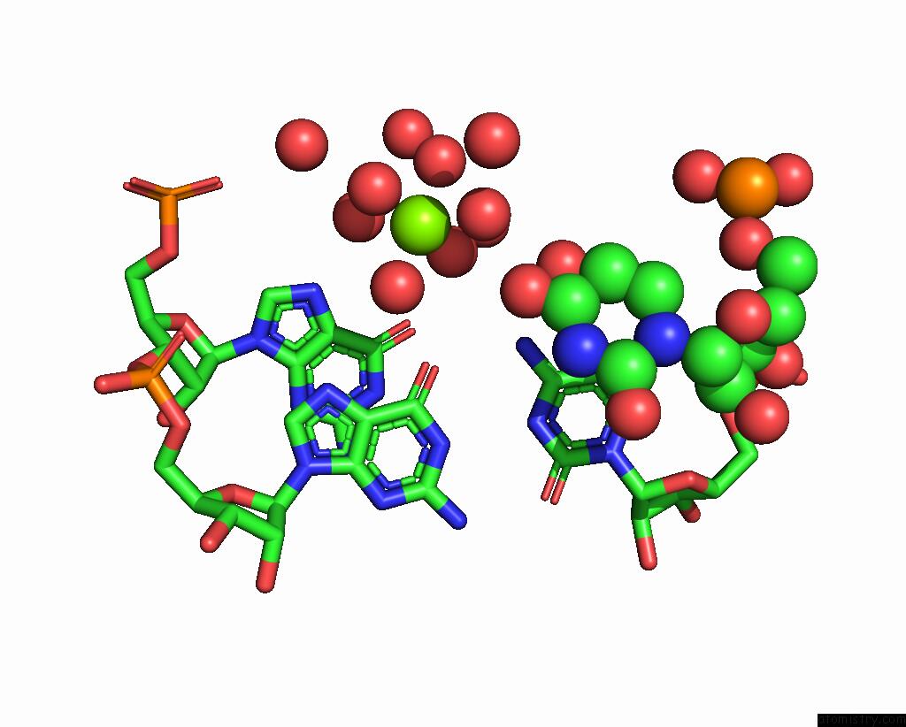
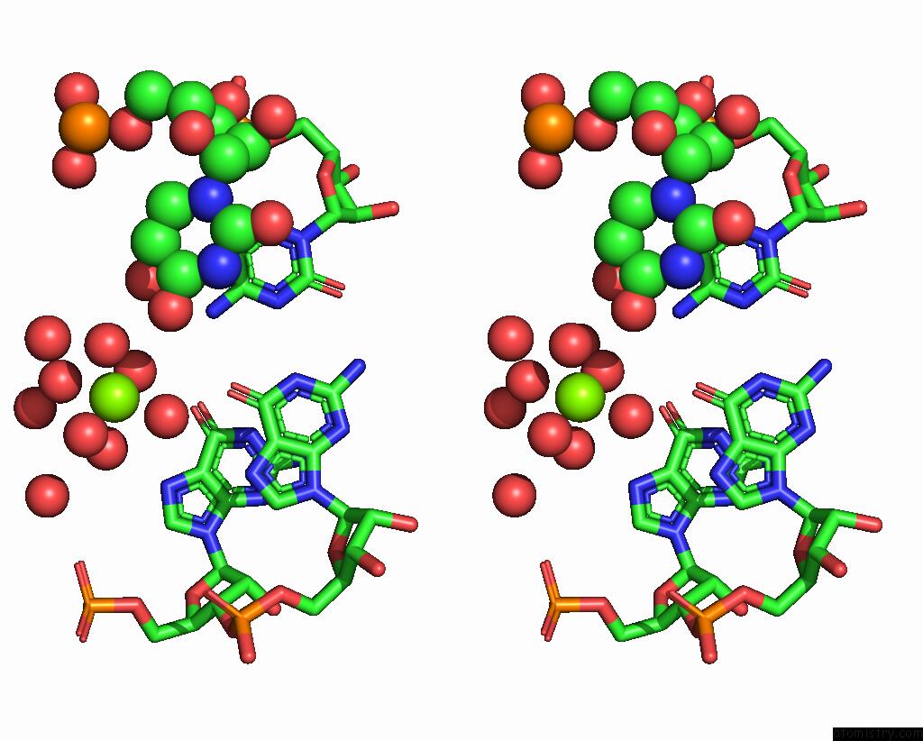
Magnesium binding site 1 out of 7 in 8xzr
Go back to
Magnesium binding site 1 out
of 7 in the Crystal Structure of Fole Riboswitch with 8-NH2 Guanine
Mono view
Stereo pair view
Mono view
Stereo pair view
A full contact list of Magnesium with other atoms in the Mg binding
site number 1 of Crystal Structure of Fole Riboswitch with 8-NH2 Guanine within 5.0Å range:
|
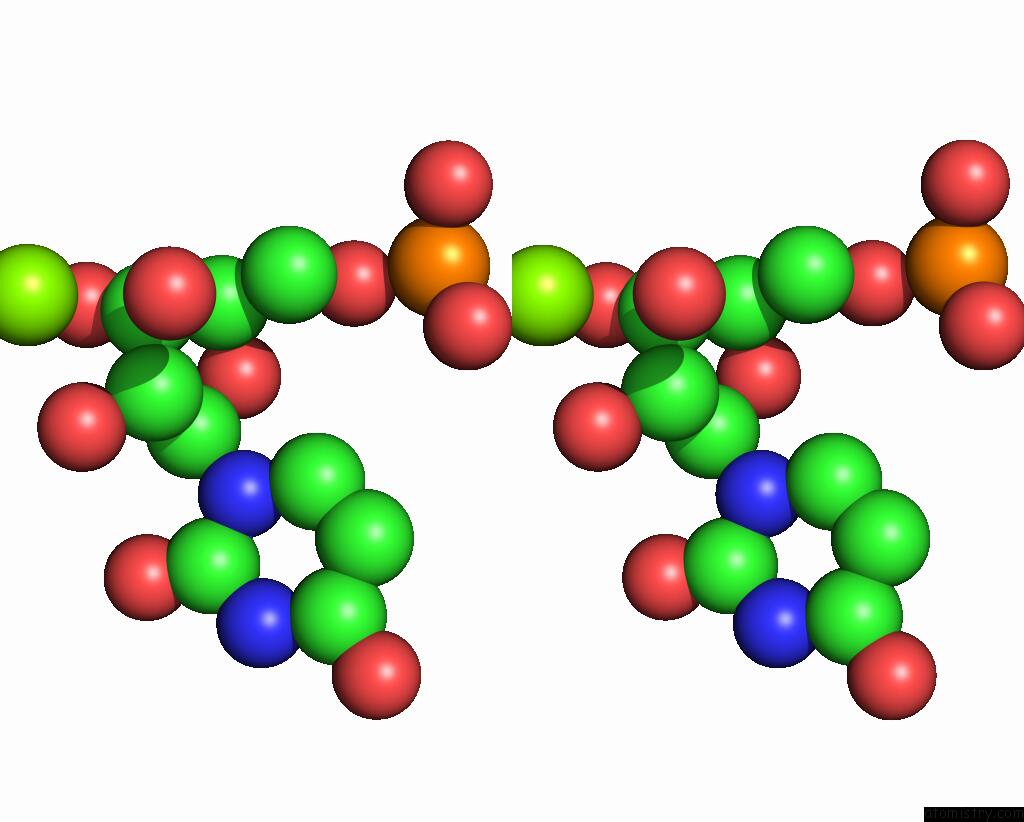
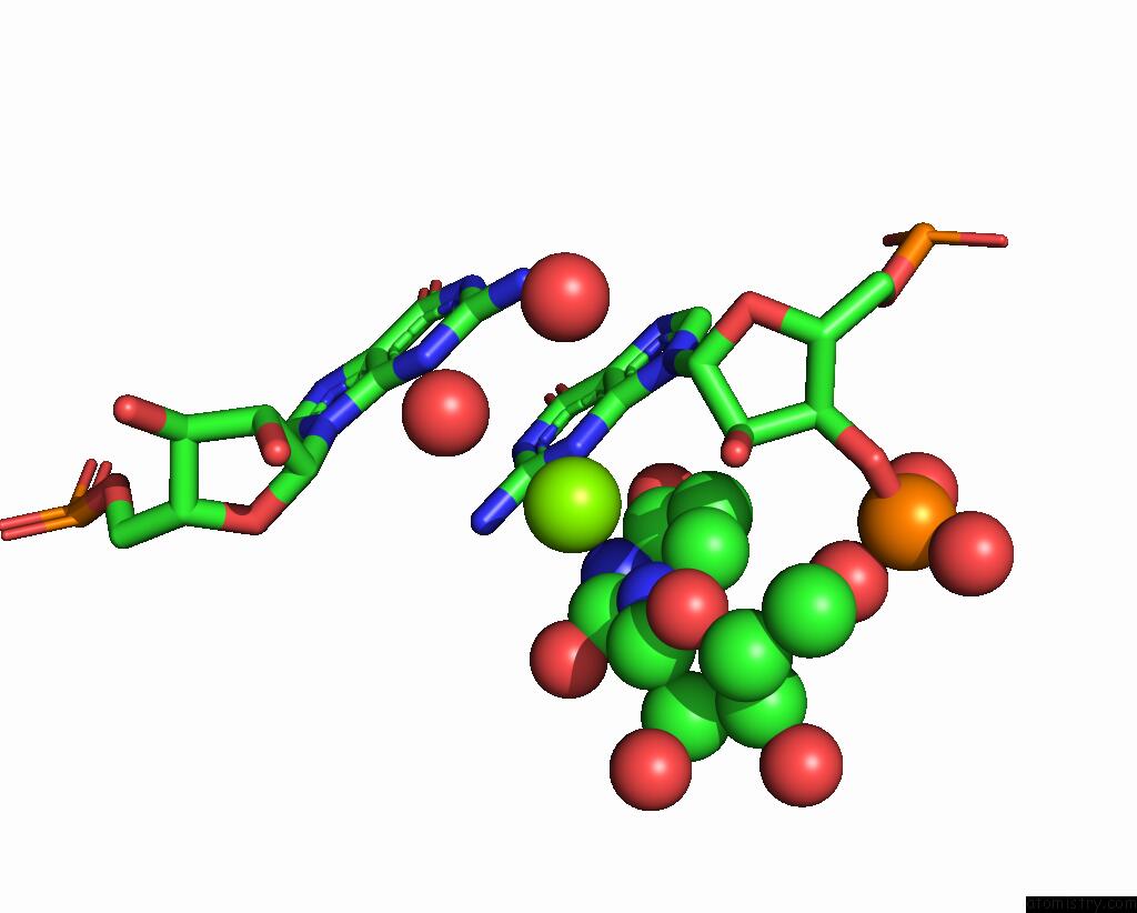
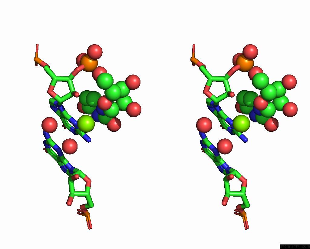
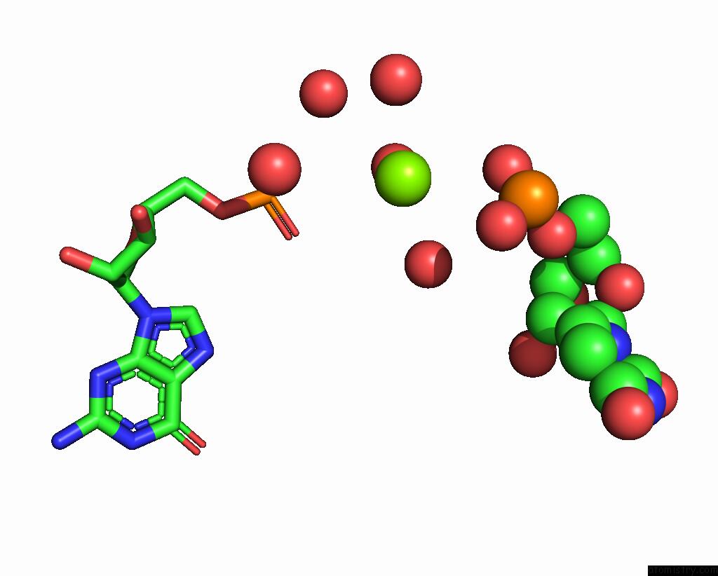
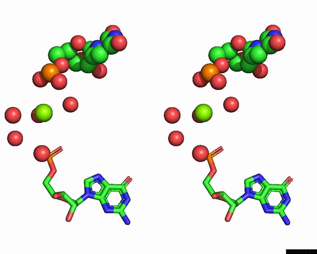
Magnesium binding site 2 out of 7 in 8xzr
Go back to
Magnesium binding site 2 out
of 7 in the Crystal Structure of Fole Riboswitch with 8-NH2 Guanine
Mono view
Stereo pair view
Mono view
Stereo pair view
A full contact list of Magnesium with other atoms in the Mg binding
site number 2 of Crystal Structure of Fole Riboswitch with 8-NH2 Guanine within 5.0Å range:
|
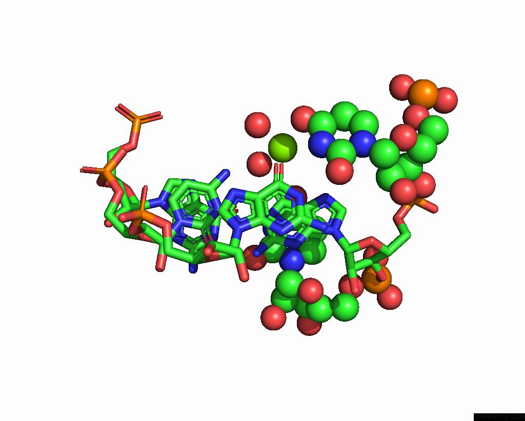
Magnesium binding site 3 out of 7 in 8xzr
Go back to
Magnesium binding site 3 out
of 7 in the Crystal Structure of Fole Riboswitch with 8-NH2 Guanine
Mono view
Stereo pair view
Mono view
Stereo pair view
A full contact list of Magnesium with other atoms in the Mg binding
site number 3 of Crystal Structure of Fole Riboswitch with 8-NH2 Guanine within 5.0Å range:
|
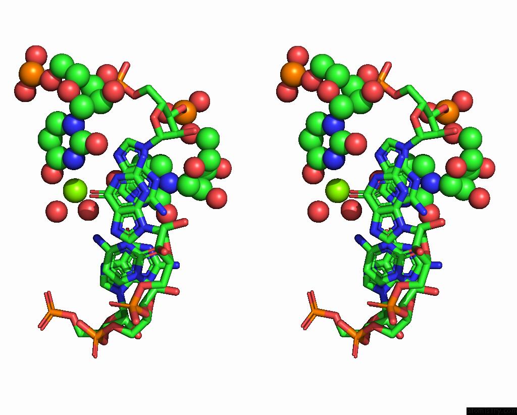
Magnesium binding site 4 out of 7 in 8xzr
Go back to
Magnesium binding site 4 out
of 7 in the Crystal Structure of Fole Riboswitch with 8-NH2 Guanine
Mono view
Stereo pair view
Mono view
Stereo pair view
A full contact list of Magnesium with other atoms in the Mg binding
site number 4 of Crystal Structure of Fole Riboswitch with 8-NH2 Guanine within 5.0Å range:
|
Magnesium binding site 5 out of 7 in 8xzr
Go back to
Magnesium binding site 5 out
of 7 in the Crystal Structure of Fole Riboswitch with 8-NH2 Guanine
Mono view
Stereo pair view
Mono view
Stereo pair view
A full contact list of Magnesium with other atoms in the Mg binding
site number 5 of Crystal Structure of Fole Riboswitch with 8-NH2 Guanine within 5.0Å range:
|
Magnesium binding site 6 out of 7 in 8xzr
Go back to
Magnesium binding site 6 out
of 7 in the Crystal Structure of Fole Riboswitch with 8-NH2 Guanine
Mono view
Stereo pair view
Mono view
Stereo pair view
A full contact list of Magnesium with other atoms in the Mg binding
site number 6 of Crystal Structure of Fole Riboswitch with 8-NH2 Guanine within 5.0Å range:
|
Magnesium binding site 7 out of 7 in 8xzr
Go back to
Magnesium binding site 7 out
of 7 in the Crystal Structure of Fole Riboswitch with 8-NH2 Guanine
Mono view
Stereo pair view
Mono view
Stereo pair view
A full contact list of Magnesium with other atoms in the Mg binding
site number 7 of Crystal Structure of Fole Riboswitch with 8-NH2 Guanine within 5.0Å range:
|
Reference:
C.Li,
X.Xu,
Z.Geng,
L.Zheng,
Q.Song,
X.Shen,
J.Wu,
J.Zhao,
H.Li,
M.He,
X.Tai,
L.Zhang,
J.Ma,
Y.Dong,
A.Ren.
Structure-Based Characterization and Compound Identification of the Wild-Type Thf Class-II Riboswitch. Nucleic Acids Res. 2024.
ISSN: ESSN 1362-4962
PubMed: 38769061
DOI: 10.1093/NAR/GKAE377
Page generated: Fri Aug 15 21:15:50 2025
ISSN: ESSN 1362-4962
PubMed: 38769061
DOI: 10.1093/NAR/GKAE377
Last articles
Mn in 2ADQMn in 2A8S
Mn in 2ADP
Mn in 2A9P
Mn in 2A9O
Mn in 2A9I
Mn in 2A8W
Mn in 2A8U
Mn in 2A8P
Mn in 2A8R