Magnesium »
PDB 8ydm-8ysl »
8yec »
Magnesium in PDB 8yec: Crystal Structure of L-Ribulose 3-Epimerase in Complex with D-Allulose
Protein crystallography data
The structure of Crystal Structure of L-Ribulose 3-Epimerase in Complex with D-Allulose, PDB code: 8yec
was solved by
M.Watanabe,
Y.Nakamichi,
S.Mine,
with X-Ray Crystallography technique. A brief refinement statistics is given in the table below:
| Resolution Low / High (Å) | 30.00 / 2.50 |
| Space group | P 1 21 1 |
| Cell size a, b, c (Å), α, β, γ (°) | 130.425, 135.131, 151.131, 90, 90.06, 90 |
| R / Rfree (%) | 20.7 / 25.5 |
Other elements in 8yec:
The structure of Crystal Structure of L-Ribulose 3-Epimerase in Complex with D-Allulose also contains other interesting chemical elements:
| Sodium | (Na) | 8 atoms |
Magnesium Binding Sites:
Pages:
>>> Page 1 <<< Page 2, Binding sites: 11 - 16;Binding sites:
The binding sites of Magnesium atom in the Crystal Structure of L-Ribulose 3-Epimerase in Complex with D-Allulose (pdb code 8yec). This binding sites where shown within 5.0 Angstroms radius around Magnesium atom.In total 16 binding sites of Magnesium where determined in the Crystal Structure of L-Ribulose 3-Epimerase in Complex with D-Allulose, PDB code: 8yec:
Jump to Magnesium binding site number: 1; 2; 3; 4; 5; 6; 7; 8; 9; 10;
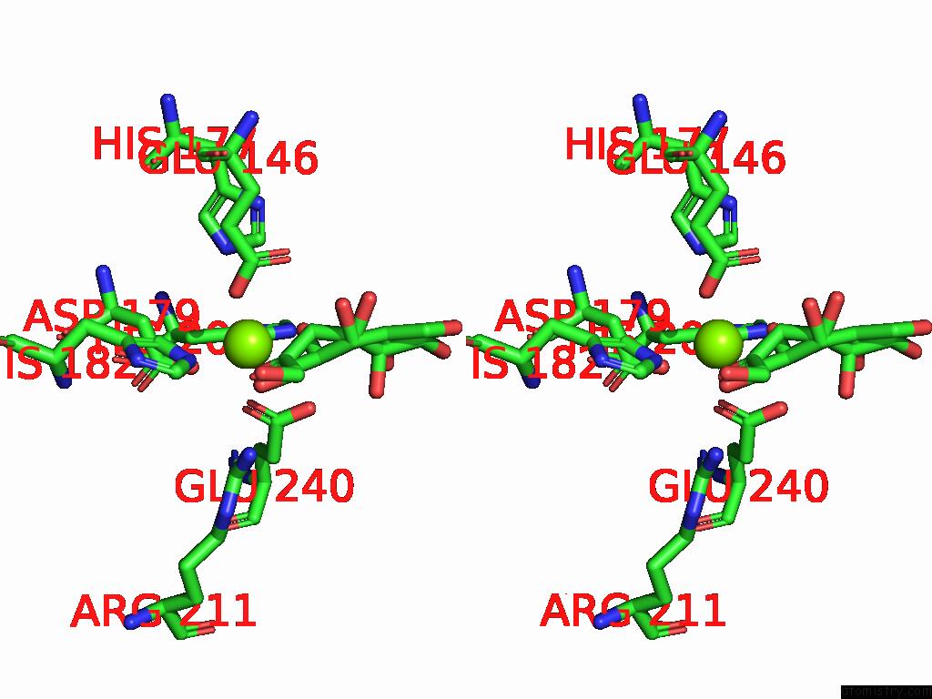
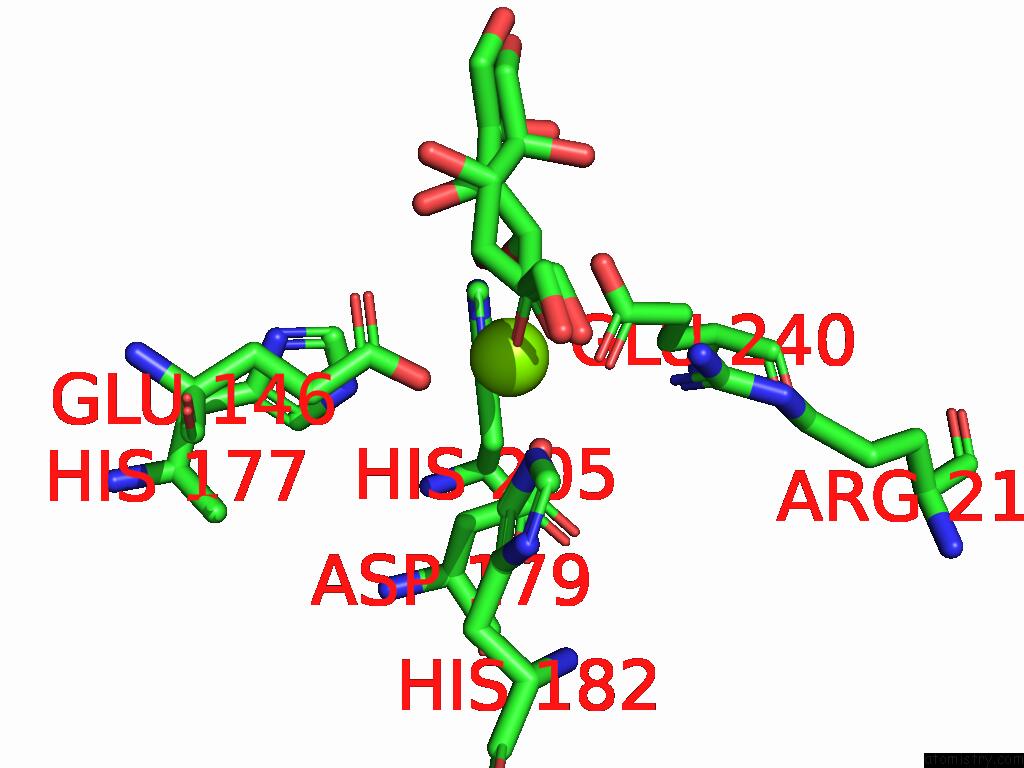
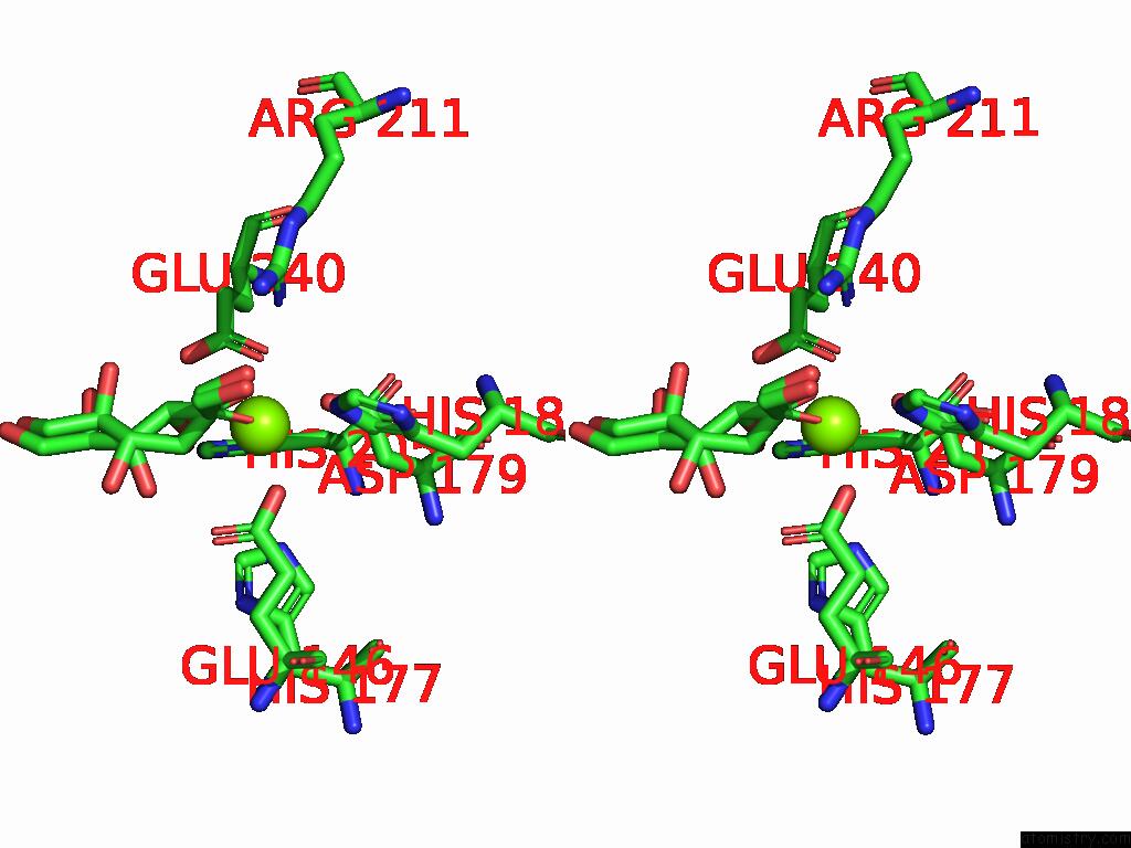
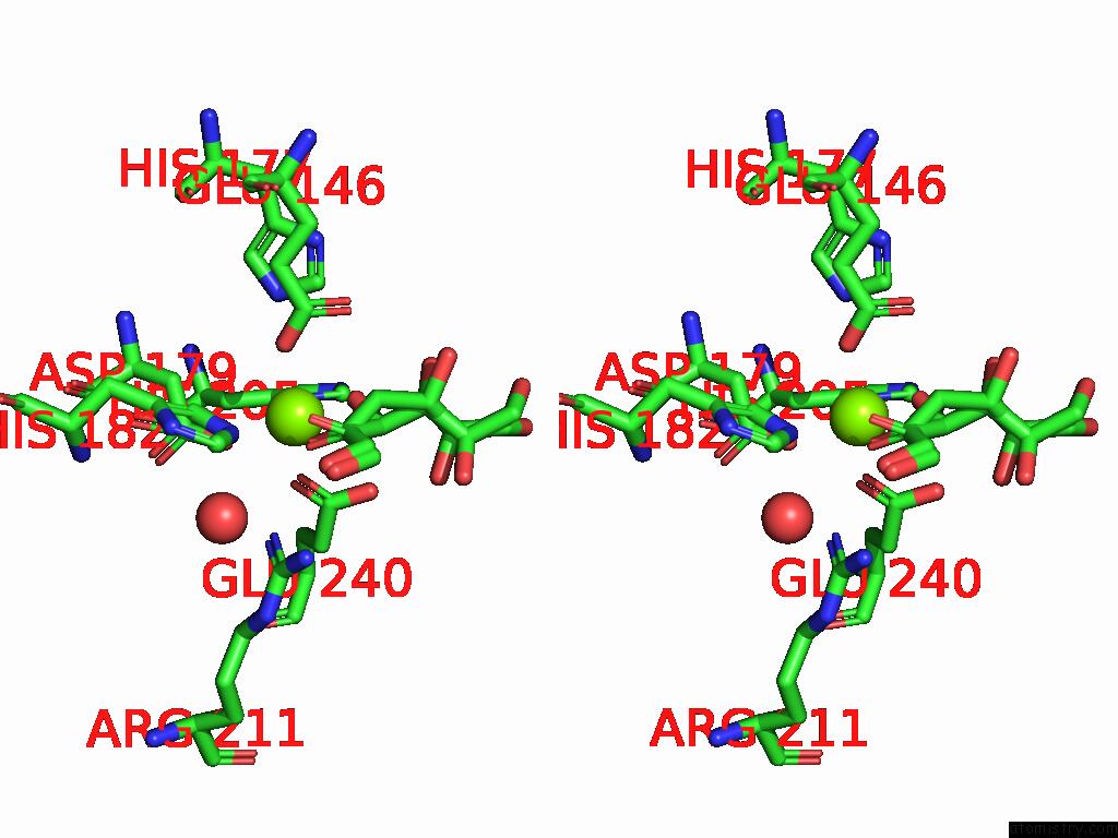
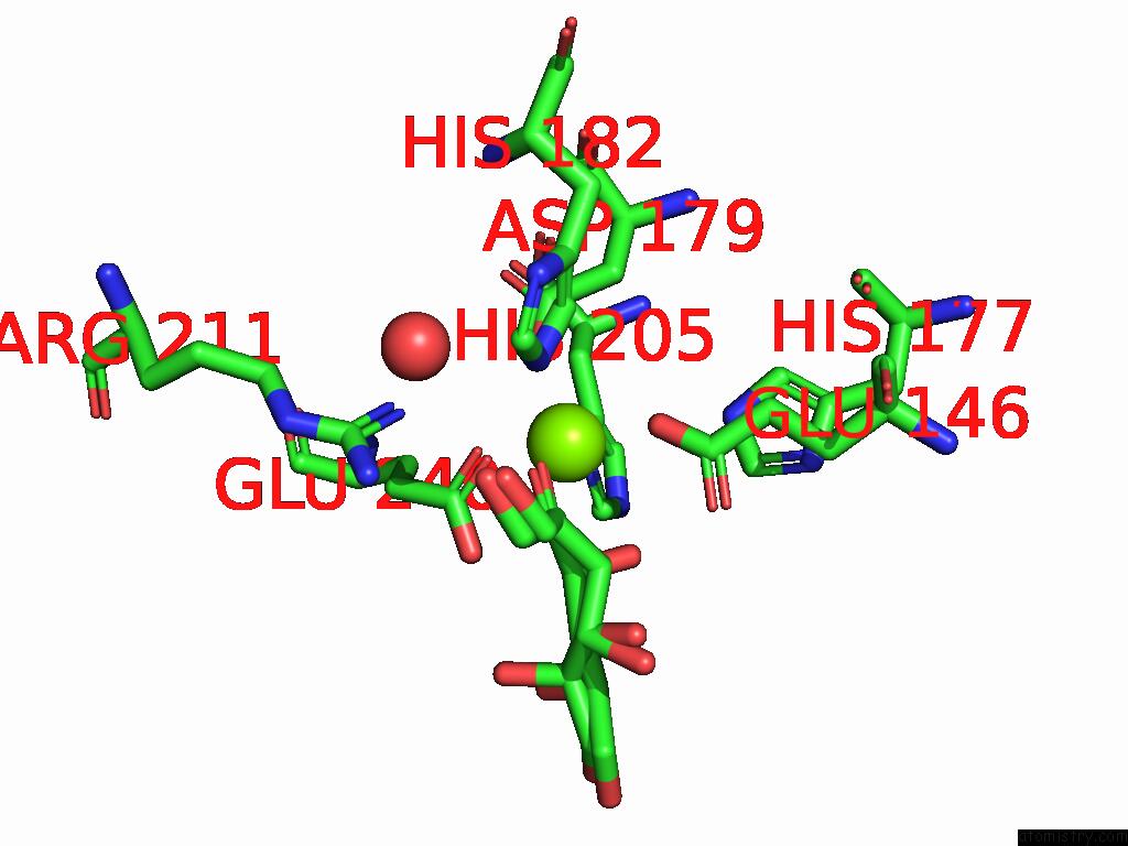
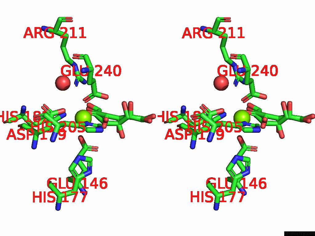
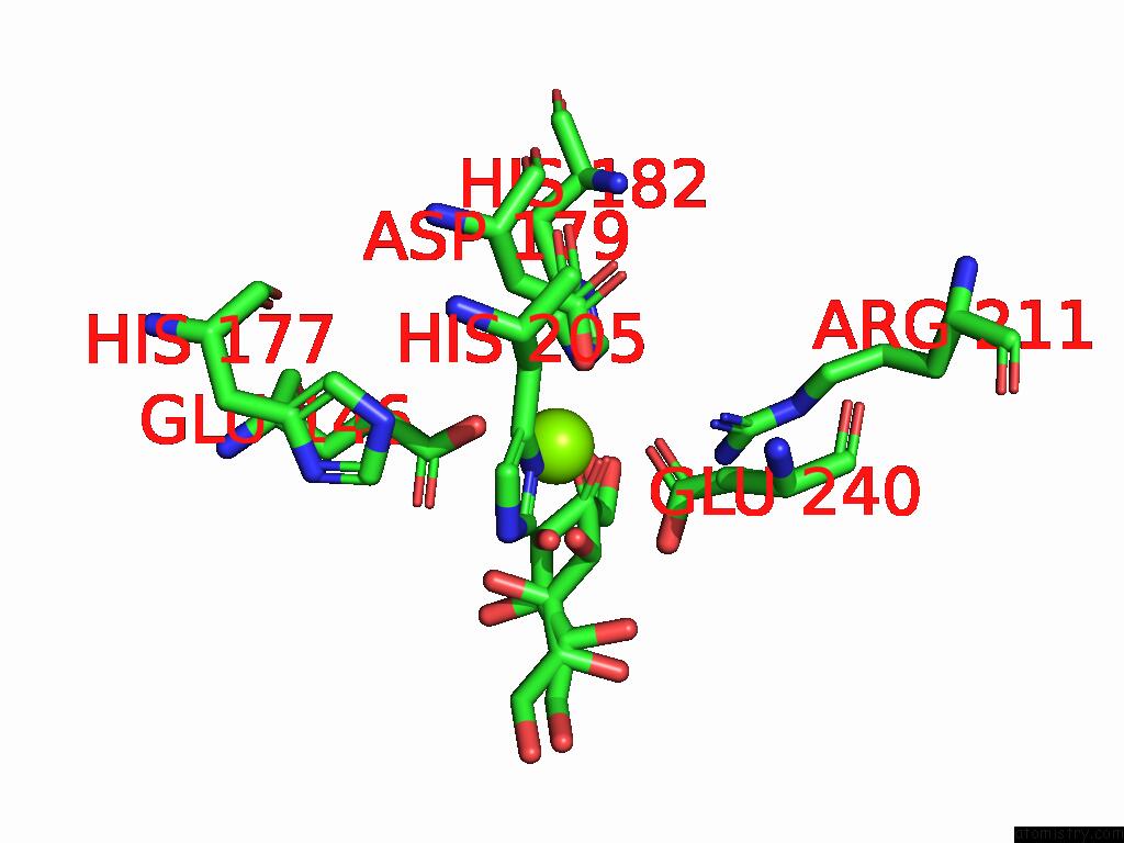
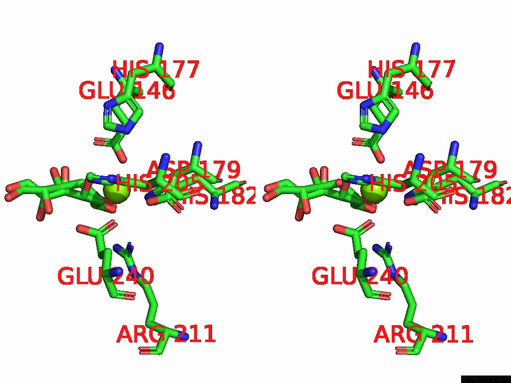
Magnesium binding site 1 out of 16 in 8yec
Go back to
Magnesium binding site 1 out
of 16 in the Crystal Structure of L-Ribulose 3-Epimerase in Complex with D-Allulose
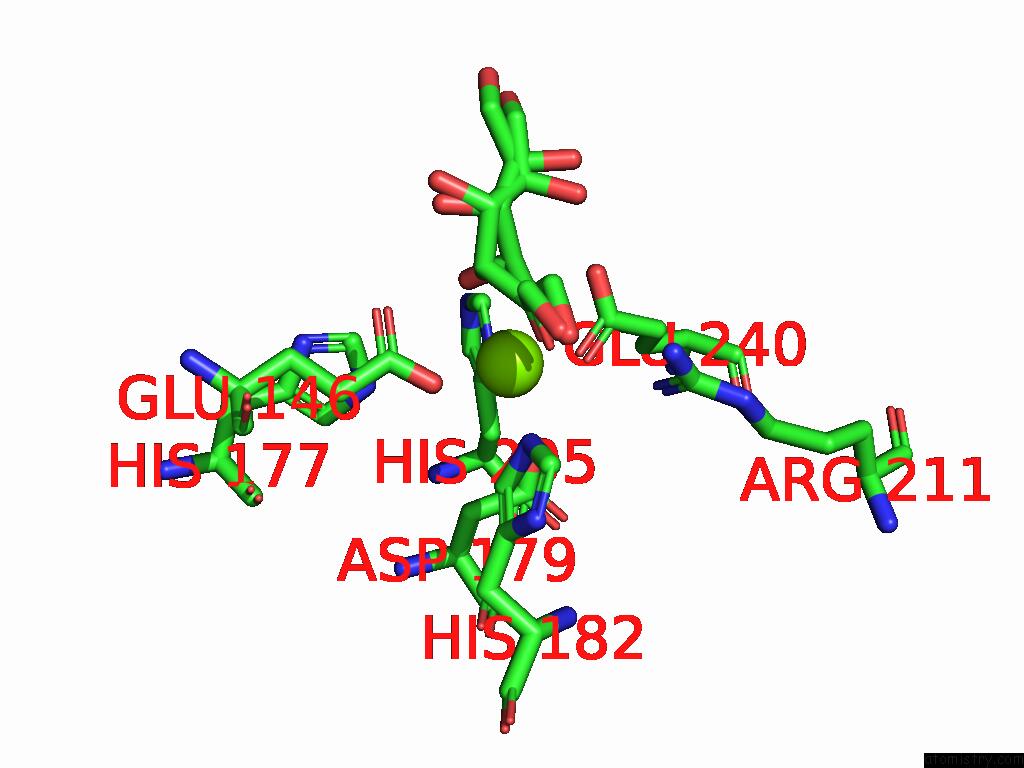
Mono view

Stereo pair view
Mono view
Stereo pair view
A full contact list of Magnesium with other atoms in the Mg binding
site number 1 of Crystal Structure of L-Ribulose 3-Epimerase in Complex with D-Allulose within 5.0Å range:
|
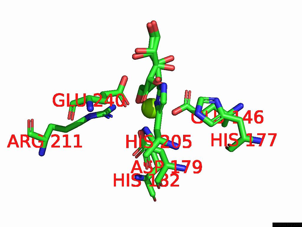
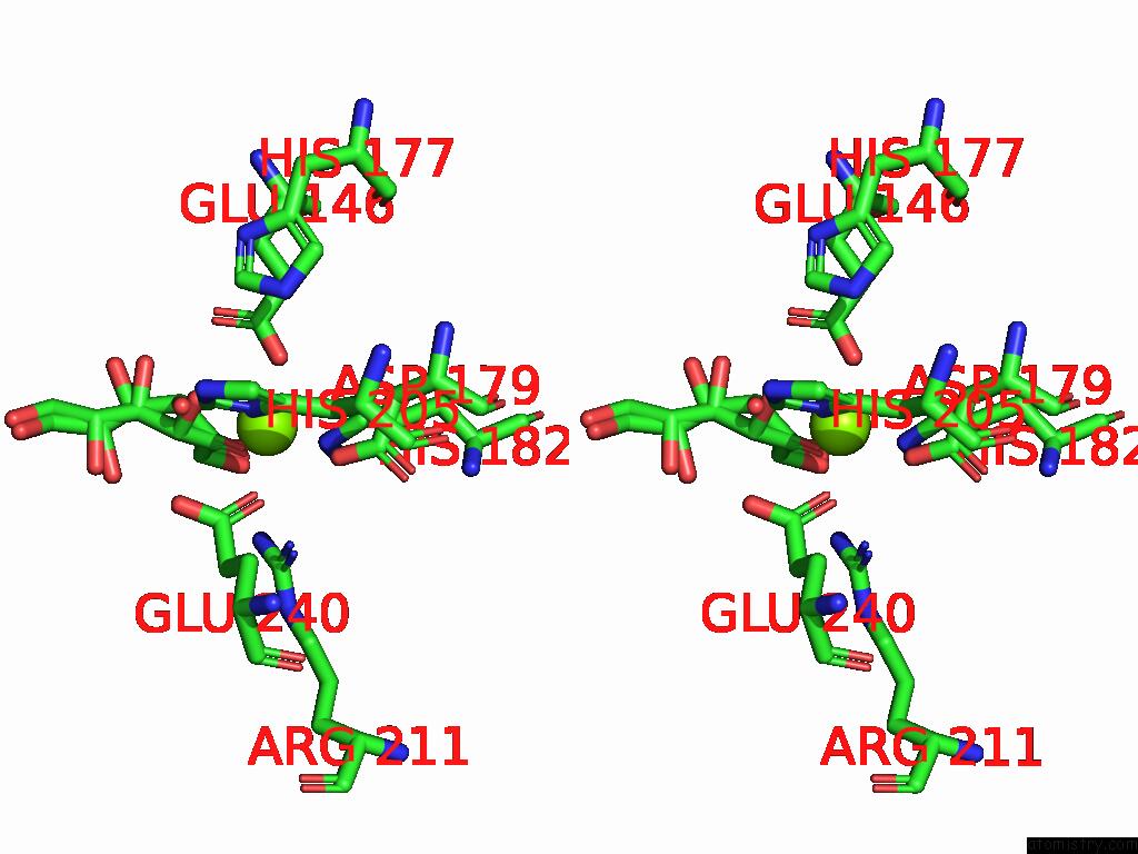
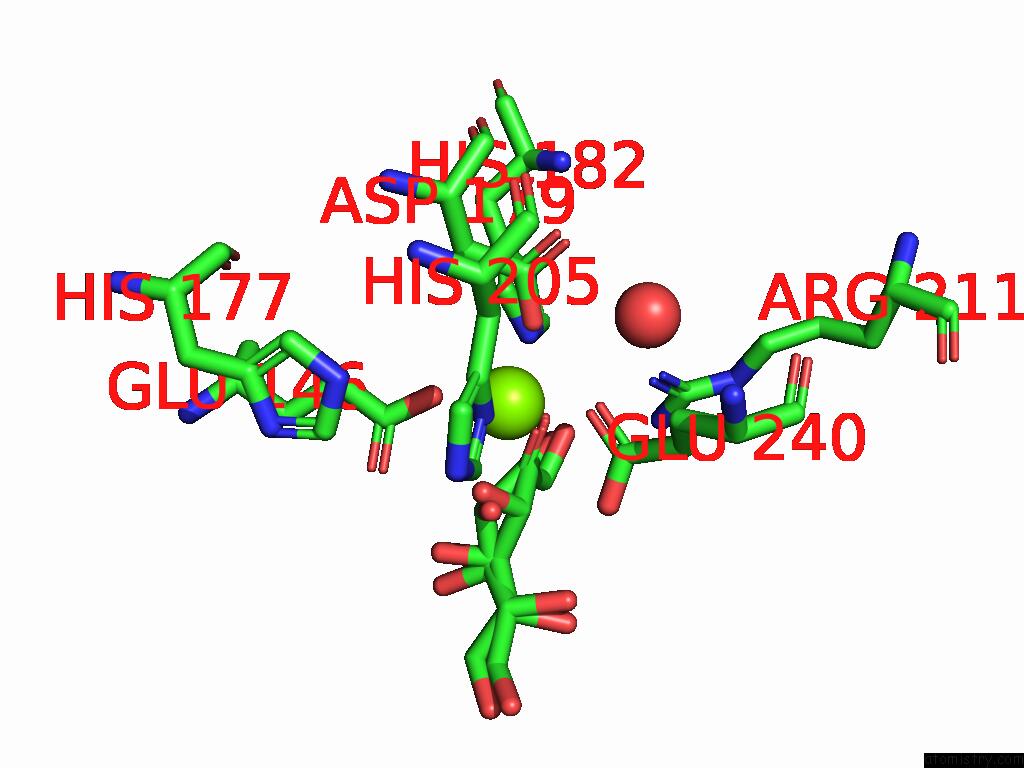
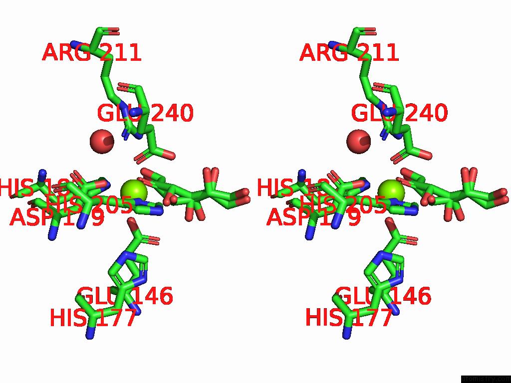
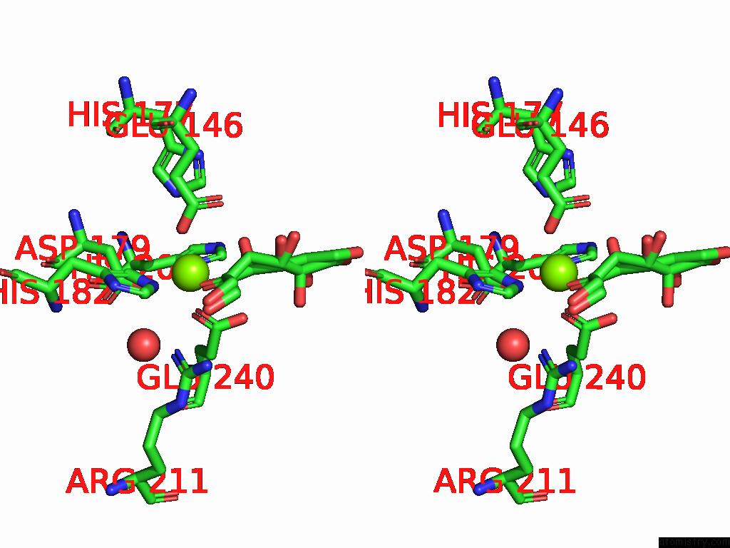
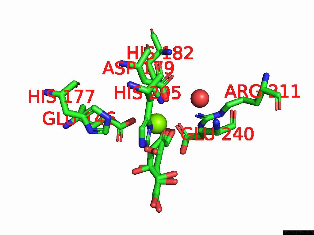
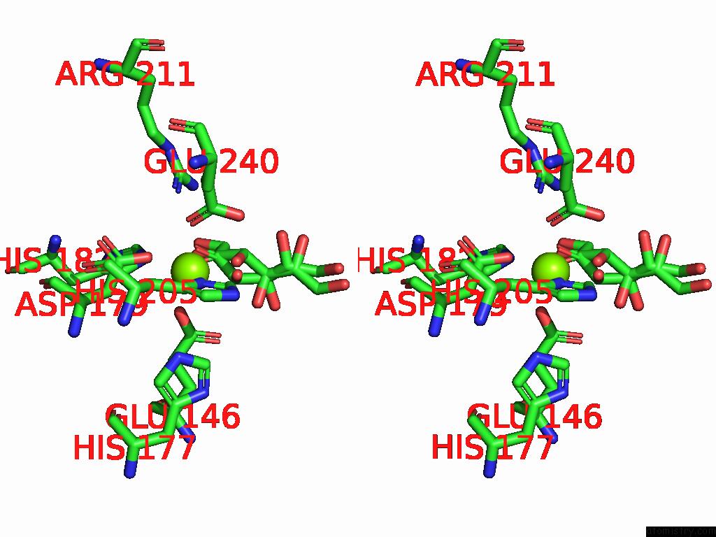
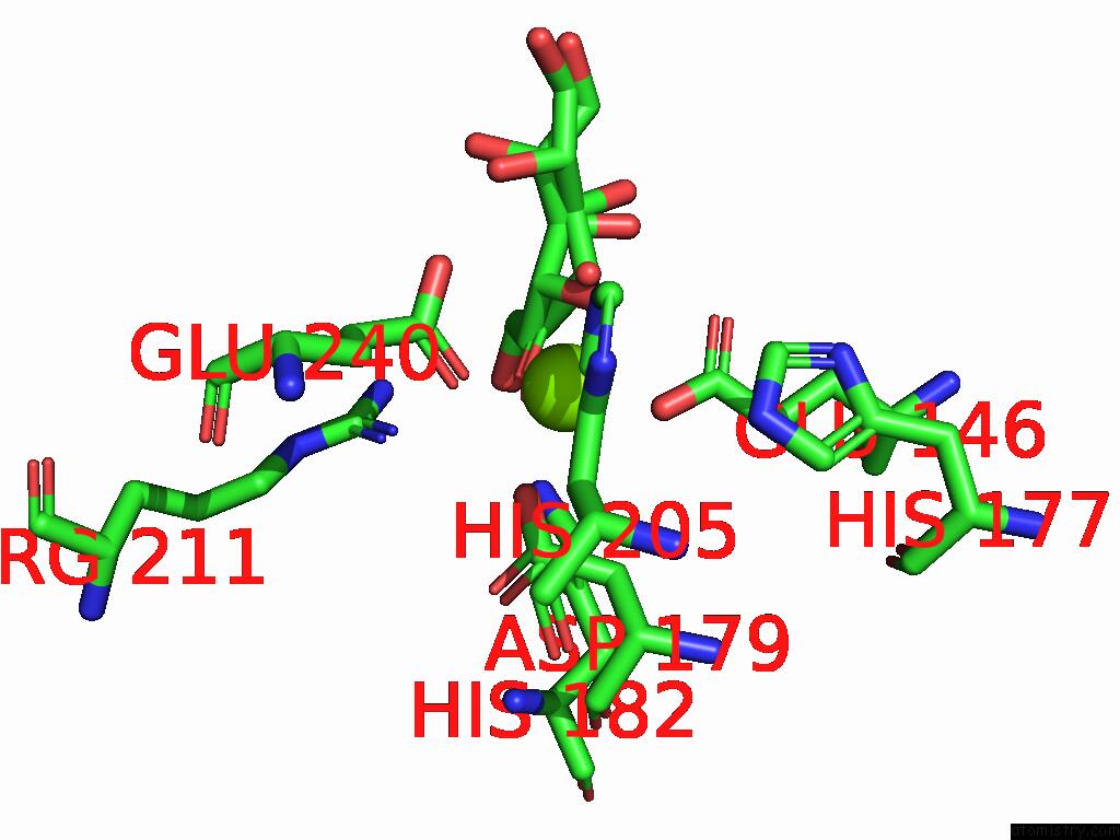
Magnesium binding site 2 out of 16 in 8yec
Go back to
Magnesium binding site 2 out
of 16 in the Crystal Structure of L-Ribulose 3-Epimerase in Complex with D-Allulose

Mono view
Stereo pair view
Mono view
Stereo pair view
A full contact list of Magnesium with other atoms in the Mg binding
site number 2 of Crystal Structure of L-Ribulose 3-Epimerase in Complex with D-Allulose within 5.0Å range:
|
Magnesium binding site 3 out of 16 in 8yec
Go back to
Magnesium binding site 3 out
of 16 in the Crystal Structure of L-Ribulose 3-Epimerase in Complex with D-Allulose
Mono view
Stereo pair view
Mono view
Stereo pair view
A full contact list of Magnesium with other atoms in the Mg binding
site number 3 of Crystal Structure of L-Ribulose 3-Epimerase in Complex with D-Allulose within 5.0Å range:
|
Magnesium binding site 4 out of 16 in 8yec
Go back to
Magnesium binding site 4 out
of 16 in the Crystal Structure of L-Ribulose 3-Epimerase in Complex with D-Allulose
Mono view
Stereo pair view
Mono view
Stereo pair view
A full contact list of Magnesium with other atoms in the Mg binding
site number 4 of Crystal Structure of L-Ribulose 3-Epimerase in Complex with D-Allulose within 5.0Å range:
|
Magnesium binding site 5 out of 16 in 8yec
Go back to
Magnesium binding site 5 out
of 16 in the Crystal Structure of L-Ribulose 3-Epimerase in Complex with D-Allulose
Mono view
Stereo pair view
Mono view
Stereo pair view
A full contact list of Magnesium with other atoms in the Mg binding
site number 5 of Crystal Structure of L-Ribulose 3-Epimerase in Complex with D-Allulose within 5.0Å range:
|
Magnesium binding site 6 out of 16 in 8yec
Go back to
Magnesium binding site 6 out
of 16 in the Crystal Structure of L-Ribulose 3-Epimerase in Complex with D-Allulose

Mono view
Stereo pair view
Mono view
Stereo pair view
A full contact list of Magnesium with other atoms in the Mg binding
site number 6 of Crystal Structure of L-Ribulose 3-Epimerase in Complex with D-Allulose within 5.0Å range:
|
Magnesium binding site 7 out of 16 in 8yec
Go back to
Magnesium binding site 7 out
of 16 in the Crystal Structure of L-Ribulose 3-Epimerase in Complex with D-Allulose
Mono view
Stereo pair view
Mono view
Stereo pair view
A full contact list of Magnesium with other atoms in the Mg binding
site number 7 of Crystal Structure of L-Ribulose 3-Epimerase in Complex with D-Allulose within 5.0Å range:
|
Magnesium binding site 8 out of 16 in 8yec
Go back to
Magnesium binding site 8 out
of 16 in the Crystal Structure of L-Ribulose 3-Epimerase in Complex with D-Allulose
Mono view
Stereo pair view
Mono view
Stereo pair view
A full contact list of Magnesium with other atoms in the Mg binding
site number 8 of Crystal Structure of L-Ribulose 3-Epimerase in Complex with D-Allulose within 5.0Å range:
|
Magnesium binding site 9 out of 16 in 8yec
Go back to
Magnesium binding site 9 out
of 16 in the Crystal Structure of L-Ribulose 3-Epimerase in Complex with D-Allulose
Mono view
Stereo pair view
Mono view
Stereo pair view
A full contact list of Magnesium with other atoms in the Mg binding
site number 9 of Crystal Structure of L-Ribulose 3-Epimerase in Complex with D-Allulose within 5.0Å range:
|
Magnesium binding site 10 out of 16 in 8yec
Go back to
Magnesium binding site 10 out
of 16 in the Crystal Structure of L-Ribulose 3-Epimerase in Complex with D-Allulose
Mono view
Stereo pair view
Mono view
Stereo pair view
A full contact list of Magnesium with other atoms in the Mg binding
site number 10 of Crystal Structure of L-Ribulose 3-Epimerase in Complex with D-Allulose within 5.0Å range:
|
Reference:
M.Watanabe,
Y.Nakamichi,
S.Mine.
Crystal Structure of L-Ribulose 3-Epimerase in Complex with D-Allulose To Be Published.
Page generated: Tue Feb 25 11:01:12 2025
Last articles
Ca in 5TIWCa in 5TJ6
Ca in 5TH9
Ca in 5TIS
Ca in 5TI8
Ca in 5THG
Ca in 5TGX
Ca in 5TFK
Ca in 5TG3
Ca in 5TGQ