Magnesium »
PDB 8yo0-8z2l »
8yud »
Magnesium in PDB 8yud: Crystal Structure of Xylose Isomerase From Streptomyces Avermitilis
Enzymatic activity of Crystal Structure of Xylose Isomerase From Streptomyces Avermitilis
All present enzymatic activity of Crystal Structure of Xylose Isomerase From Streptomyces Avermitilis:
5.3.1.5;
5.3.1.5;
Protein crystallography data
The structure of Crystal Structure of Xylose Isomerase From Streptomyces Avermitilis, PDB code: 8yud
was solved by
K.H.Nam,
with X-Ray Crystallography technique. A brief refinement statistics is given in the table below:
| Resolution Low / High (Å) | 34.21 / 2.81 |
| Space group | P 32 2 1 |
| Cell size a, b, c (Å), α, β, γ (°) | 129.135, 129.135, 233.058, 90, 90, 120 |
| R / Rfree (%) | 18.7 / 24.3 |
Magnesium Binding Sites:
Pages:
>>> Page 1 <<< Page 2, Binding sites: 11 - 12;Binding sites:
The binding sites of Magnesium atom in the Crystal Structure of Xylose Isomerase From Streptomyces Avermitilis (pdb code 8yud). This binding sites where shown within 5.0 Angstroms radius around Magnesium atom.In total 12 binding sites of Magnesium where determined in the Crystal Structure of Xylose Isomerase From Streptomyces Avermitilis, PDB code: 8yud:
Jump to Magnesium binding site number: 1; 2; 3; 4; 5; 6; 7; 8; 9; 10;
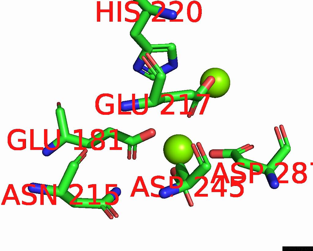
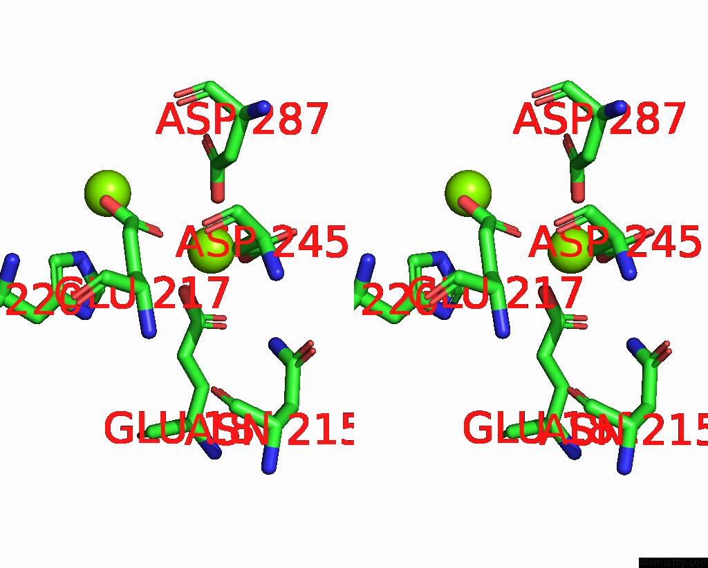
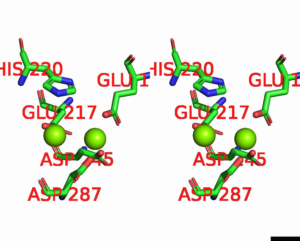
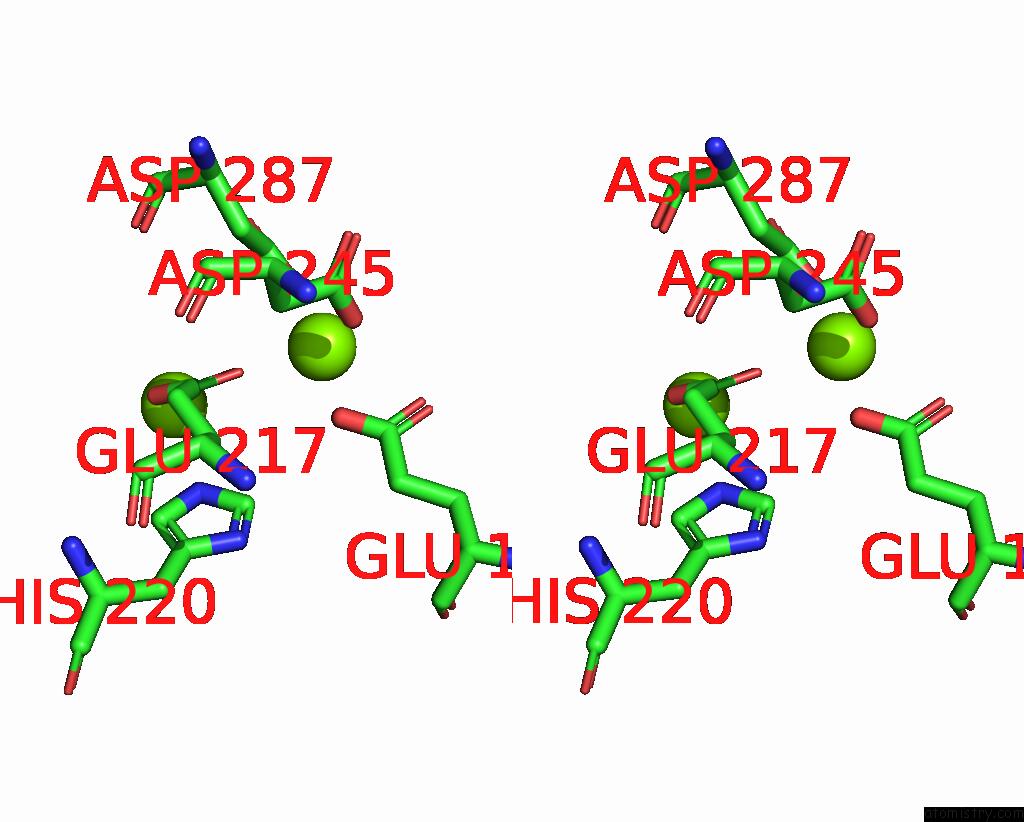
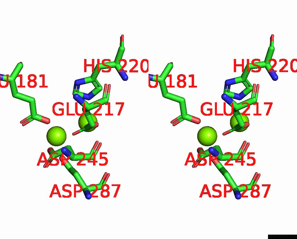
Magnesium binding site 1 out of 12 in 8yud
Go back to
Magnesium binding site 1 out
of 12 in the Crystal Structure of Xylose Isomerase From Streptomyces Avermitilis
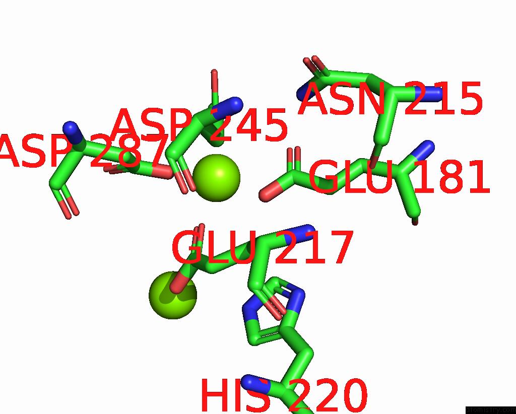
Mono view
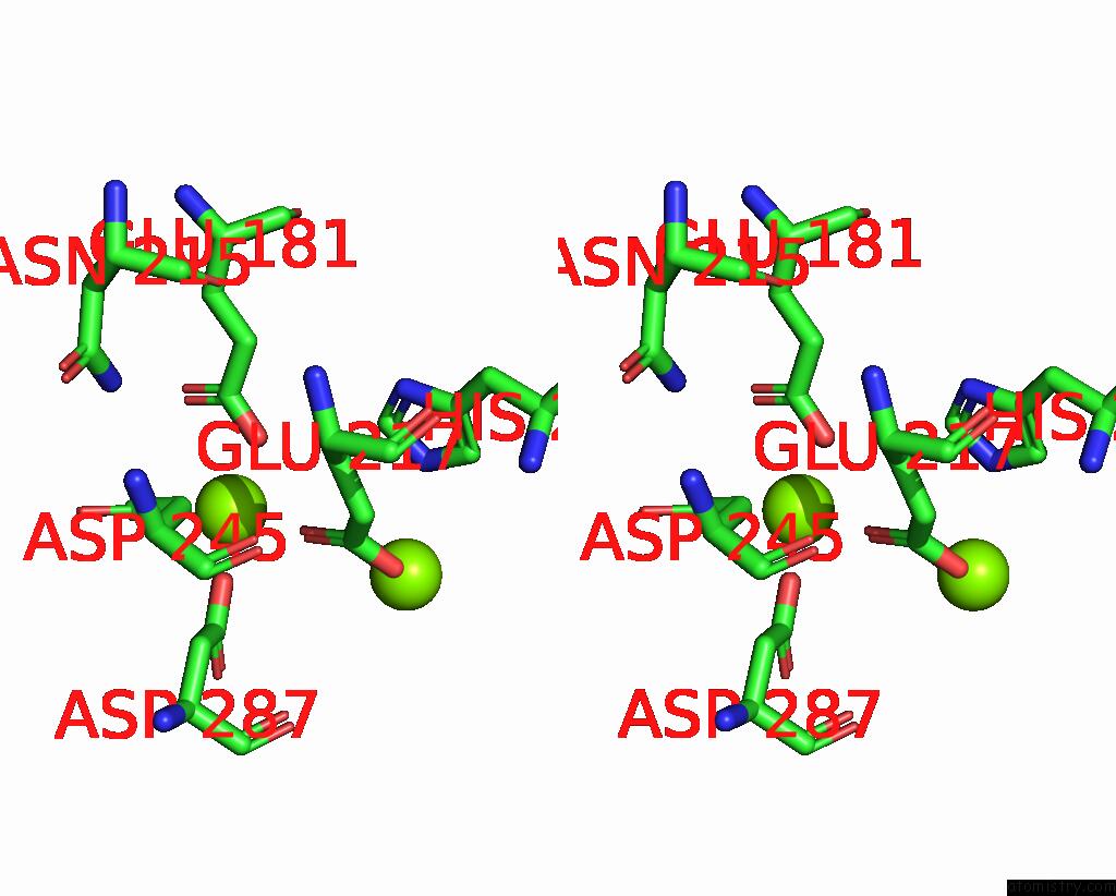
Stereo pair view
Mono view
Stereo pair view
A full contact list of Magnesium with other atoms in the Mg binding
site number 1 of Crystal Structure of Xylose Isomerase From Streptomyces Avermitilis within 5.0Å range:
|
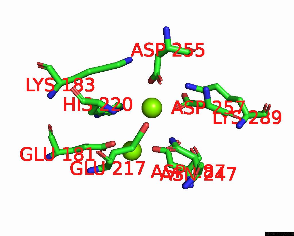
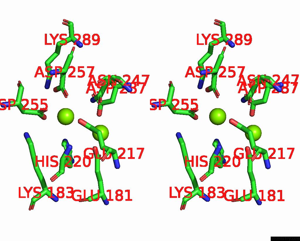
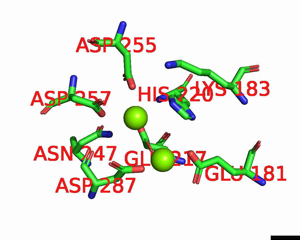
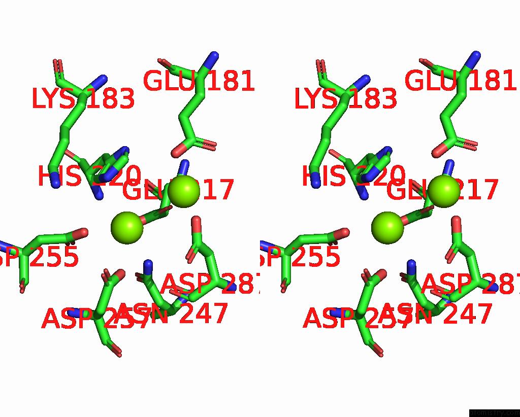
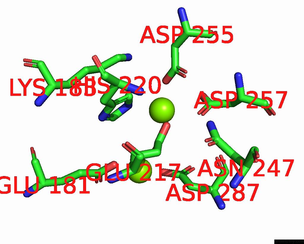
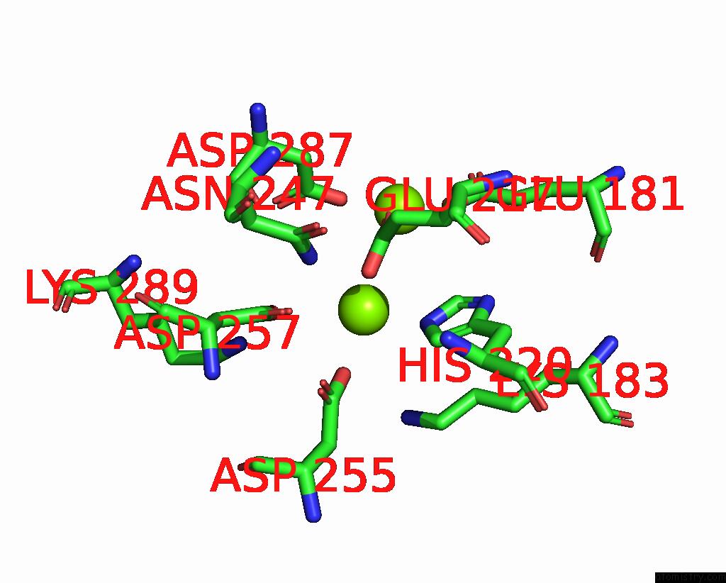
Magnesium binding site 2 out of 12 in 8yud
Go back to
Magnesium binding site 2 out
of 12 in the Crystal Structure of Xylose Isomerase From Streptomyces Avermitilis
Mono view
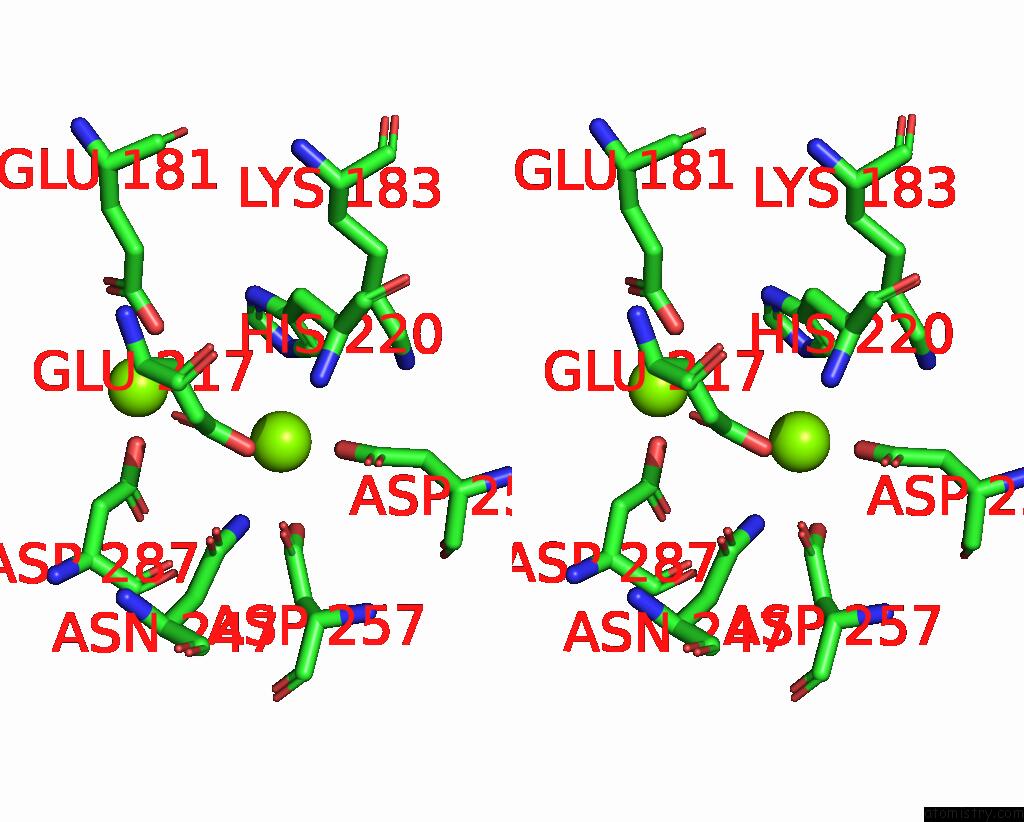
Stereo pair view
Mono view
Stereo pair view
A full contact list of Magnesium with other atoms in the Mg binding
site number 2 of Crystal Structure of Xylose Isomerase From Streptomyces Avermitilis within 5.0Å range:
|
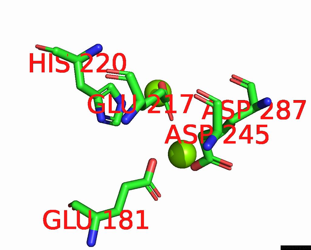
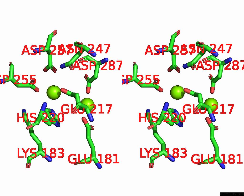
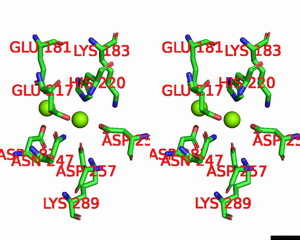
Magnesium binding site 3 out of 12 in 8yud
Go back to
Magnesium binding site 3 out
of 12 in the Crystal Structure of Xylose Isomerase From Streptomyces Avermitilis
Mono view
Stereo pair view
Mono view
Stereo pair view
A full contact list of Magnesium with other atoms in the Mg binding
site number 3 of Crystal Structure of Xylose Isomerase From Streptomyces Avermitilis within 5.0Å range:
|
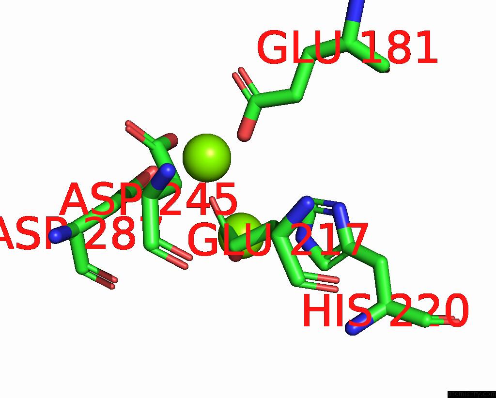
Magnesium binding site 4 out of 12 in 8yud
Go back to
Magnesium binding site 4 out
of 12 in the Crystal Structure of Xylose Isomerase From Streptomyces Avermitilis
Mono view
Stereo pair view
Mono view
Stereo pair view
A full contact list of Magnesium with other atoms in the Mg binding
site number 4 of Crystal Structure of Xylose Isomerase From Streptomyces Avermitilis within 5.0Å range:
|
Magnesium binding site 5 out of 12 in 8yud
Go back to
Magnesium binding site 5 out
of 12 in the Crystal Structure of Xylose Isomerase From Streptomyces Avermitilis

Mono view
Stereo pair view
Mono view
Stereo pair view
A full contact list of Magnesium with other atoms in the Mg binding
site number 5 of Crystal Structure of Xylose Isomerase From Streptomyces Avermitilis within 5.0Å range:
|
Magnesium binding site 6 out of 12 in 8yud
Go back to
Magnesium binding site 6 out
of 12 in the Crystal Structure of Xylose Isomerase From Streptomyces Avermitilis
Mono view
Stereo pair view
Mono view
Stereo pair view
A full contact list of Magnesium with other atoms in the Mg binding
site number 6 of Crystal Structure of Xylose Isomerase From Streptomyces Avermitilis within 5.0Å range:
|
Magnesium binding site 7 out of 12 in 8yud
Go back to
Magnesium binding site 7 out
of 12 in the Crystal Structure of Xylose Isomerase From Streptomyces Avermitilis
Mono view
Stereo pair view
Mono view
Stereo pair view
A full contact list of Magnesium with other atoms in the Mg binding
site number 7 of Crystal Structure of Xylose Isomerase From Streptomyces Avermitilis within 5.0Å range:
|
Magnesium binding site 8 out of 12 in 8yud
Go back to
Magnesium binding site 8 out
of 12 in the Crystal Structure of Xylose Isomerase From Streptomyces Avermitilis
Mono view
Stereo pair view
Mono view
Stereo pair view
A full contact list of Magnesium with other atoms in the Mg binding
site number 8 of Crystal Structure of Xylose Isomerase From Streptomyces Avermitilis within 5.0Å range:
|
Magnesium binding site 9 out of 12 in 8yud
Go back to
Magnesium binding site 9 out
of 12 in the Crystal Structure of Xylose Isomerase From Streptomyces Avermitilis
Mono view
Stereo pair view
Mono view
Stereo pair view
A full contact list of Magnesium with other atoms in the Mg binding
site number 9 of Crystal Structure of Xylose Isomerase From Streptomyces Avermitilis within 5.0Å range:
|
Magnesium binding site 10 out of 12 in 8yud
Go back to
Magnesium binding site 10 out
of 12 in the Crystal Structure of Xylose Isomerase From Streptomyces Avermitilis
Mono view
Stereo pair view
Mono view
Stereo pair view
A full contact list of Magnesium with other atoms in the Mg binding
site number 10 of Crystal Structure of Xylose Isomerase From Streptomyces Avermitilis within 5.0Å range:
|
Reference:
K.H.Nam,
K.H.Nam.
N/A N/A.
Page generated: Fri Aug 15 21:38:16 2025
Last articles
Mn in 3CQWMn in 3CM5
Mn in 3CNA
Mn in 3CK2
Mn in 3CKQ
Mn in 3CKN
Mn in 3C2M
Mn in 3C5M
Mn in 3C3T
Mn in 3C2L