Magnesium »
PDB 8zj7-8zwq »
8zuq »
Magnesium in PDB 8zuq: Crystal Structure of the F99S/M153T/V163A/T203I/E222Q Variant of Gfp at pH 8.5
Protein crystallography data
The structure of Crystal Structure of the F99S/M153T/V163A/T203I/E222Q Variant of Gfp at pH 8.5, PDB code: 8zuq
was solved by
R.Takeda,
K.Takeda,
with X-Ray Crystallography technique. A brief refinement statistics is given in the table below:
| Resolution Low / High (Å) | 45.93 / 1.48 |
| Space group | P 21 21 21 |
| Cell size a, b, c (Å), α, β, γ (°) | 50.498, 62.452, 67.783, 90, 90, 90 |
| R / Rfree (%) | 15.6 / 19.3 |
Magnesium Binding Sites:
The binding sites of Magnesium atom in the Crystal Structure of the F99S/M153T/V163A/T203I/E222Q Variant of Gfp at pH 8.5
(pdb code 8zuq). This binding sites where shown within
5.0 Angstroms radius around Magnesium atom.
In total 2 binding sites of Magnesium where determined in the Crystal Structure of the F99S/M153T/V163A/T203I/E222Q Variant of Gfp at pH 8.5, PDB code: 8zuq:
Jump to Magnesium binding site number: 1; 2;
In total 2 binding sites of Magnesium where determined in the Crystal Structure of the F99S/M153T/V163A/T203I/E222Q Variant of Gfp at pH 8.5, PDB code: 8zuq:
Jump to Magnesium binding site number: 1; 2;
Magnesium binding site 1 out of 2 in 8zuq
Go back to
Magnesium binding site 1 out
of 2 in the Crystal Structure of the F99S/M153T/V163A/T203I/E222Q Variant of Gfp at pH 8.5
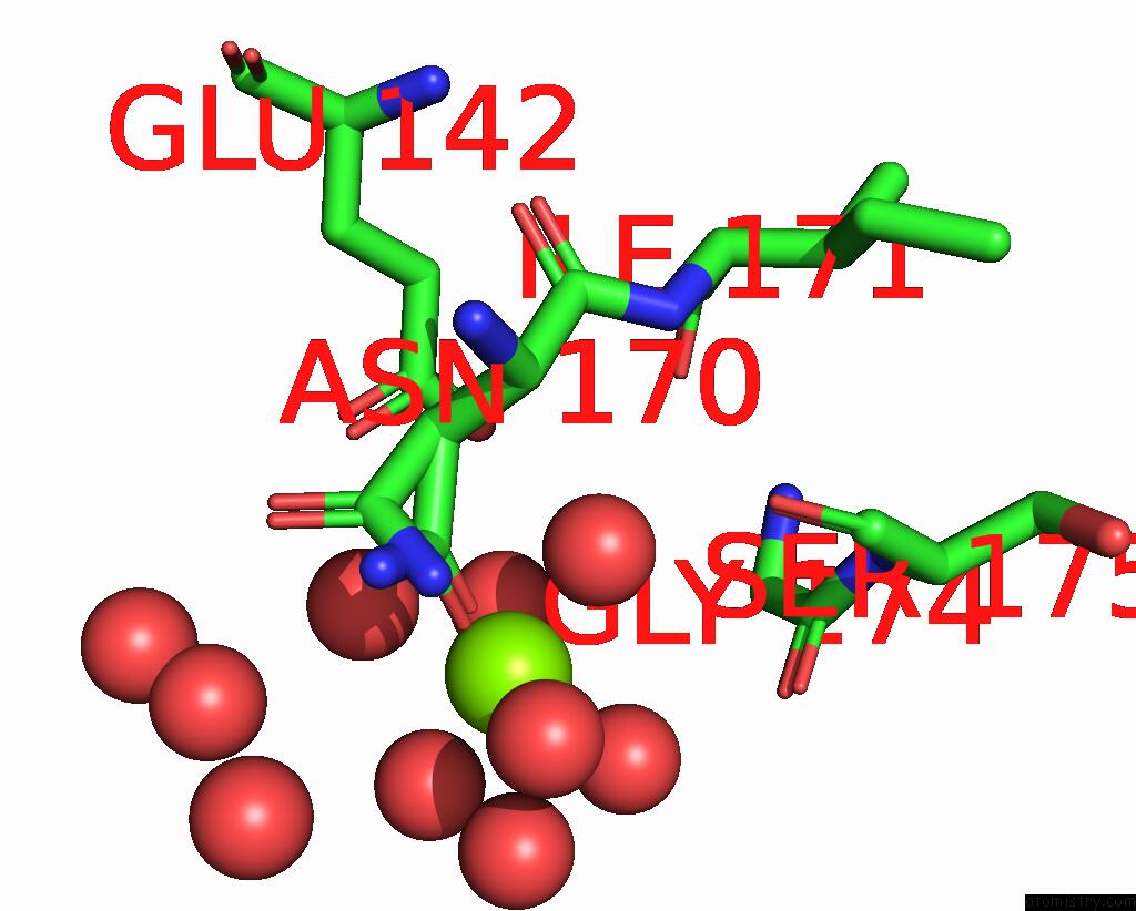
Mono view
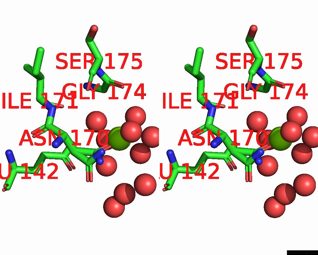
Stereo pair view
Mono view
Stereo pair view
A full contact list of Magnesium with other atoms in the Mg binding
site number 1 of Crystal Structure of the F99S/M153T/V163A/T203I/E222Q Variant of Gfp at pH 8.5 within 5.0Å range:
|
Magnesium binding site 2 out of 2 in 8zuq
Go back to
Magnesium binding site 2 out
of 2 in the Crystal Structure of the F99S/M153T/V163A/T203I/E222Q Variant of Gfp at pH 8.5
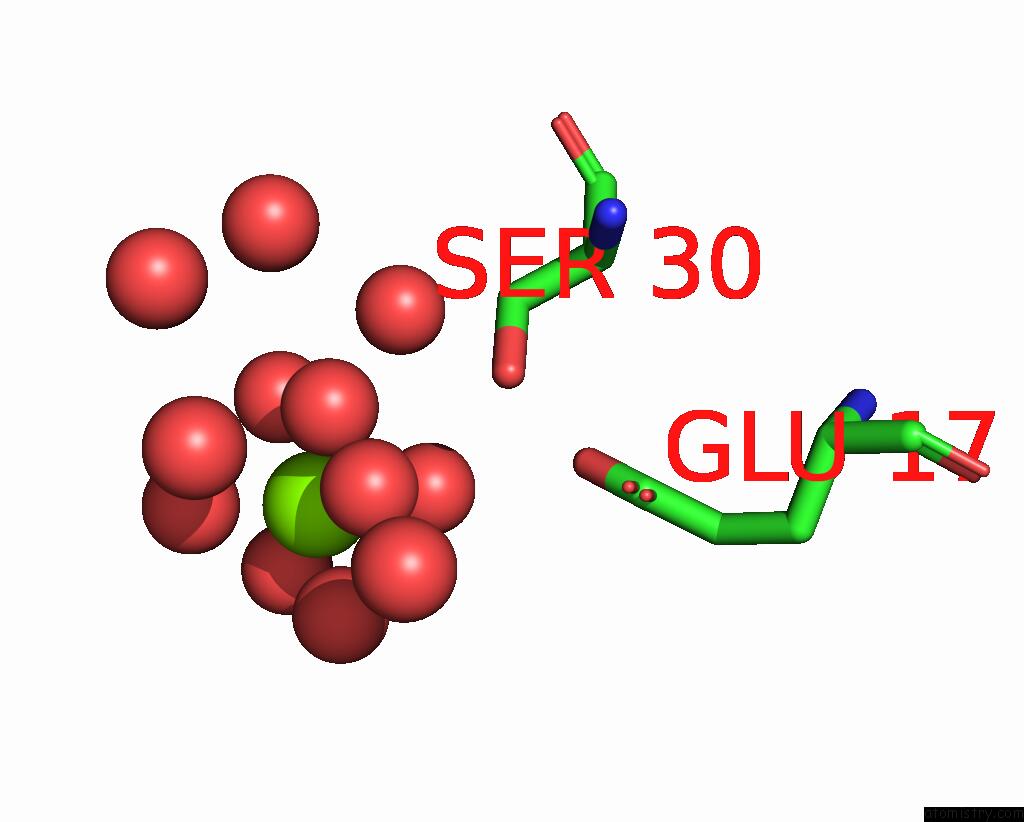
Mono view
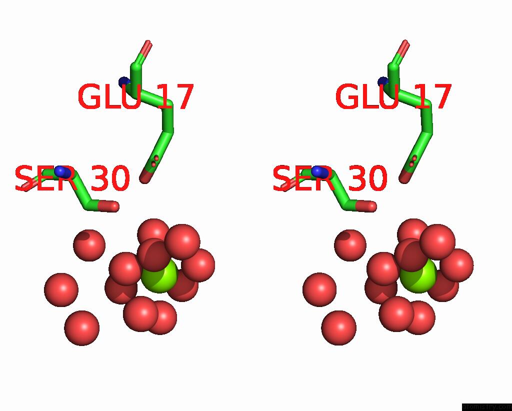
Stereo pair view
Mono view
Stereo pair view
A full contact list of Magnesium with other atoms in the Mg binding
site number 2 of Crystal Structure of the F99S/M153T/V163A/T203I/E222Q Variant of Gfp at pH 8.5 within 5.0Å range:
|
Reference:
R.Takeda,
E.Tsutsumi,
K.Okatsu,
S.Fukai,
K.Takeda.
Structural Characterization of Green Fluorescent Protein in the I-State. Sci Rep V. 14 22832 2024.
ISSN: ESSN 2045-2322
PubMed: 39353998
DOI: 10.1038/S41598-024-73696-Y
Page generated: Fri Aug 15 22:54:20 2025
ISSN: ESSN 2045-2322
PubMed: 39353998
DOI: 10.1038/S41598-024-73696-Y
Last articles
Mg in 9ENAMg in 9EMI
Mg in 9EJN
Mg in 9EM9
Mg in 9EM1
Mg in 9EI4
Mg in 9EJM
Mg in 9E0J
Mg in 9ED0
Mg in 9EH5