Magnesium »
PDB 9bz9-9c87 »
9c7j »
Magnesium in PDB 9c7j: Crystal Structure of Caryolan-1-Ol Synthase Complexed with 2- Fluorofarnesyl Diphosphate
Enzymatic activity of Crystal Structure of Caryolan-1-Ol Synthase Complexed with 2- Fluorofarnesyl Diphosphate
All present enzymatic activity of Crystal Structure of Caryolan-1-Ol Synthase Complexed with 2- Fluorofarnesyl Diphosphate:
4.2.1.138; 4.2.3.89;
4.2.1.138; 4.2.3.89;
Protein crystallography data
The structure of Crystal Structure of Caryolan-1-Ol Synthase Complexed with 2- Fluorofarnesyl Diphosphate, PDB code: 9c7j
was solved by
R.Prem Kumar,
B.Y.Black,
D.D.Oprian,
with X-Ray Crystallography technique. A brief refinement statistics is given in the table below:
| Resolution Low / High (Å) | 19.99 / 2.65 |
| Space group | P 1 21 1 |
| Cell size a, b, c (Å), α, β, γ (°) | 68.648, 89.606, 114.814, 90, 90.71, 90 |
| R / Rfree (%) | 20.4 / 24.9 |
Other elements in 9c7j:
The structure of Crystal Structure of Caryolan-1-Ol Synthase Complexed with 2- Fluorofarnesyl Diphosphate also contains other interesting chemical elements:
| Fluorine | (F) | 4 atoms |
Magnesium Binding Sites:
The binding sites of Magnesium atom in the Crystal Structure of Caryolan-1-Ol Synthase Complexed with 2- Fluorofarnesyl Diphosphate
(pdb code 9c7j). This binding sites where shown within
5.0 Angstroms radius around Magnesium atom.
In total 3 binding sites of Magnesium where determined in the Crystal Structure of Caryolan-1-Ol Synthase Complexed with 2- Fluorofarnesyl Diphosphate, PDB code: 9c7j:
Jump to Magnesium binding site number: 1; 2; 3;
In total 3 binding sites of Magnesium where determined in the Crystal Structure of Caryolan-1-Ol Synthase Complexed with 2- Fluorofarnesyl Diphosphate, PDB code: 9c7j:
Jump to Magnesium binding site number: 1; 2; 3;
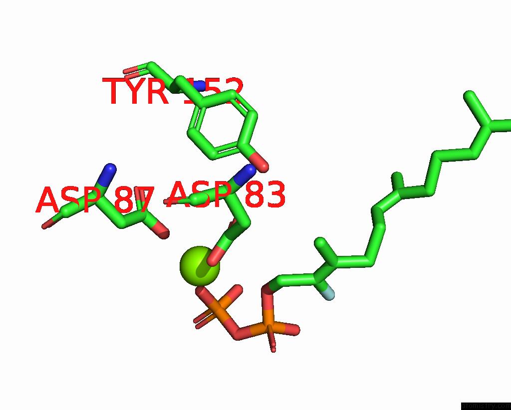
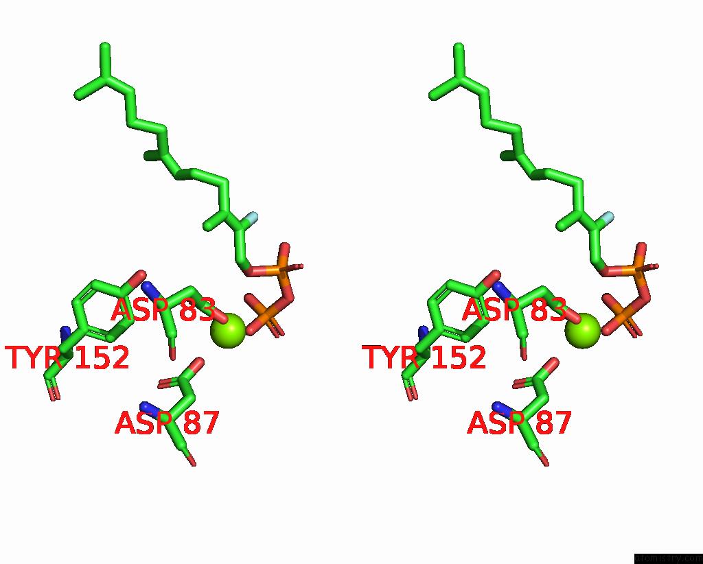
Magnesium binding site 1 out of 3 in 9c7j
Go back to
Magnesium binding site 1 out
of 3 in the Crystal Structure of Caryolan-1-Ol Synthase Complexed with 2- Fluorofarnesyl Diphosphate
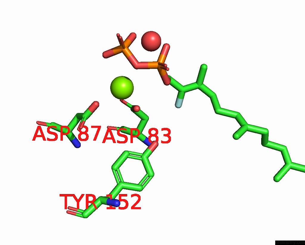
Mono view
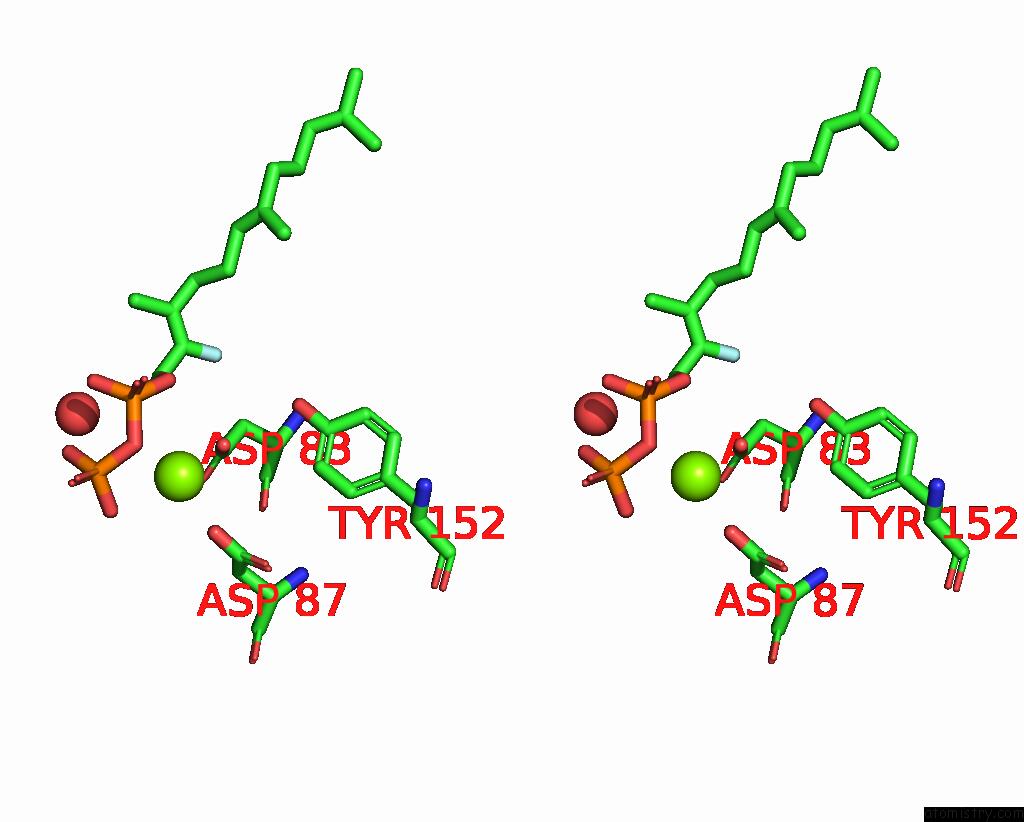
Stereo pair view
Mono view
Stereo pair view
A full contact list of Magnesium with other atoms in the Mg binding
site number 1 of Crystal Structure of Caryolan-1-Ol Synthase Complexed with 2- Fluorofarnesyl Diphosphate within 5.0Å range:
|
Magnesium binding site 2 out of 3 in 9c7j
Go back to
Magnesium binding site 2 out
of 3 in the Crystal Structure of Caryolan-1-Ol Synthase Complexed with 2- Fluorofarnesyl Diphosphate
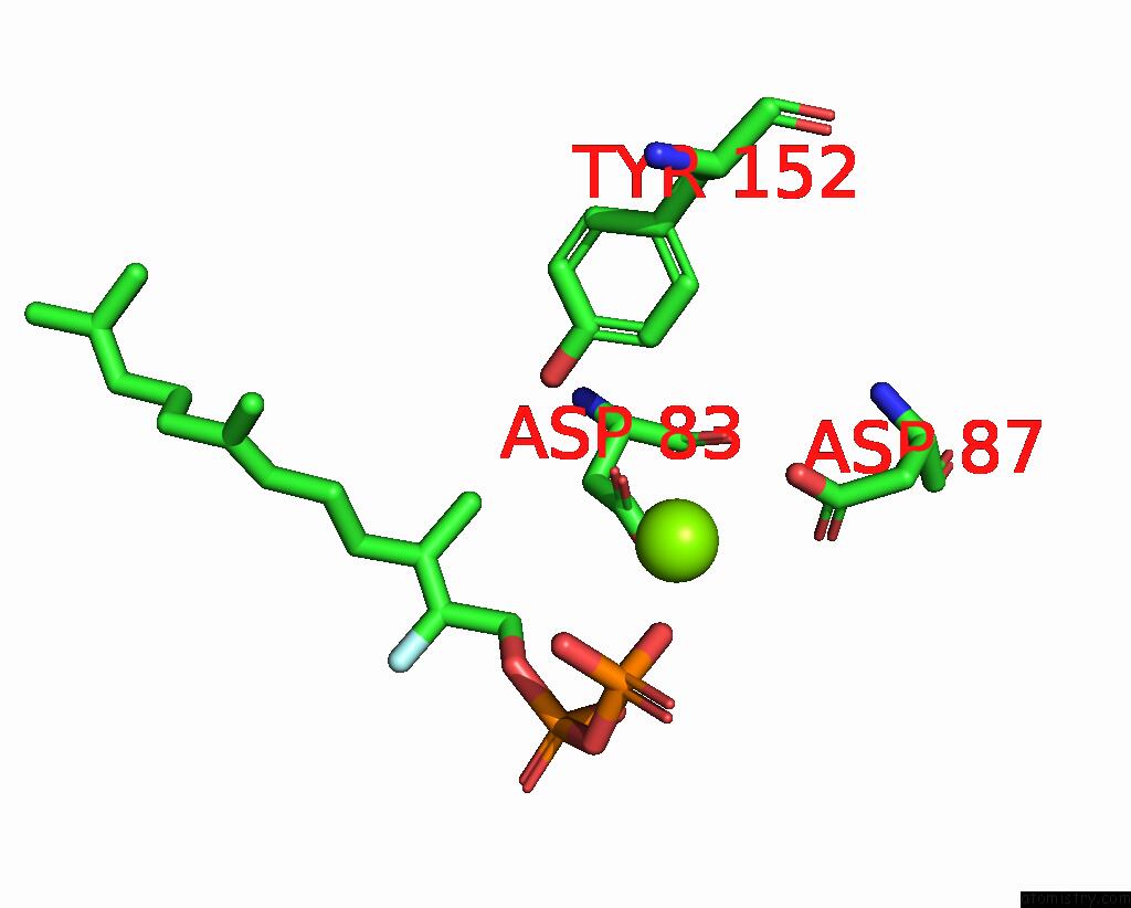
Mono view
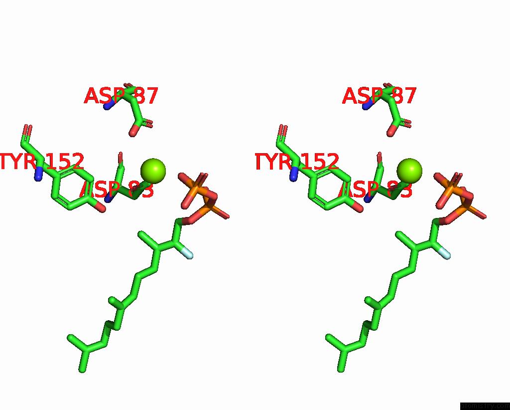
Stereo pair view
Mono view
Stereo pair view
A full contact list of Magnesium with other atoms in the Mg binding
site number 2 of Crystal Structure of Caryolan-1-Ol Synthase Complexed with 2- Fluorofarnesyl Diphosphate within 5.0Å range:
|
Magnesium binding site 3 out of 3 in 9c7j
Go back to
Magnesium binding site 3 out
of 3 in the Crystal Structure of Caryolan-1-Ol Synthase Complexed with 2- Fluorofarnesyl Diphosphate
Mono view
Stereo pair view
Mono view
Stereo pair view
A full contact list of Magnesium with other atoms in the Mg binding
site number 3 of Crystal Structure of Caryolan-1-Ol Synthase Complexed with 2- Fluorofarnesyl Diphosphate within 5.0Å range:
|
Reference:
R.P.Kumar,
J.O.Matos,
B.Y.Black,
W.H.Ellenburg,
J.Chen,
M.Patterson,
J.A.Gehtman,
D.L.Theobald,
I.J.Krauss,
D.D.Oprian.
Crystal Structure of Caryolan-1-Ol Synthase, A Sesquiterpene Synthase Catalyzing An Initial Anti-Markovnikov Cyclization Reaction. Biochemistry 2024.
ISSN: ISSN 0006-2960
PubMed: 39400323
DOI: 10.1021/ACS.BIOCHEM.4C00547
Page generated: Fri Aug 15 23:43:30 2025
ISSN: ISSN 0006-2960
PubMed: 39400323
DOI: 10.1021/ACS.BIOCHEM.4C00547
Last articles
Mn in 5RY8Mn in 5RB7
Mn in 5RB6
Mn in 5RB5
Mn in 5RB4
Mn in 5RB3
Mn in 5RB2
Mn in 5RB1
Mn in 5RB0
Mn in 5RAZ