Magnesium »
PDB 9c1a-9chw »
9cfu »
Magnesium in PDB 9cfu: Cryo-Em Structure of Myosin-1C Bound to F-Actin in the Adp-A State
Magnesium Binding Sites:
The binding sites of Magnesium atom in the Cryo-Em Structure of Myosin-1C Bound to F-Actin in the Adp-A State
(pdb code 9cfu). This binding sites where shown within
5.0 Angstroms radius around Magnesium atom.
In total 4 binding sites of Magnesium where determined in the Cryo-Em Structure of Myosin-1C Bound to F-Actin in the Adp-A State, PDB code: 9cfu:
Jump to Magnesium binding site number: 1; 2; 3; 4;
In total 4 binding sites of Magnesium where determined in the Cryo-Em Structure of Myosin-1C Bound to F-Actin in the Adp-A State, PDB code: 9cfu:
Jump to Magnesium binding site number: 1; 2; 3; 4;
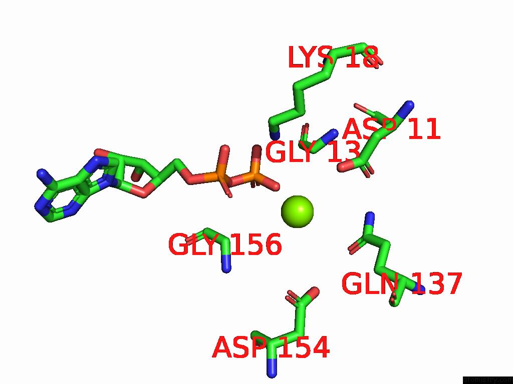
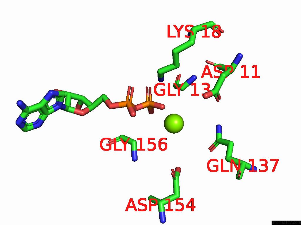
Magnesium binding site 1 out of 4 in 9cfu
Go back to
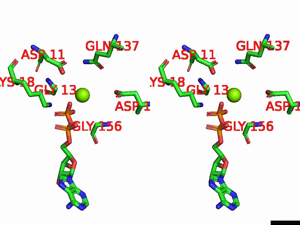
Magnesium binding site 1 out
of 4 in the Cryo-Em Structure of Myosin-1C Bound to F-Actin in the Adp-A State
Mono view

Stereo pair view
Mono view
Stereo pair view
A full contact list of Magnesium with other atoms in the Mg binding
site number 1 of Cryo-Em Structure of Myosin-1C Bound to F-Actin in the Adp-A State within 5.0Å range:
|
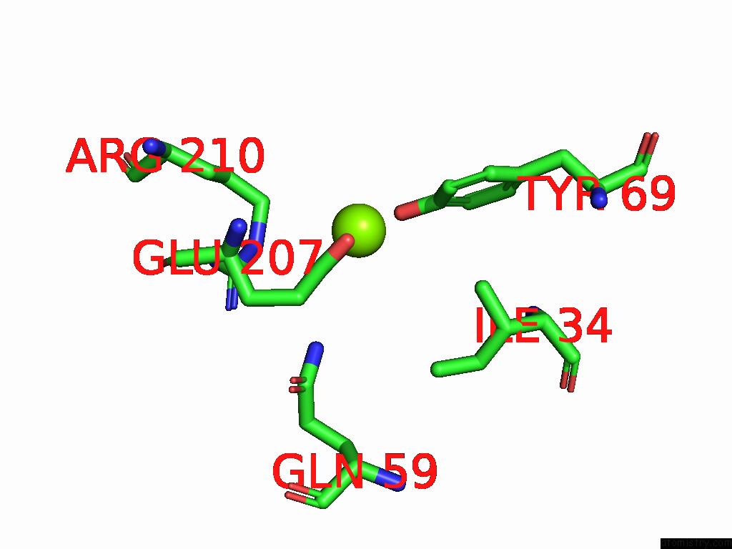
Magnesium binding site 2 out of 4 in 9cfu
Go back to
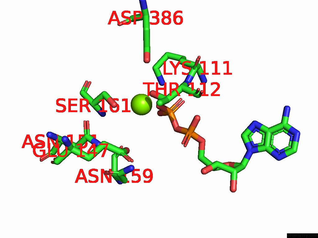
Magnesium binding site 2 out
of 4 in the Cryo-Em Structure of Myosin-1C Bound to F-Actin in the Adp-A State
Mono view
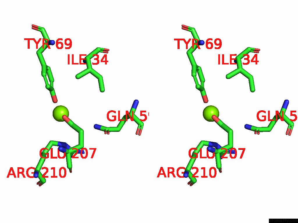
Stereo pair view
Mono view
Stereo pair view
A full contact list of Magnesium with other atoms in the Mg binding
site number 2 of Cryo-Em Structure of Myosin-1C Bound to F-Actin in the Adp-A State within 5.0Å range:
|
Magnesium binding site 3 out of 4 in 9cfu
Go back to
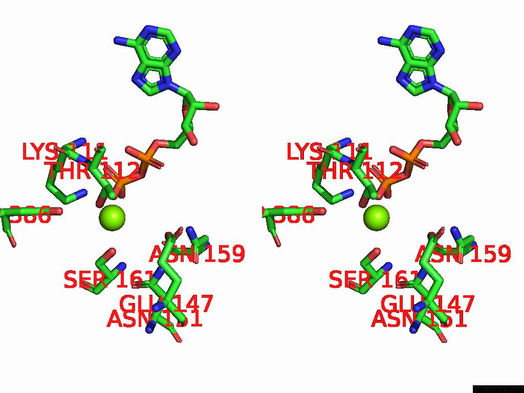
Magnesium binding site 3 out
of 4 in the Cryo-Em Structure of Myosin-1C Bound to F-Actin in the Adp-A State
Mono view
Stereo pair view
Mono view
Stereo pair view
A full contact list of Magnesium with other atoms in the Mg binding
site number 3 of Cryo-Em Structure of Myosin-1C Bound to F-Actin in the Adp-A State within 5.0Å range:
|
Magnesium binding site 4 out of 4 in 9cfu
Go back to
Magnesium binding site 4 out
of 4 in the Cryo-Em Structure of Myosin-1C Bound to F-Actin in the Adp-A State
Mono view
Stereo pair view
Mono view
Stereo pair view
A full contact list of Magnesium with other atoms in the Mg binding
site number 4 of Cryo-Em Structure of Myosin-1C Bound to F-Actin in the Adp-A State within 5.0Å range:
|
Reference:
S.S.Chavali,
C.V.Sindelar,
M.E.Ostap.
High Resolution Structures of Myosin-Ic Reveal A Unique Actin-Binding Orientation, Adp Release Pathway, and Power Stroke Trajectory To Be Published.
Page generated: Tue Feb 25 11:15:52 2025
Last articles
Ca in 5S4NCa in 5S4M
Ca in 5S4L
Ca in 5QJ2
Ca in 5QJ3
Ca in 5PTP
Ca in 5PB6
Ca in 5PB5
Ca in 5PB4
Ca in 5PB3