Magnesium »
PDB 9dq0-9enb »
9drs »
Magnesium in PDB 9drs: Crystal Structure of M. Tuberculosis Phers-Trna Complex Bound to Inhibitor D-116
Enzymatic activity of Crystal Structure of M. Tuberculosis Phers-Trna Complex Bound to Inhibitor D-116
All present enzymatic activity of Crystal Structure of M. Tuberculosis Phers-Trna Complex Bound to Inhibitor D-116:
6.1.1.20;
6.1.1.20;
Protein crystallography data
The structure of Crystal Structure of M. Tuberculosis Phers-Trna Complex Bound to Inhibitor D-116, PDB code: 9drs
was solved by
P.Gade,
C.Chang,
B.Forte,
J.Wower,
I.H.Gilbert,
B.Baragana,
K.Michalska,
A.Joachimiak,
Center For Structural Biology Of Infectious Diseases(Csbid),
with X-Ray Crystallography technique. A brief refinement statistics is given in the table below:
| Resolution Low / High (Å) | 46.77 / 2.35 |
| Space group | P 1 21 1 |
| Cell size a, b, c (Å), α, β, γ (°) | 146.676, 64.106, 188.334, 90, 111.21, 90 |
| R / Rfree (%) | 20.1 / 25 |
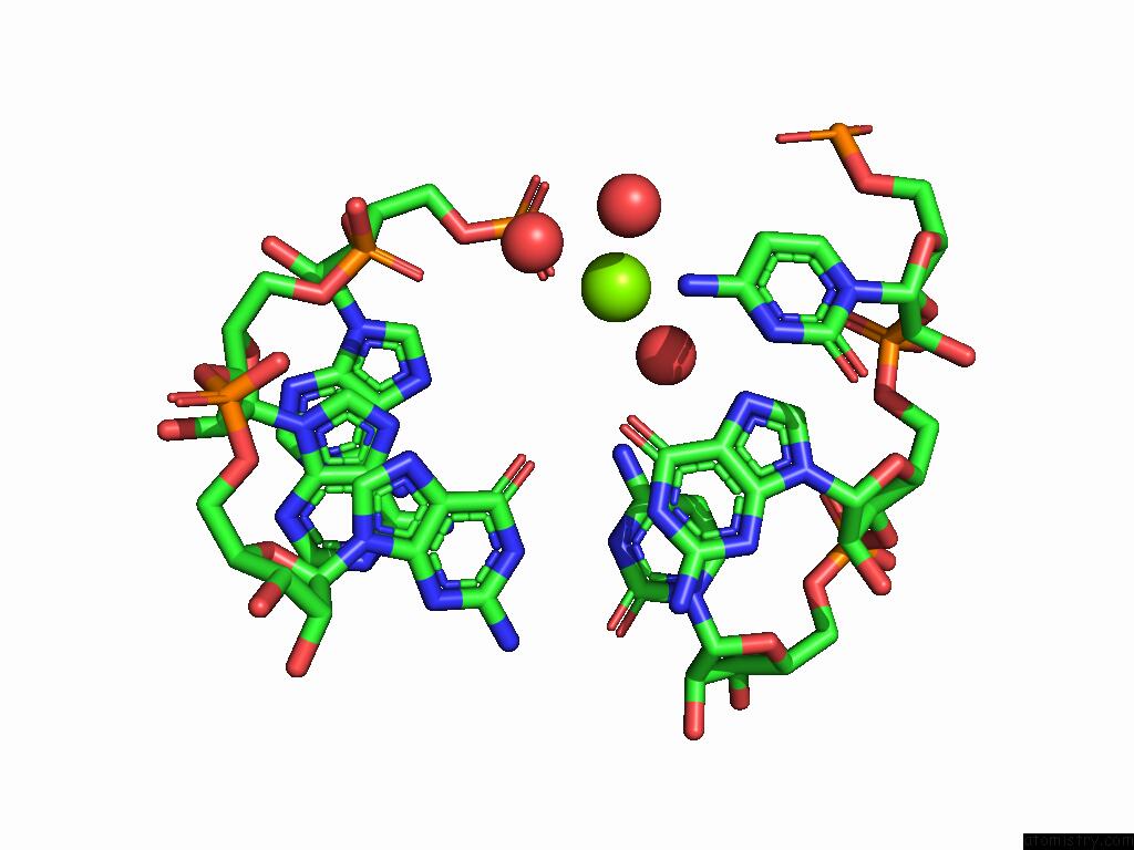
Magnesium Binding Sites:
The binding sites of Magnesium atom in the Crystal Structure of M. Tuberculosis Phers-Trna Complex Bound to Inhibitor D-116
(pdb code 9drs). This binding sites where shown within
5.0 Angstroms radius around Magnesium atom.
In total 7 binding sites of Magnesium where determined in the Crystal Structure of M. Tuberculosis Phers-Trna Complex Bound to Inhibitor D-116, PDB code: 9drs:
Jump to Magnesium binding site number: 1; 2; 3; 4; 5; 6; 7;
In total 7 binding sites of Magnesium where determined in the Crystal Structure of M. Tuberculosis Phers-Trna Complex Bound to Inhibitor D-116, PDB code: 9drs:
Jump to Magnesium binding site number: 1; 2; 3; 4; 5; 6; 7;
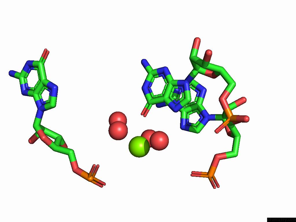
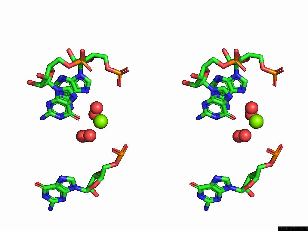
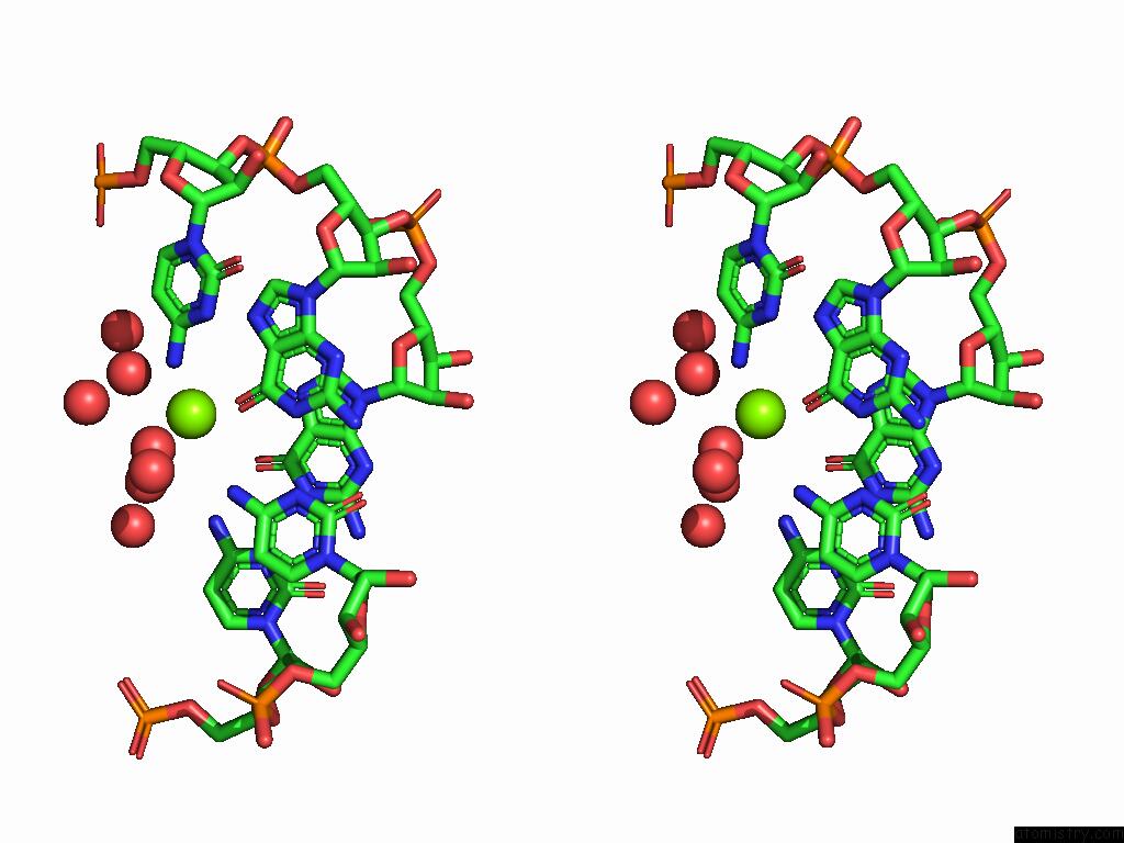
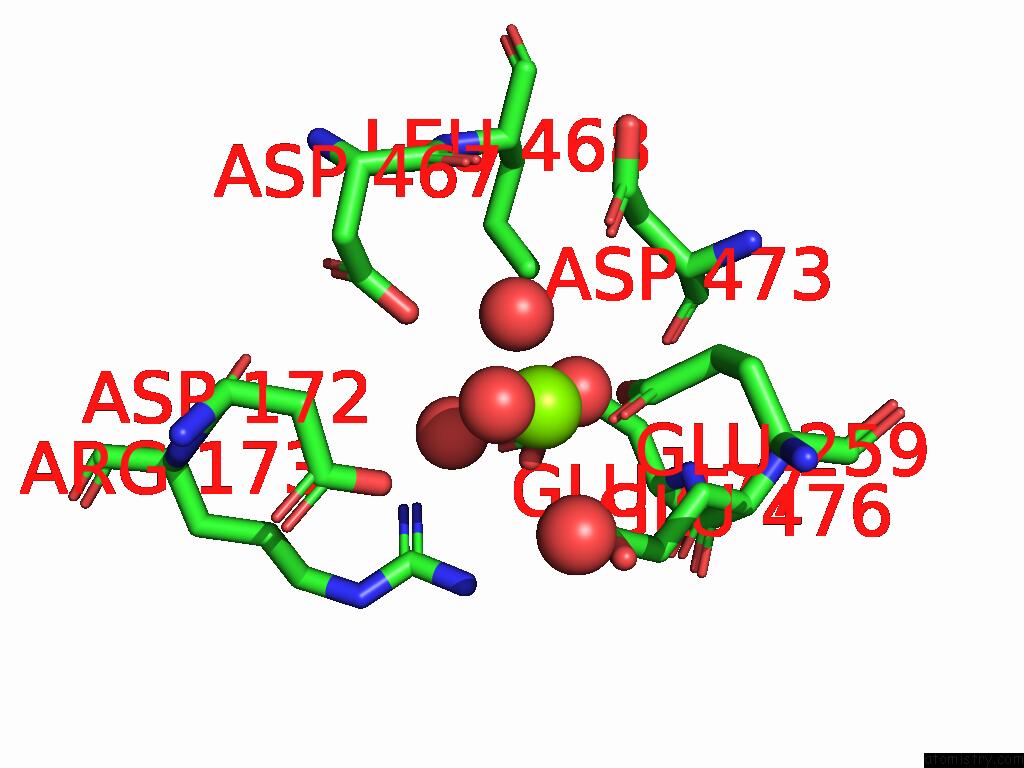
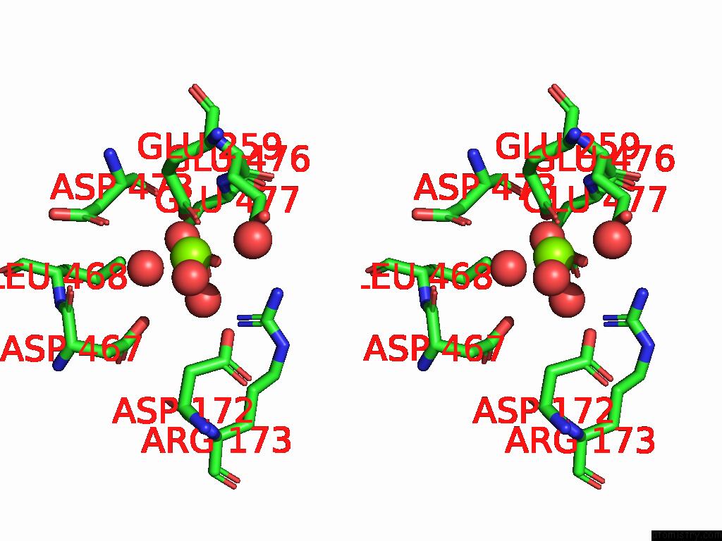
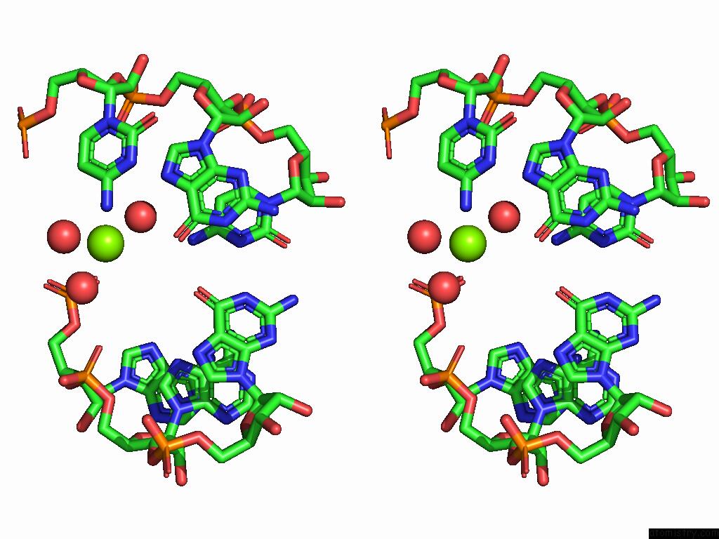
Magnesium binding site 1 out of 7 in 9drs
Go back to
Magnesium binding site 1 out
of 7 in the Crystal Structure of M. Tuberculosis Phers-Trna Complex Bound to Inhibitor D-116
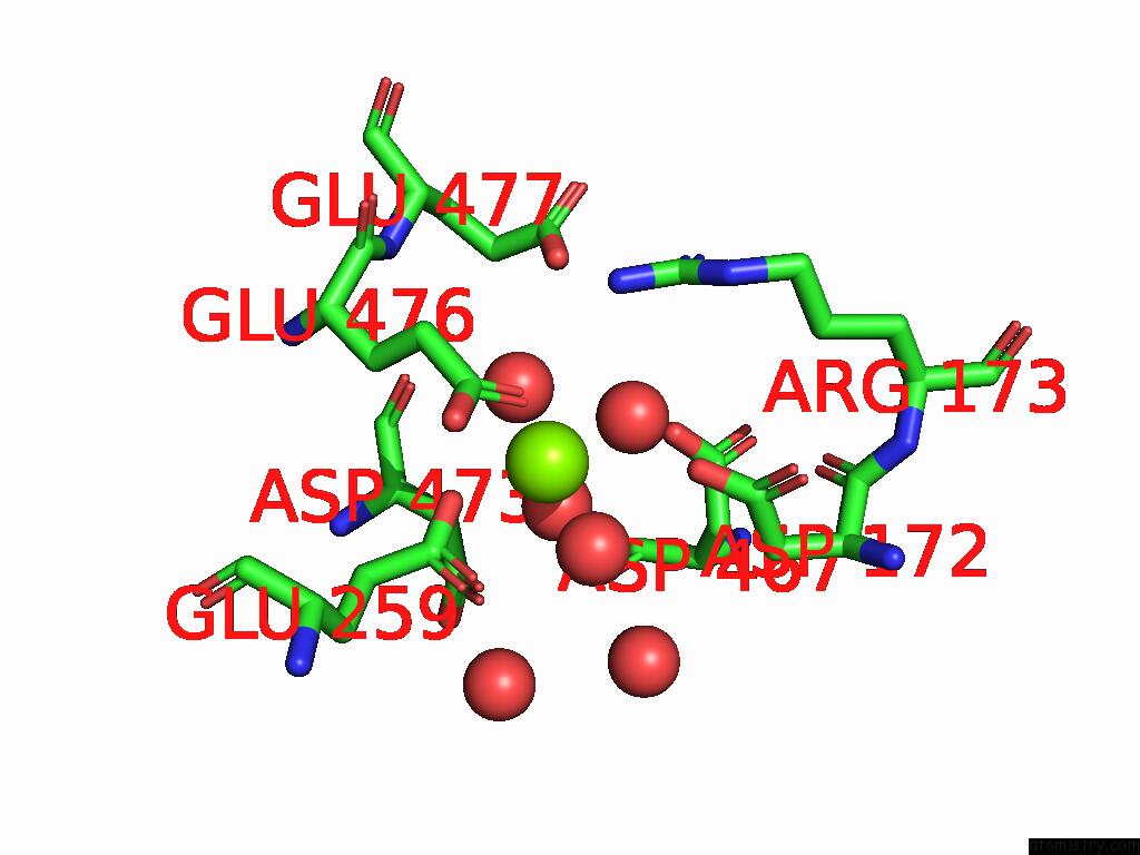
Mono view
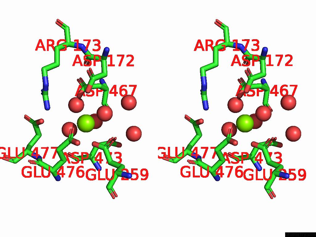
Stereo pair view
Mono view
Stereo pair view
A full contact list of Magnesium with other atoms in the Mg binding
site number 1 of Crystal Structure of M. Tuberculosis Phers-Trna Complex Bound to Inhibitor D-116 within 5.0Å range:
|
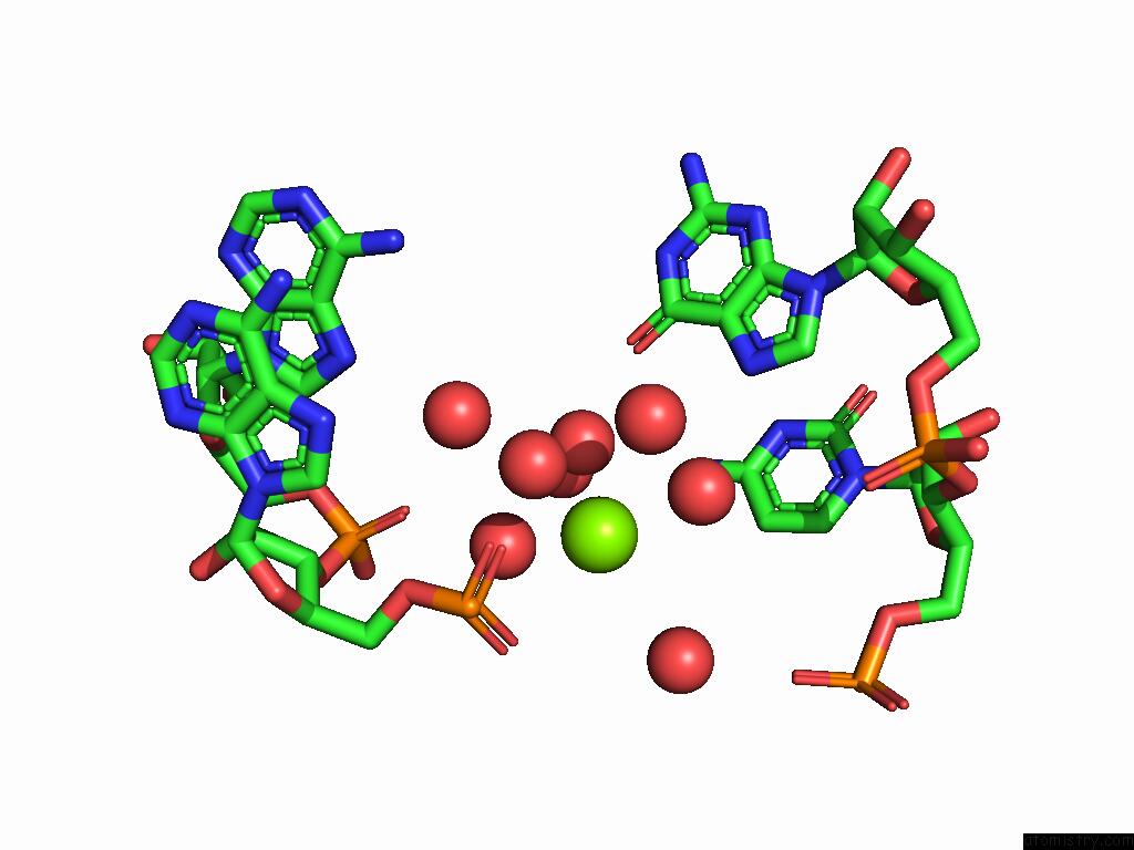
Magnesium binding site 2 out of 7 in 9drs
Go back to
Magnesium binding site 2 out
of 7 in the Crystal Structure of M. Tuberculosis Phers-Trna Complex Bound to Inhibitor D-116
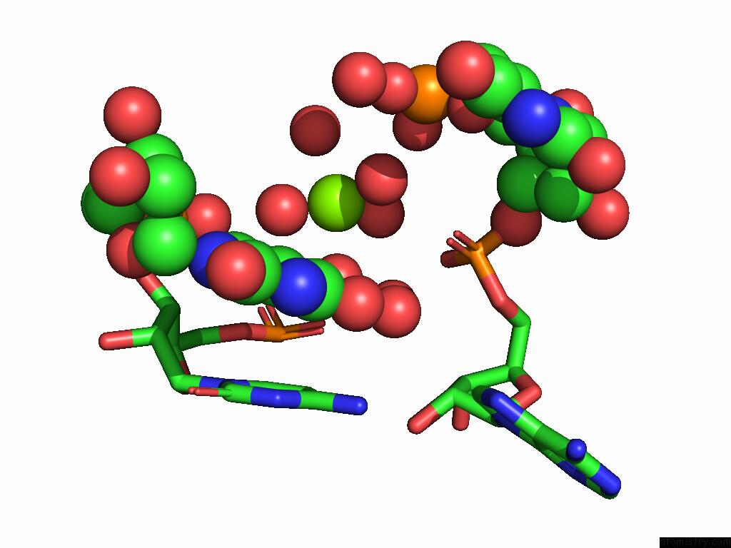
Mono view
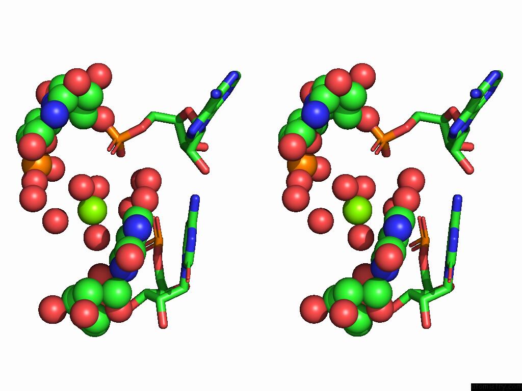
Stereo pair view
Mono view
Stereo pair view
A full contact list of Magnesium with other atoms in the Mg binding
site number 2 of Crystal Structure of M. Tuberculosis Phers-Trna Complex Bound to Inhibitor D-116 within 5.0Å range:
|
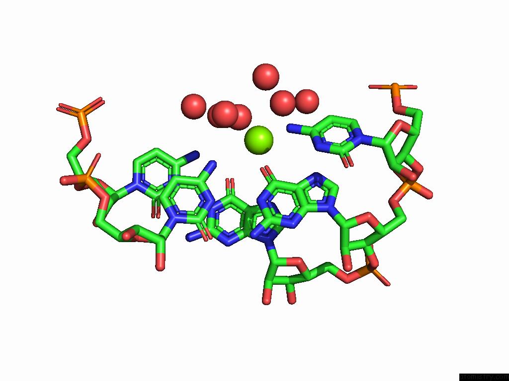
Magnesium binding site 3 out of 7 in 9drs
Go back to
Magnesium binding site 3 out
of 7 in the Crystal Structure of M. Tuberculosis Phers-Trna Complex Bound to Inhibitor D-116
Mono view
Stereo pair view
Mono view
Stereo pair view
A full contact list of Magnesium with other atoms in the Mg binding
site number 3 of Crystal Structure of M. Tuberculosis Phers-Trna Complex Bound to Inhibitor D-116 within 5.0Å range:
|
Magnesium binding site 4 out of 7 in 9drs
Go back to
Magnesium binding site 4 out
of 7 in the Crystal Structure of M. Tuberculosis Phers-Trna Complex Bound to Inhibitor D-116
Mono view

Stereo pair view
Mono view
Stereo pair view
A full contact list of Magnesium with other atoms in the Mg binding
site number 4 of Crystal Structure of M. Tuberculosis Phers-Trna Complex Bound to Inhibitor D-116 within 5.0Å range:
|
Magnesium binding site 5 out of 7 in 9drs
Go back to
Magnesium binding site 5 out
of 7 in the Crystal Structure of M. Tuberculosis Phers-Trna Complex Bound to Inhibitor D-116
Mono view
Stereo pair view
Mono view
Stereo pair view
A full contact list of Magnesium with other atoms in the Mg binding
site number 5 of Crystal Structure of M. Tuberculosis Phers-Trna Complex Bound to Inhibitor D-116 within 5.0Å range:
|
Magnesium binding site 6 out of 7 in 9drs
Go back to
Magnesium binding site 6 out
of 7 in the Crystal Structure of M. Tuberculosis Phers-Trna Complex Bound to Inhibitor D-116
Mono view
Stereo pair view
Mono view
Stereo pair view
A full contact list of Magnesium with other atoms in the Mg binding
site number 6 of Crystal Structure of M. Tuberculosis Phers-Trna Complex Bound to Inhibitor D-116 within 5.0Å range:
|
Magnesium binding site 7 out of 7 in 9drs
Go back to
Magnesium binding site 7 out
of 7 in the Crystal Structure of M. Tuberculosis Phers-Trna Complex Bound to Inhibitor D-116
Mono view
Stereo pair view
Mono view
Stereo pair view
A full contact list of Magnesium with other atoms in the Mg binding
site number 7 of Crystal Structure of M. Tuberculosis Phers-Trna Complex Bound to Inhibitor D-116 within 5.0Å range:
|
Reference:
P.Gade,
C.Chang,
D.S.Pryde,
D.Fletcher,
S.Niven,
L.G.Magalhaes,
D.Robinson,
J.Saini,
P.E.Ibrahim,
B.Forte,
J.Wower,
M.J.Bodkin,
B.Baragana,
I.H.Gilbert,
K.Michalska,
A.Joachimiak.
Different Chemical Scaffolds Bind to L-Phe Site in Mycobacterium Tuberculosis Phe-Trna Synthetase Eur.J.Med.Chem. 17335 2025.
ISSN: ISSN 0223-5234
DOI: 10.1016/J.EJMECH.2025.117335
Page generated: Tue Feb 25 11:17:35 2025
ISSN: ISSN 0223-5234
DOI: 10.1016/J.EJMECH.2025.117335
Last articles
Cl in 5WQ3Cl in 5WRO
Cl in 5WRB
Cl in 5WRA
Cl in 5WR9
Cl in 5WO3
Cl in 5WO4
Cl in 5WPN
Cl in 5WO2
Cl in 5WO1