Magnesium »
PDB 9he9-9ikf »
9i7l »
Magnesium in PDB 9i7l: Xylose Isomerase Collected at 50C Using Time-Resolved Serial Synchrotron Crystallography with Glucose at 180 Seconds
Enzymatic activity of Xylose Isomerase Collected at 50C Using Time-Resolved Serial Synchrotron Crystallography with Glucose at 180 Seconds
All present enzymatic activity of Xylose Isomerase Collected at 50C Using Time-Resolved Serial Synchrotron Crystallography with Glucose at 180 Seconds:
5.3.1.5;
5.3.1.5;
Protein crystallography data
The structure of Xylose Isomerase Collected at 50C Using Time-Resolved Serial Synchrotron Crystallography with Glucose at 180 Seconds, PDB code: 9i7l
was solved by
E.C.Schulz,
A.Prester,
D.V.Stetten,
G.Gore,
C.E.Hatton,
K.Bartels,
J.P.Leimkohl,
H.Schikora,
H.M.Ginn,
F.Tellkamp,
P.Mehrabi,
with X-Ray Crystallography technique. A brief refinement statistics is given in the table below:
| Resolution Low / High (Å) | 71.49 / 1.70 |
| Space group | I 2 2 2 |
| Cell size a, b, c (Å), α, β, γ (°) | 94.2, 103.05, 99.25, 90, 90, 90 |
| R / Rfree (%) | 16.9 / 20.7 |
Magnesium Binding Sites:
The binding sites of Magnesium atom in the Xylose Isomerase Collected at 50C Using Time-Resolved Serial Synchrotron Crystallography with Glucose at 180 Seconds
(pdb code 9i7l). This binding sites where shown within
5.0 Angstroms radius around Magnesium atom.
In total 3 binding sites of Magnesium where determined in the Xylose Isomerase Collected at 50C Using Time-Resolved Serial Synchrotron Crystallography with Glucose at 180 Seconds, PDB code: 9i7l:
Jump to Magnesium binding site number: 1; 2; 3;
In total 3 binding sites of Magnesium where determined in the Xylose Isomerase Collected at 50C Using Time-Resolved Serial Synchrotron Crystallography with Glucose at 180 Seconds, PDB code: 9i7l:
Jump to Magnesium binding site number: 1; 2; 3;
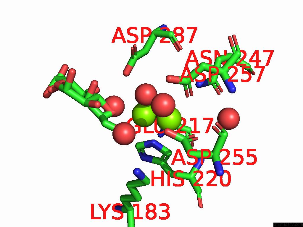
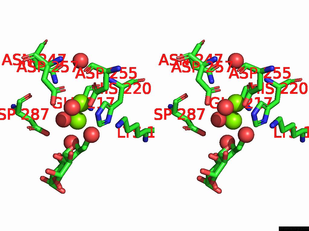
Magnesium binding site 1 out of 3 in 9i7l
Go back to
Magnesium binding site 1 out
of 3 in the Xylose Isomerase Collected at 50C Using Time-Resolved Serial Synchrotron Crystallography with Glucose at 180 Seconds
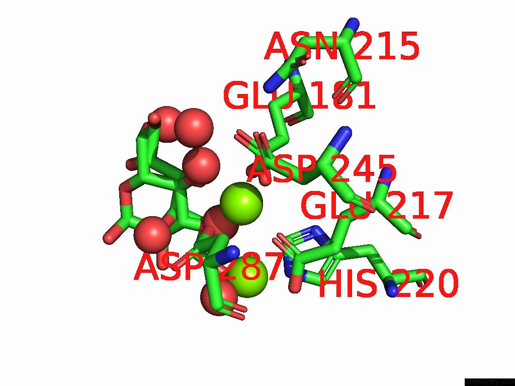
Mono view
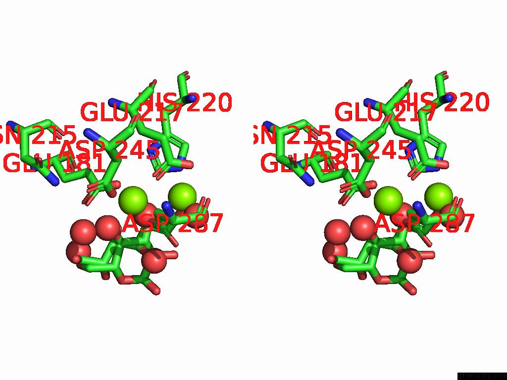
Stereo pair view
Mono view
Stereo pair view
A full contact list of Magnesium with other atoms in the Mg binding
site number 1 of Xylose Isomerase Collected at 50C Using Time-Resolved Serial Synchrotron Crystallography with Glucose at 180 Seconds within 5.0Å range:
|
Magnesium binding site 2 out of 3 in 9i7l
Go back to
Magnesium binding site 2 out
of 3 in the Xylose Isomerase Collected at 50C Using Time-Resolved Serial Synchrotron Crystallography with Glucose at 180 Seconds
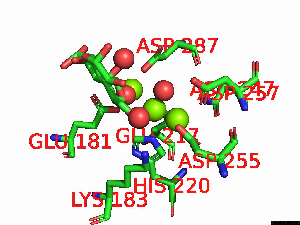
Mono view
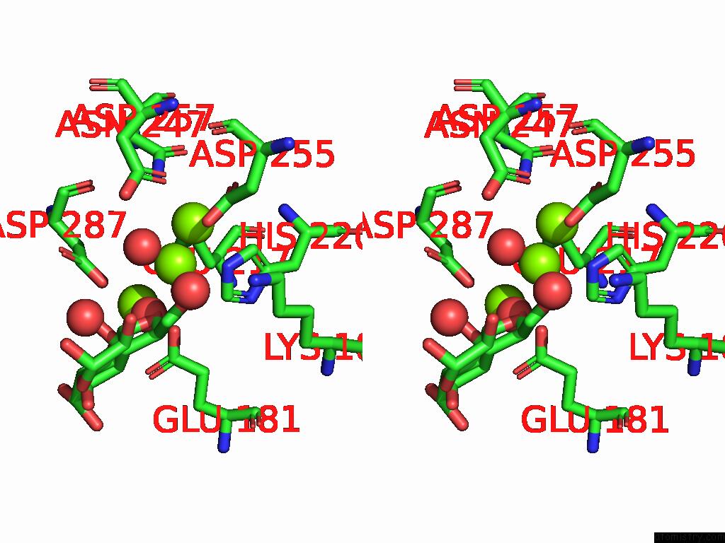
Stereo pair view
Mono view
Stereo pair view
A full contact list of Magnesium with other atoms in the Mg binding
site number 2 of Xylose Isomerase Collected at 50C Using Time-Resolved Serial Synchrotron Crystallography with Glucose at 180 Seconds within 5.0Å range:
|
Magnesium binding site 3 out of 3 in 9i7l
Go back to
Magnesium binding site 3 out
of 3 in the Xylose Isomerase Collected at 50C Using Time-Resolved Serial Synchrotron Crystallography with Glucose at 180 Seconds
Mono view
Stereo pair view
Mono view
Stereo pair view
A full contact list of Magnesium with other atoms in the Mg binding
site number 3 of Xylose Isomerase Collected at 50C Using Time-Resolved Serial Synchrotron Crystallography with Glucose at 180 Seconds within 5.0Å range:
|
Reference:
E.C.Schulz,
A.Prester,
D.Von Stetten,
G.Gore,
C.E.Hatton,
K.Bartels,
J.P.Leimkohl,
H.Schikora,
H.M.Ginn,
F.Tellkamp,
P.Mehrabi.
Probing the Modulation of Enzyme Kinetics By Multi-Temperature, Time-Resolved Serial Crystallography. Nat Commun V. 16 6553 2025.
ISSN: ESSN 2041-1723
PubMed: 40670369
DOI: 10.1038/S41467-025-61631-2
Page generated: Sat Aug 23 06:02:48 2025
ISSN: ESSN 2041-1723
PubMed: 40670369
DOI: 10.1038/S41467-025-61631-2
Last articles
Mn in 9LJUMn in 9LJW
Mn in 9LJS
Mn in 9LJR
Mn in 9LJT
Mn in 9LJV
Mg in 9UA2
Mg in 9R96
Mg in 9VM1
Mg in 9P01