Magnesium »
PDB 9h5p-9ik0 »
9ij6 »
Magnesium in PDB 9ij6: Crystal Structure of the Complex of Erythrose-4-Phosphate Dehydrogenase From Acinetobacter Baumannii with Adenosine Phosphate at 2.40 A Resolution.
Protein crystallography data
The structure of Crystal Structure of the Complex of Erythrose-4-Phosphate Dehydrogenase From Acinetobacter Baumannii with Adenosine Phosphate at 2.40 A Resolution., PDB code: 9ij6
was solved by
V.Viswanathan,
A.Kumari,
A.Singh,
A.Kumar,
P.Sharma,
S.Chopra,
J.Jeyakanthan,
S.Sharma,
C.I.Raje,
T.P.Singh,
with X-Ray Crystallography technique. A brief refinement statistics is given in the table below:
| Resolution Low / High (Å) | 83.65 / 2.40 |
| Space group | C 2 2 21 |
| Cell size a, b, c (Å), α, β, γ (°) | 147.578, 167.012, 155.058, 90, 90, 90 |
| R / Rfree (%) | 17 / 22 |
Magnesium Binding Sites:
The binding sites of Magnesium atom in the Crystal Structure of the Complex of Erythrose-4-Phosphate Dehydrogenase From Acinetobacter Baumannii with Adenosine Phosphate at 2.40 A Resolution.
(pdb code 9ij6). This binding sites where shown within
5.0 Angstroms radius around Magnesium atom.
In total 5 binding sites of Magnesium where determined in the Crystal Structure of the Complex of Erythrose-4-Phosphate Dehydrogenase From Acinetobacter Baumannii with Adenosine Phosphate at 2.40 A Resolution., PDB code: 9ij6:
Jump to Magnesium binding site number: 1; 2; 3; 4; 5;
In total 5 binding sites of Magnesium where determined in the Crystal Structure of the Complex of Erythrose-4-Phosphate Dehydrogenase From Acinetobacter Baumannii with Adenosine Phosphate at 2.40 A Resolution., PDB code: 9ij6:
Jump to Magnesium binding site number: 1; 2; 3; 4; 5;
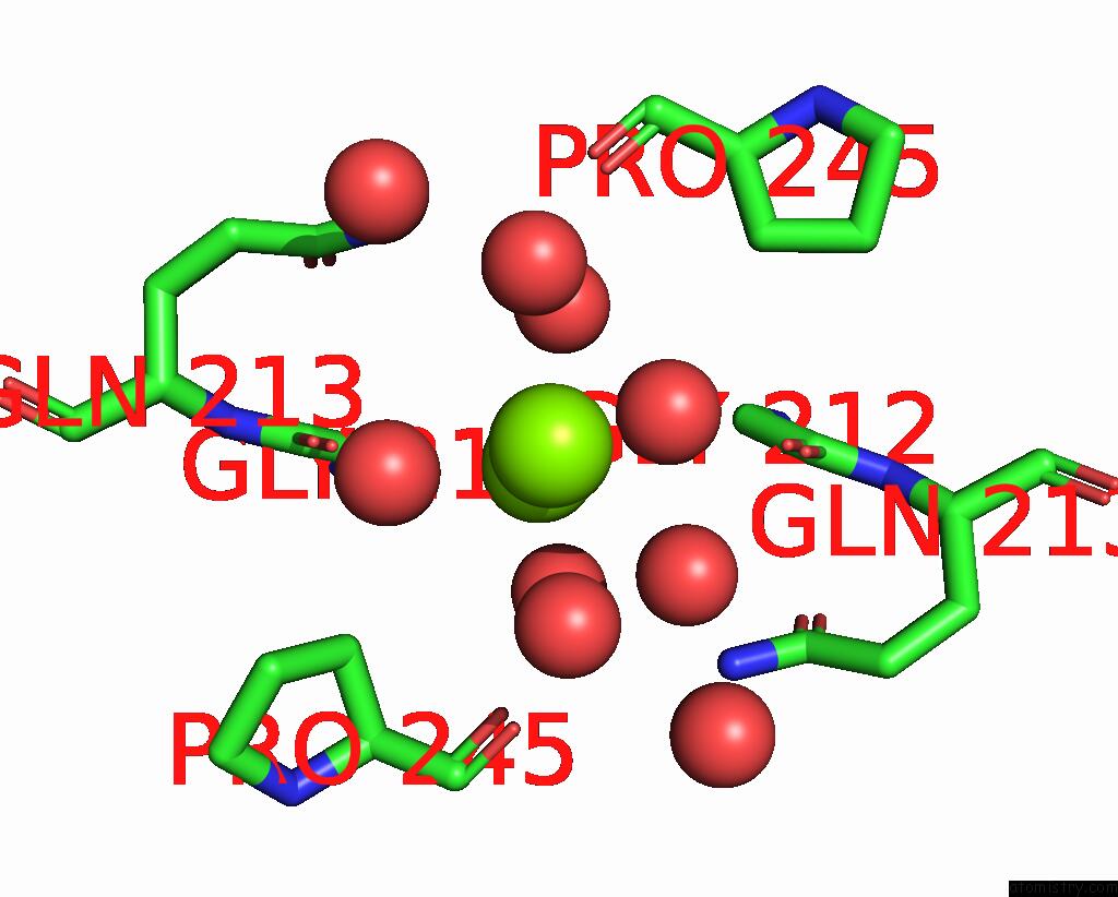
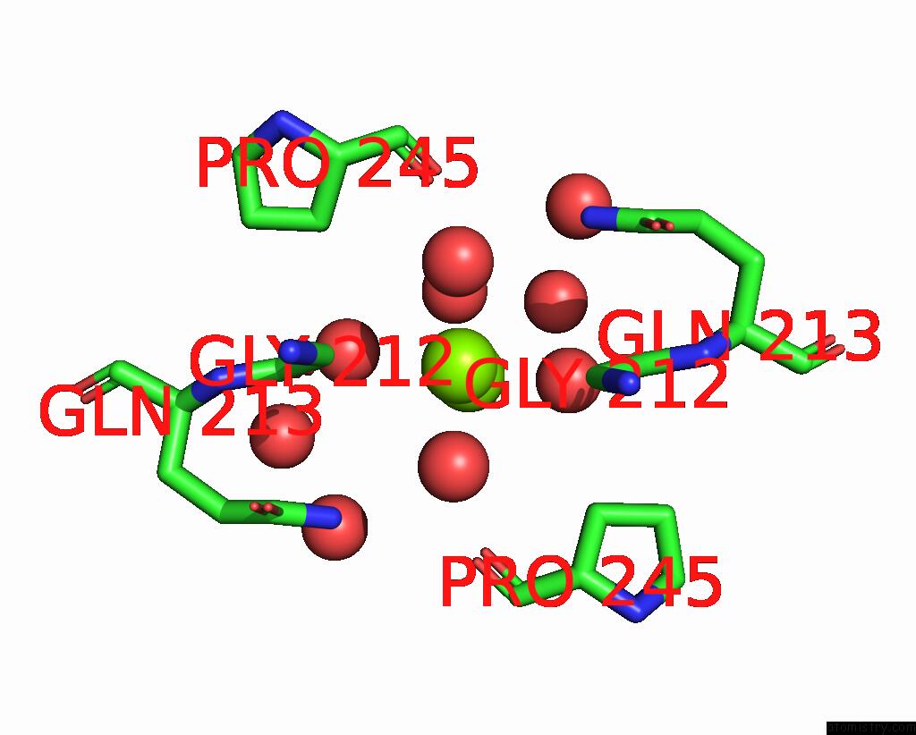
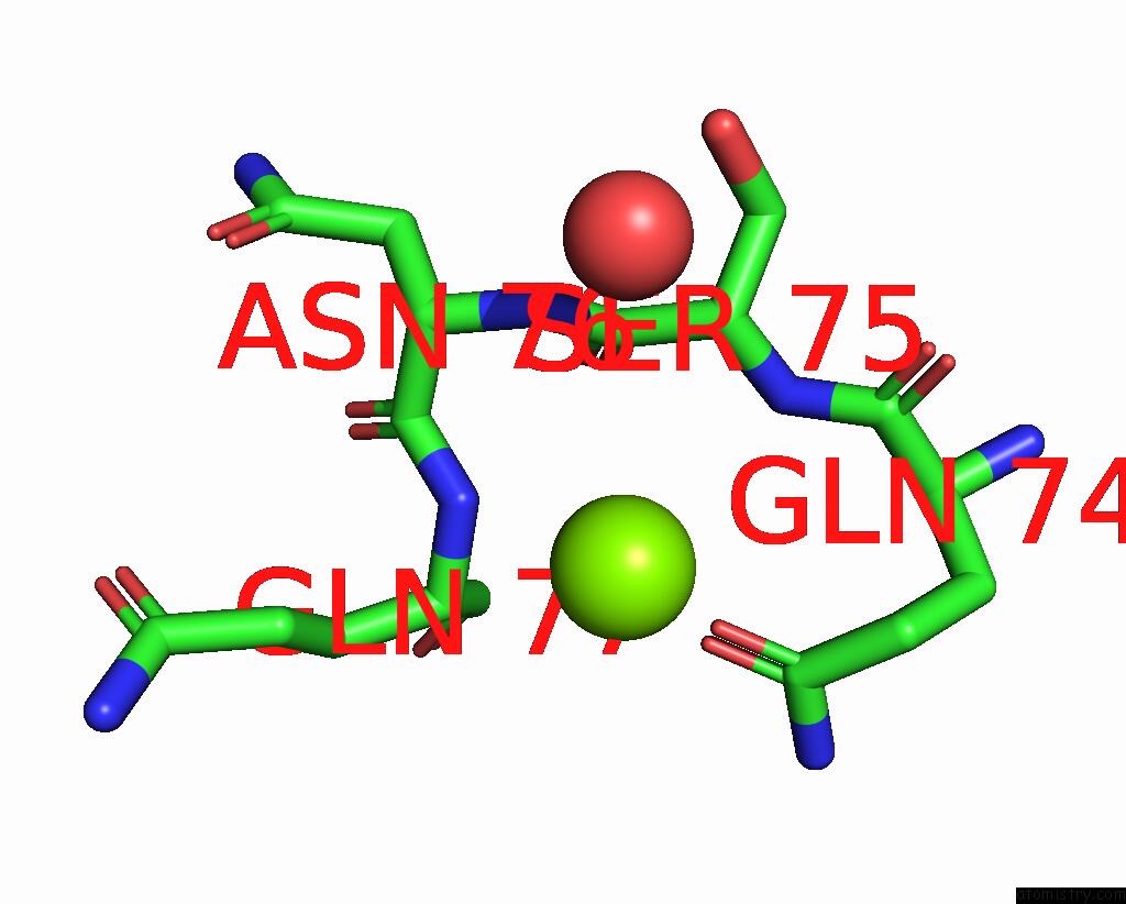
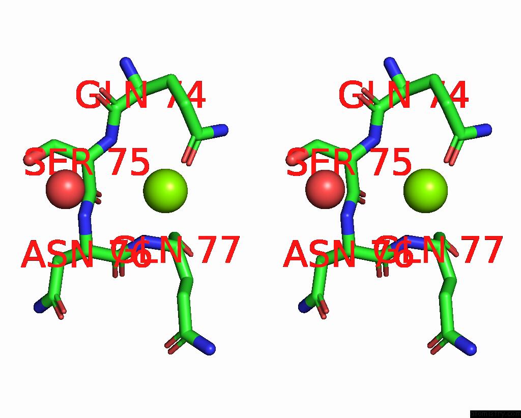
Magnesium binding site 1 out of 5 in 9ij6
Go back to
Magnesium binding site 1 out
of 5 in the Crystal Structure of the Complex of Erythrose-4-Phosphate Dehydrogenase From Acinetobacter Baumannii with Adenosine Phosphate at 2.40 A Resolution.
Mono view
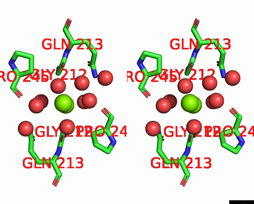
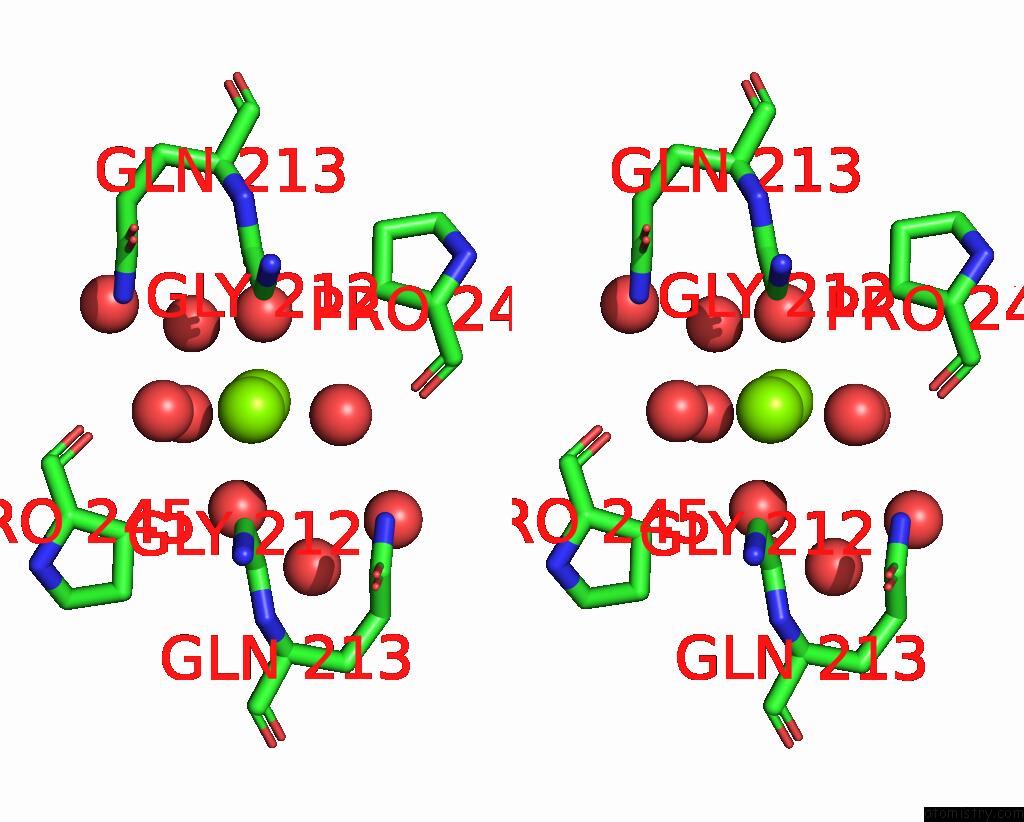
Stereo pair view
Mono view
Stereo pair view
A full contact list of Magnesium with other atoms in the Mg binding
site number 1 of Crystal Structure of the Complex of Erythrose-4-Phosphate Dehydrogenase From Acinetobacter Baumannii with Adenosine Phosphate at 2.40 A Resolution. within 5.0Å range:
|
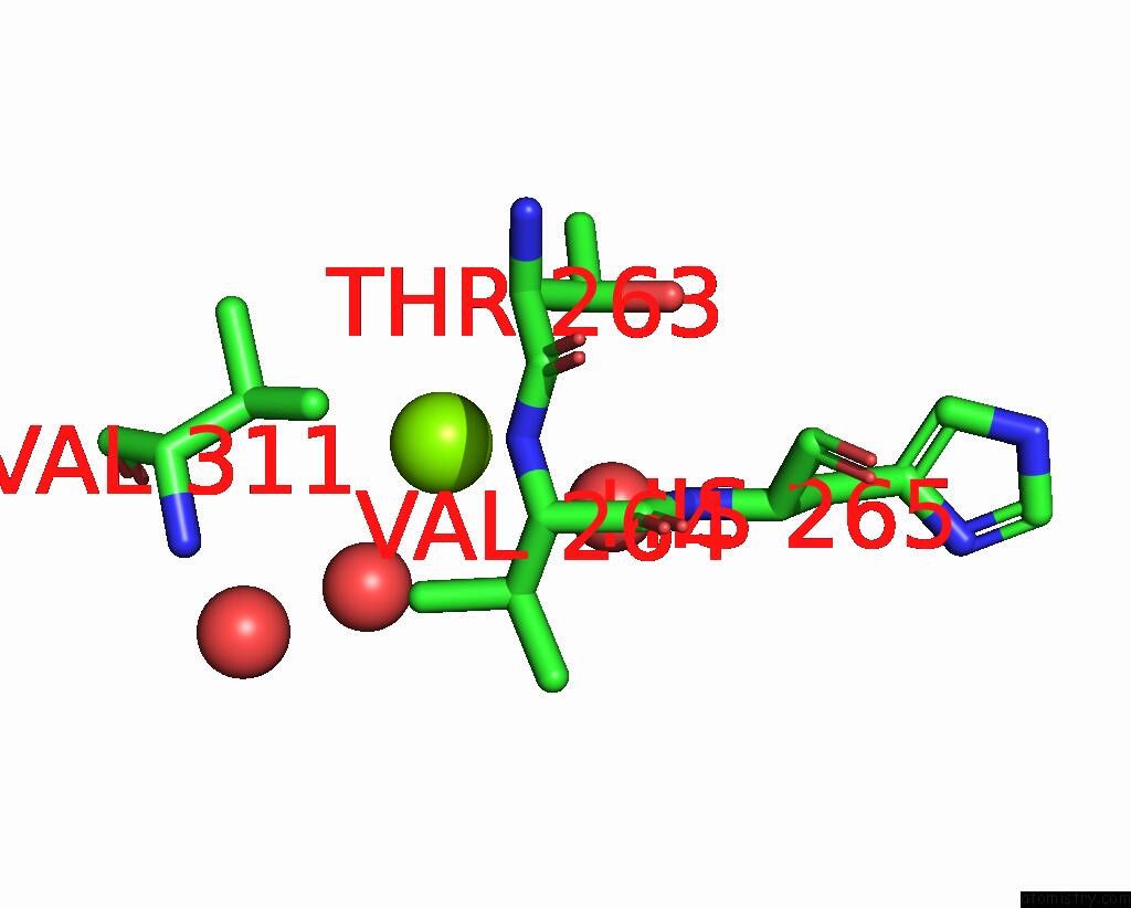
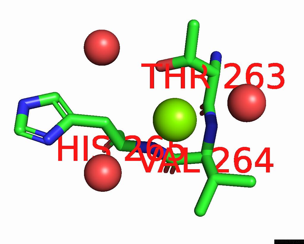
Magnesium binding site 2 out of 5 in 9ij6
Go back to
Magnesium binding site 2 out
of 5 in the Crystal Structure of the Complex of Erythrose-4-Phosphate Dehydrogenase From Acinetobacter Baumannii with Adenosine Phosphate at 2.40 A Resolution.
Mono view
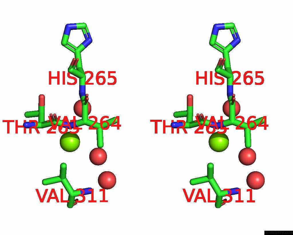
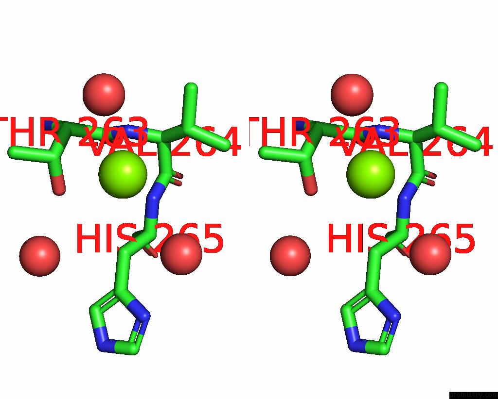
Stereo pair view
Mono view
Stereo pair view
A full contact list of Magnesium with other atoms in the Mg binding
site number 2 of Crystal Structure of the Complex of Erythrose-4-Phosphate Dehydrogenase From Acinetobacter Baumannii with Adenosine Phosphate at 2.40 A Resolution. within 5.0Å range:
|
Magnesium binding site 3 out of 5 in 9ij6
Go back to
Magnesium binding site 3 out
of 5 in the Crystal Structure of the Complex of Erythrose-4-Phosphate Dehydrogenase From Acinetobacter Baumannii with Adenosine Phosphate at 2.40 A Resolution.
Mono view
Stereo pair view
Mono view
Stereo pair view
A full contact list of Magnesium with other atoms in the Mg binding
site number 3 of Crystal Structure of the Complex of Erythrose-4-Phosphate Dehydrogenase From Acinetobacter Baumannii with Adenosine Phosphate at 2.40 A Resolution. within 5.0Å range:
|
Magnesium binding site 4 out of 5 in 9ij6
Go back to
Magnesium binding site 4 out
of 5 in the Crystal Structure of the Complex of Erythrose-4-Phosphate Dehydrogenase From Acinetobacter Baumannii with Adenosine Phosphate at 2.40 A Resolution.
Mono view
Stereo pair view
Mono view
Stereo pair view
A full contact list of Magnesium with other atoms in the Mg binding
site number 4 of Crystal Structure of the Complex of Erythrose-4-Phosphate Dehydrogenase From Acinetobacter Baumannii with Adenosine Phosphate at 2.40 A Resolution. within 5.0Å range:
|
Magnesium binding site 5 out of 5 in 9ij6
Go back to
Magnesium binding site 5 out
of 5 in the Crystal Structure of the Complex of Erythrose-4-Phosphate Dehydrogenase From Acinetobacter Baumannii with Adenosine Phosphate at 2.40 A Resolution.
Mono view
Stereo pair view
Mono view
Stereo pair view
A full contact list of Magnesium with other atoms in the Mg binding
site number 5 of Crystal Structure of the Complex of Erythrose-4-Phosphate Dehydrogenase From Acinetobacter Baumannii with Adenosine Phosphate at 2.40 A Resolution. within 5.0Å range:
|
Reference:
V.Viswanathan,
A.Kumari,
A.Singh,
A.Kumar,
P.Sharma,
S.Chopra,
J.Jeyakanthan,
S.Sharma,
C.I.Raje,
T.P.Singh.
Crystal Structure of the Complex of Erythrose-4-Phosphate Dehydrogenase From Acinetobacter Baumannii with Adenosine Phosphate at 2.40 A Resolution. To Be Published.
Page generated: Sat Aug 16 04:11:45 2025
Last articles
Mn in 5YVSMn in 5YVR
Mn in 5YVM
Mn in 5YTZ
Mn in 5YTY
Mn in 5YM3
Mn in 5YLW
Mn in 5YL3
Mn in 5Y0D
Mn in 5YH1