Magnesium »
PDB 1eo3-1f6t »
1f61 »
Magnesium in PDB 1f61: Crystal Structure of Isocitrate Lyase From Mycobacterium Tuberculosis
Enzymatic activity of Crystal Structure of Isocitrate Lyase From Mycobacterium Tuberculosis
All present enzymatic activity of Crystal Structure of Isocitrate Lyase From Mycobacterium Tuberculosis:
4.1.3.1;
4.1.3.1;
Protein crystallography data
The structure of Crystal Structure of Isocitrate Lyase From Mycobacterium Tuberculosis, PDB code: 1f61
was solved by
V.Sharma,
S.Sharma,
K.H.Hoener Zu Bentrup,
J.D.Mckinney,
D.G.Russell,
W.R.Jacobs Jr.,
J.C.Sacchettini,
Tb Structural Genomics Consortium(Tbsgc),
with X-Ray Crystallography technique. A brief refinement statistics is given in the table below:
| Resolution Low / High (Å) | 20.00 / 2.00 |
| Space group | P 65 2 2 |
| Cell size a, b, c (Å), α, β, γ (°) | 130.245, 130.245, 288.405, 90.00, 90.00, 120.00 |
| R / Rfree (%) | 16.5 / 19.1 |
Magnesium Binding Sites:
The binding sites of Magnesium atom in the Crystal Structure of Isocitrate Lyase From Mycobacterium Tuberculosis
(pdb code 1f61). This binding sites where shown within
5.0 Angstroms radius around Magnesium atom.
In total 2 binding sites of Magnesium where determined in the Crystal Structure of Isocitrate Lyase From Mycobacterium Tuberculosis, PDB code: 1f61:
Jump to Magnesium binding site number: 1; 2;
In total 2 binding sites of Magnesium where determined in the Crystal Structure of Isocitrate Lyase From Mycobacterium Tuberculosis, PDB code: 1f61:
Jump to Magnesium binding site number: 1; 2;
Magnesium binding site 1 out of 2 in 1f61
Go back to
Magnesium binding site 1 out
of 2 in the Crystal Structure of Isocitrate Lyase From Mycobacterium Tuberculosis

Mono view
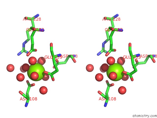
Stereo pair view
Mono view
Stereo pair view
A full contact list of Magnesium with other atoms in the Mg binding
site number 1 of Crystal Structure of Isocitrate Lyase From Mycobacterium Tuberculosis within 5.0Å range:
|
Magnesium binding site 2 out of 2 in 1f61
Go back to
Magnesium binding site 2 out
of 2 in the Crystal Structure of Isocitrate Lyase From Mycobacterium Tuberculosis

Mono view
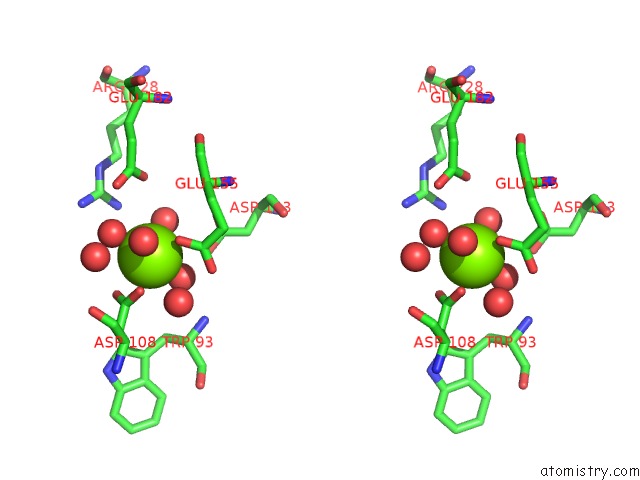
Stereo pair view
Mono view
Stereo pair view
A full contact list of Magnesium with other atoms in the Mg binding
site number 2 of Crystal Structure of Isocitrate Lyase From Mycobacterium Tuberculosis within 5.0Å range:
|
Reference:
V.Sharma,
S.Sharma,
K.Hoener Zu Bentrup,
J.D.Mckinney,
D.G.Russell,
W.R.Jacobs Jr.,
J.C.Sacchettini.
Structure of Isocitrate Lyase, A Persistence Factor of Mycobacterium Tuberculosis. Nat.Struct.Biol. V. 7 663 2000.
ISSN: ISSN 1072-8368
PubMed: 10932251
DOI: 10.1038/77964
Page generated: Sat Aug 9 20:53:18 2025
ISSN: ISSN 1072-8368
PubMed: 10932251
DOI: 10.1038/77964
Last articles
Mg in 4ZTZMg in 4ZTU
Mg in 4ZU3
Mg in 4ZSE
Mg in 4ZTF
Mg in 4ZTJ
Mg in 4ZR8
Mg in 4ZRT
Mg in 4ZQF
Mg in 4ZQG