Magnesium »
PDB 1fp6-1g7t »
1fq1 »
Magnesium in PDB 1fq1: Crystal Structure of Kinase Associated Phosphatase (Kap) in Complex with Phospho-CDK2
Enzymatic activity of Crystal Structure of Kinase Associated Phosphatase (Kap) in Complex with Phospho-CDK2
All present enzymatic activity of Crystal Structure of Kinase Associated Phosphatase (Kap) in Complex with Phospho-CDK2:
2.7.11.22; 3.1.3.48;
2.7.11.22; 3.1.3.48;
Protein crystallography data
The structure of Crystal Structure of Kinase Associated Phosphatase (Kap) in Complex with Phospho-CDK2, PDB code: 1fq1
was solved by
H.Song,
N.Hanlon,
N.R.Brown,
M.E.M.Noble,
L.N.Johnson,
D.Barford,
with X-Ray Crystallography technique. A brief refinement statistics is given in the table below:
| Resolution Low / High (Å) | 20.00 / 3.00 |
| Space group | P 32 2 1 |
| Cell size a, b, c (Å), α, β, γ (°) | 134.450, 134.450, 65.800, 90.00, 90.00, 120.00 |
| R / Rfree (%) | 23.5 / 31.3 |
Magnesium Binding Sites:
The binding sites of Magnesium atom in the Crystal Structure of Kinase Associated Phosphatase (Kap) in Complex with Phospho-CDK2
(pdb code 1fq1). This binding sites where shown within
5.0 Angstroms radius around Magnesium atom.
In total only one binding site of Magnesium was determined in the Crystal Structure of Kinase Associated Phosphatase (Kap) in Complex with Phospho-CDK2, PDB code: 1fq1:
In total only one binding site of Magnesium was determined in the Crystal Structure of Kinase Associated Phosphatase (Kap) in Complex with Phospho-CDK2, PDB code: 1fq1:
Magnesium binding site 1 out of 1 in 1fq1
Go back to
Magnesium binding site 1 out
of 1 in the Crystal Structure of Kinase Associated Phosphatase (Kap) in Complex with Phospho-CDK2
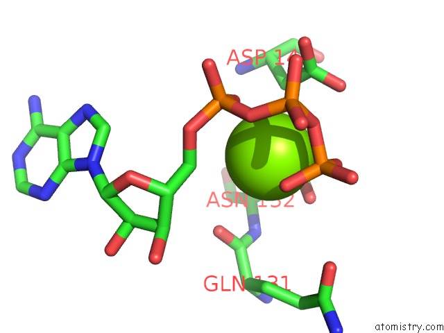
Mono view
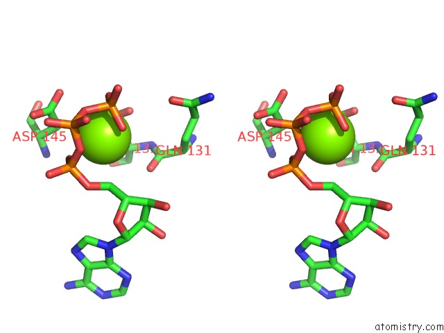
Stereo pair view
Mono view
Stereo pair view
A full contact list of Magnesium with other atoms in the Mg binding
site number 1 of Crystal Structure of Kinase Associated Phosphatase (Kap) in Complex with Phospho-CDK2 within 5.0Å range:
|
Reference:
H.Song,
N.Hanlon,
N.R.Brown,
M.E.Noble,
L.N.Johnson,
D.Barford.
Phosphoprotein-Protein Interactions Revealed By the Crystal Structure of Kinase-Associated Phosphatase in Complex with PHOSPHOCDK2. Mol.Cell V. 7 615 2001.
ISSN: ISSN 1097-2765
PubMed: 11463386
DOI: 10.1016/S1097-2765(01)00208-8
Page generated: Tue Aug 13 03:34:49 2024
ISSN: ISSN 1097-2765
PubMed: 11463386
DOI: 10.1016/S1097-2765(01)00208-8
Last articles
Cl in 3CJDCl in 3CJ3
Cl in 3CIW
Cl in 3CIX
Cl in 3CIO
Cl in 3CIN
Cl in 3CFQ
Cl in 3CGY
Cl in 3CHV
Cl in 3CI0