Magnesium »
PDB 1g87-1gpm »
1geg »
Magnesium in PDB 1geg: Cryatal Structure Analysis of Meso-2,3-Butanediol Dehydrogenase
Enzymatic activity of Cryatal Structure Analysis of Meso-2,3-Butanediol Dehydrogenase
All present enzymatic activity of Cryatal Structure Analysis of Meso-2,3-Butanediol Dehydrogenase:
1.1.1.5;
1.1.1.5;
Protein crystallography data
The structure of Cryatal Structure Analysis of Meso-2,3-Butanediol Dehydrogenase, PDB code: 1geg
was solved by
M.Otagiri,
G.Kurisu,
S.Ui,
M.Kusunoki,
with X-Ray Crystallography technique. A brief refinement statistics is given in the table below:
| Resolution Low / High (Å) | 40.00 / 1.70 |
| Space group | P 1 21 1 |
| Cell size a, b, c (Å), α, β, γ (°) | 69.160, 109.780, 127.280, 90.00, 102.29, 90.00 |
| R / Rfree (%) | 19.3 / 20.9 |
Magnesium Binding Sites:
The binding sites of Magnesium atom in the Cryatal Structure Analysis of Meso-2,3-Butanediol Dehydrogenase
(pdb code 1geg). This binding sites where shown within
5.0 Angstroms radius around Magnesium atom.
In total 4 binding sites of Magnesium where determined in the Cryatal Structure Analysis of Meso-2,3-Butanediol Dehydrogenase, PDB code: 1geg:
Jump to Magnesium binding site number: 1; 2; 3; 4;
In total 4 binding sites of Magnesium where determined in the Cryatal Structure Analysis of Meso-2,3-Butanediol Dehydrogenase, PDB code: 1geg:
Jump to Magnesium binding site number: 1; 2; 3; 4;
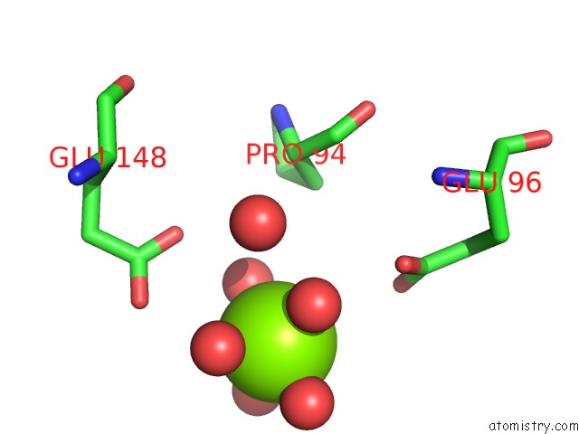
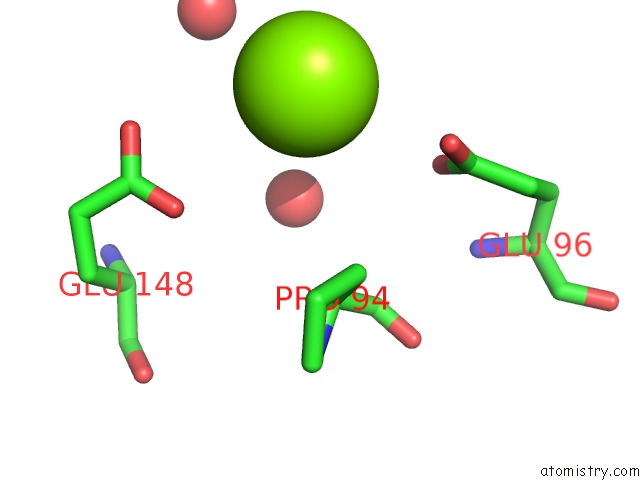
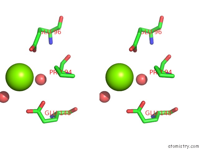
Magnesium binding site 1 out of 4 in 1geg
Go back to
Magnesium binding site 1 out
of 4 in the Cryatal Structure Analysis of Meso-2,3-Butanediol Dehydrogenase
Mono view
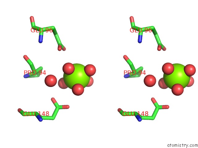
Stereo pair view
Mono view
Stereo pair view
A full contact list of Magnesium with other atoms in the Mg binding
site number 1 of Cryatal Structure Analysis of Meso-2,3-Butanediol Dehydrogenase within 5.0Å range:
|
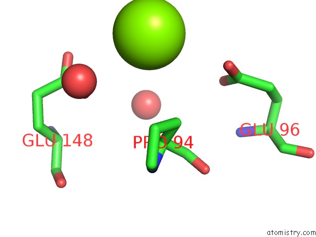
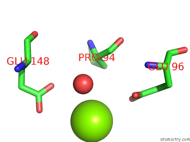
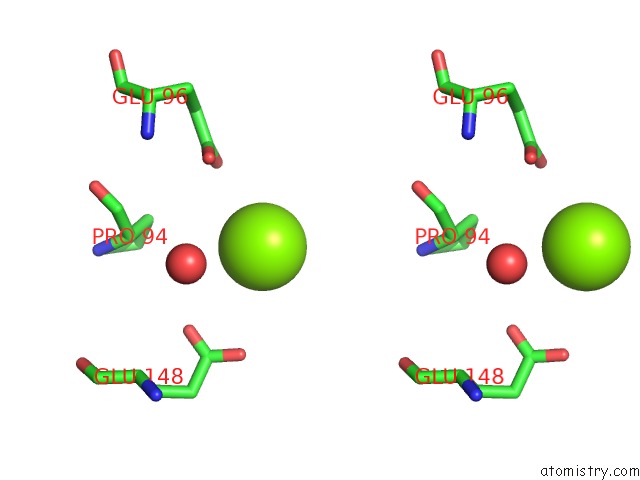
Magnesium binding site 2 out of 4 in 1geg
Go back to
Magnesium binding site 2 out
of 4 in the Cryatal Structure Analysis of Meso-2,3-Butanediol Dehydrogenase
Mono view
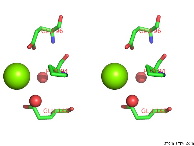
Stereo pair view
Mono view
Stereo pair view
A full contact list of Magnesium with other atoms in the Mg binding
site number 2 of Cryatal Structure Analysis of Meso-2,3-Butanediol Dehydrogenase within 5.0Å range:
|
Magnesium binding site 3 out of 4 in 1geg
Go back to
Magnesium binding site 3 out
of 4 in the Cryatal Structure Analysis of Meso-2,3-Butanediol Dehydrogenase
Mono view
Stereo pair view
Mono view
Stereo pair view
A full contact list of Magnesium with other atoms in the Mg binding
site number 3 of Cryatal Structure Analysis of Meso-2,3-Butanediol Dehydrogenase within 5.0Å range:
|
Magnesium binding site 4 out of 4 in 1geg
Go back to
Magnesium binding site 4 out
of 4 in the Cryatal Structure Analysis of Meso-2,3-Butanediol Dehydrogenase
Mono view
Stereo pair view
Mono view
Stereo pair view
A full contact list of Magnesium with other atoms in the Mg binding
site number 4 of Cryatal Structure Analysis of Meso-2,3-Butanediol Dehydrogenase within 5.0Å range:
|
Reference:
M.Otagiri,
G.Kurisu,
S.Ui,
Y.Takusagawa,
M.Ohkuma,
T.Kudo,
M.Kusunoki.
Crystal Structure of Meso-2,3-Butanediol Dehydrogenase in A Complex with Nad+ and Inhibitor Mercaptoethanol at 1.7 A Resolution For Understanding of Chiral Substrate Recognition Mechanisms. J.Biochem. V. 129 205 2001.
ISSN: ISSN 0021-924X
PubMed: 11173520
DOI: 10.1093/OXFORDJOURNALS.JBCHEM.A002845
Page generated: Tue Aug 13 03:43:49 2024
ISSN: ISSN 0021-924X
PubMed: 11173520
DOI: 10.1093/OXFORDJOURNALS.JBCHEM.A002845
Last articles
K in 8CGIK in 8CG1
K in 8CFY
K in 8CG2
K in 8CFX
K in 8CG0
K in 8CFW
K in 8CFV
K in 8CFU
K in 8CFT