Magnesium »
PDB 1gq9-1h7q »
1h6p »
Magnesium in PDB 1h6p: Dimeristion Domain From Human TRF2
Protein crystallography data
The structure of Dimeristion Domain From Human TRF2, PDB code: 1h6p
was solved by
L.Chapman,
L.Fairall,
D.Rhodes,
with X-Ray Crystallography technique. A brief refinement statistics is given in the table below:
| Resolution Low / High (Å) | 23.72 / 2.20 |
| Space group | C 1 2 1 |
| Cell size a, b, c (Å), α, β, γ (°) | 117.041, 80.284, 52.816, 90.00, 101.95, 90.00 |
| R / Rfree (%) | 23.5 / 26.8 |
Magnesium Binding Sites:
The binding sites of Magnesium atom in the Dimeristion Domain From Human TRF2
(pdb code 1h6p). This binding sites where shown within
5.0 Angstroms radius around Magnesium atom.
In total only one binding site of Magnesium was determined in the Dimeristion Domain From Human TRF2, PDB code: 1h6p:
In total only one binding site of Magnesium was determined in the Dimeristion Domain From Human TRF2, PDB code: 1h6p:
Magnesium binding site 1 out of 1 in 1h6p
Go back to
Magnesium binding site 1 out
of 1 in the Dimeristion Domain From Human TRF2
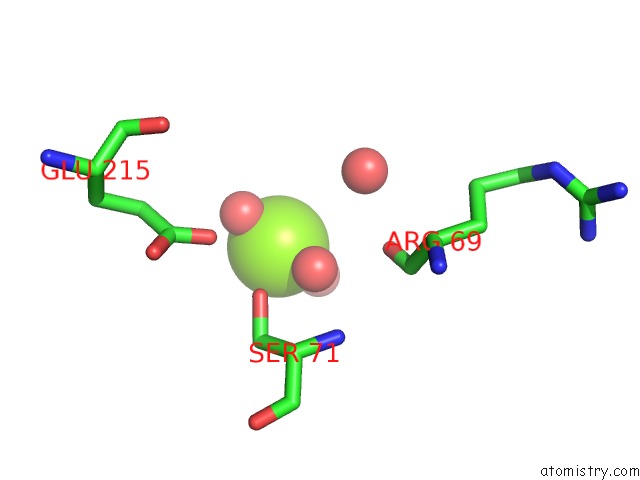
Mono view
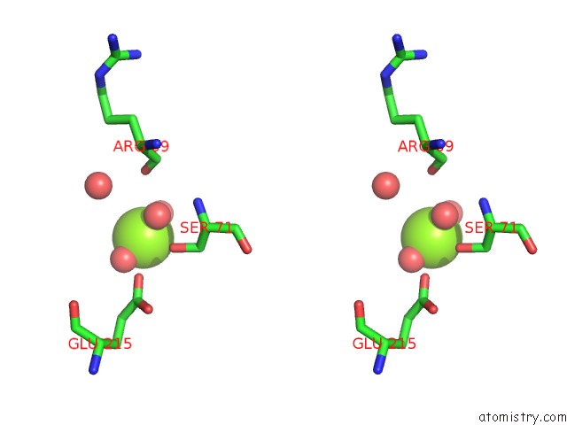
Stereo pair view
Mono view
Stereo pair view
A full contact list of Magnesium with other atoms in the Mg binding
site number 1 of Dimeristion Domain From Human TRF2 within 5.0Å range:
|
Reference:
L.Fairall,
L.Chapman,
H.Moss,
T.De Lange,
D.Rhodes.
Structure of the Trfh Dimerization Domain of the Human Telomere Proteins TRF1 and TRF2 Mol.Cell V. 8 351 2001.
ISSN: ISSN 1097-2765
PubMed: 11545737
DOI: 10.1016/S1097-2765(01)00321-5
Page generated: Tue Aug 13 03:53:11 2024
ISSN: ISSN 1097-2765
PubMed: 11545737
DOI: 10.1016/S1097-2765(01)00321-5
Last articles
Fe in 4GEDFe in 4G99
Fe in 4G98
Fe in 4G8W
Fe in 4G8U
Fe in 4G8P
Fe in 4G8H
Fe in 4G7U
Fe in 4G7T
Fe in 4G7S