Magnesium »
PDB 1it7-1jbz »
1iv3 »
Magnesium in PDB 1iv3: Structure of 2C-Methyl-D-Erythritol-2,4-Cyclodiphosphate Synthase (Bound Form Mg Atoms)
Enzymatic activity of Structure of 2C-Methyl-D-Erythritol-2,4-Cyclodiphosphate Synthase (Bound Form Mg Atoms)
All present enzymatic activity of Structure of 2C-Methyl-D-Erythritol-2,4-Cyclodiphosphate Synthase (Bound Form Mg Atoms):
4.6.1.12;
4.6.1.12;
Protein crystallography data
The structure of Structure of 2C-Methyl-D-Erythritol-2,4-Cyclodiphosphate Synthase (Bound Form Mg Atoms), PDB code: 1iv3
was solved by
H.Kishida,
T.Wada,
S.Unzai,
T.Kuzuyama,
T.Terada,
M.Sirouzu,
S.Yokoyama,
J.R.H.Tame,
S.-Y.Park,
Riken Structuralgenomics/Proteomics Initiative (Rsgi),
with X-Ray Crystallography technique. A brief refinement statistics is given in the table below:
| Resolution Low / High (Å) | 20.00 / 1.52 |
| Space group | P 41 21 2 |
| Cell size a, b, c (Å), α, β, γ (°) | 106.179, 106.179, 148.811, 90.00, 90.00, 90.00 |
| R / Rfree (%) | 20.2 / 25.7 |
Magnesium Binding Sites:
The binding sites of Magnesium atom in the Structure of 2C-Methyl-D-Erythritol-2,4-Cyclodiphosphate Synthase (Bound Form Mg Atoms)
(pdb code 1iv3). This binding sites where shown within
5.0 Angstroms radius around Magnesium atom.
In total 6 binding sites of Magnesium where determined in the Structure of 2C-Methyl-D-Erythritol-2,4-Cyclodiphosphate Synthase (Bound Form Mg Atoms), PDB code: 1iv3:
Jump to Magnesium binding site number: 1; 2; 3; 4; 5; 6;
In total 6 binding sites of Magnesium where determined in the Structure of 2C-Methyl-D-Erythritol-2,4-Cyclodiphosphate Synthase (Bound Form Mg Atoms), PDB code: 1iv3:
Jump to Magnesium binding site number: 1; 2; 3; 4; 5; 6;
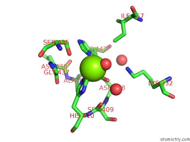
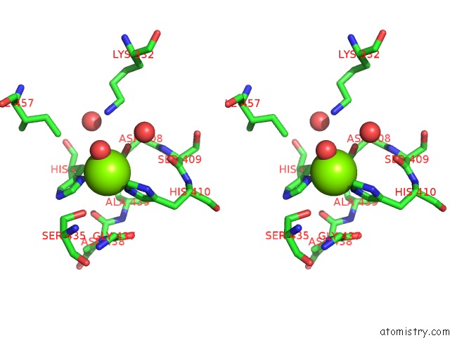
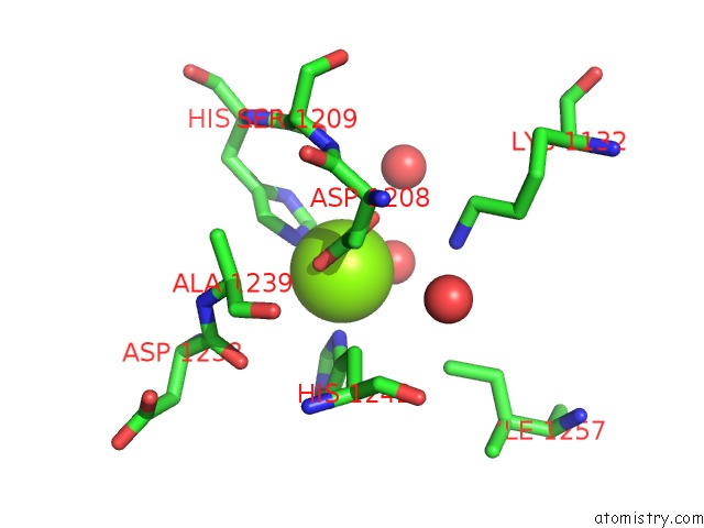
Magnesium binding site 1 out of 6 in 1iv3
Go back to
Magnesium binding site 1 out
of 6 in the Structure of 2C-Methyl-D-Erythritol-2,4-Cyclodiphosphate Synthase (Bound Form Mg Atoms)
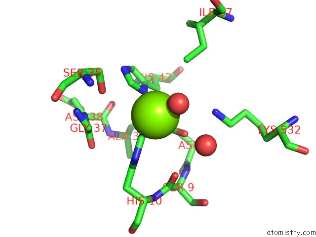
Mono view
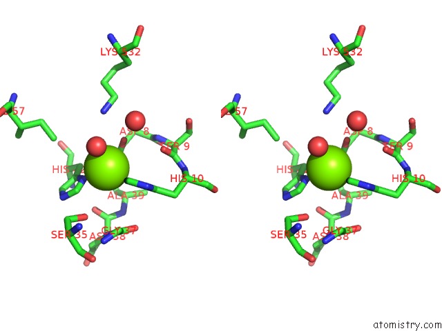
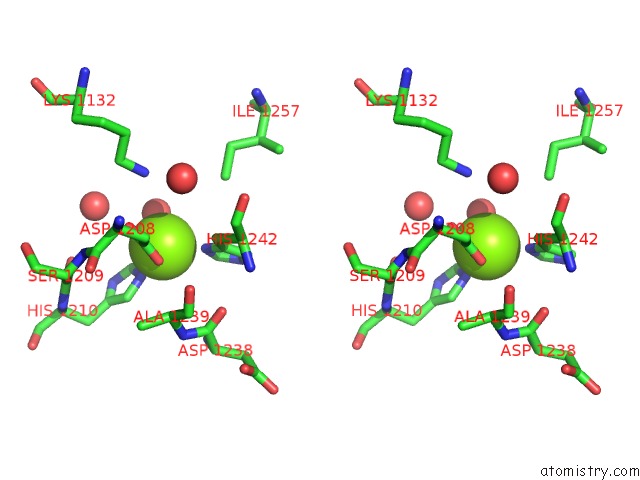
Stereo pair view
Mono view
Stereo pair view
A full contact list of Magnesium with other atoms in the Mg binding
site number 1 of Structure of 2C-Methyl-D-Erythritol-2,4-Cyclodiphosphate Synthase (Bound Form Mg Atoms) within 5.0Å range:
|
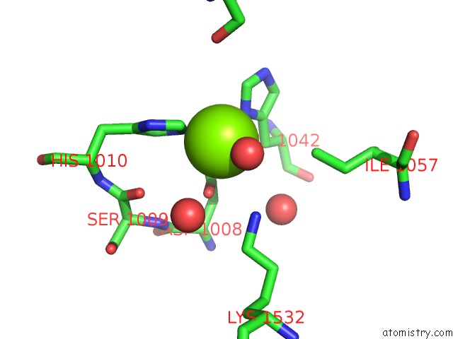
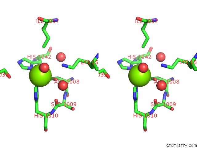
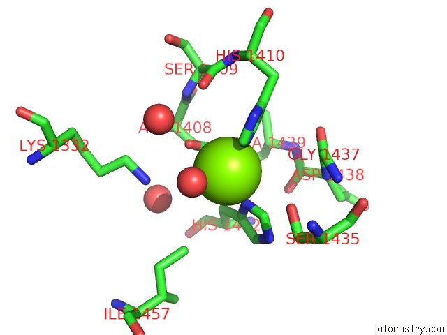
Magnesium binding site 2 out of 6 in 1iv3
Go back to
Magnesium binding site 2 out
of 6 in the Structure of 2C-Methyl-D-Erythritol-2,4-Cyclodiphosphate Synthase (Bound Form Mg Atoms)
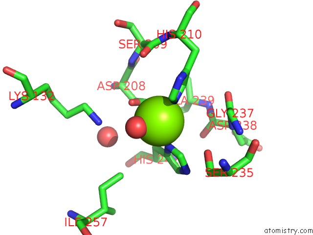
Mono view
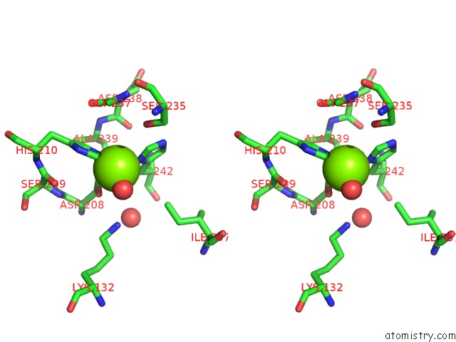
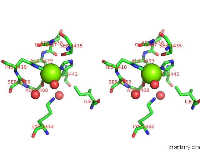
Stereo pair view
Mono view
Stereo pair view
A full contact list of Magnesium with other atoms in the Mg binding
site number 2 of Structure of 2C-Methyl-D-Erythritol-2,4-Cyclodiphosphate Synthase (Bound Form Mg Atoms) within 5.0Å range:
|
Magnesium binding site 3 out of 6 in 1iv3
Go back to
Magnesium binding site 3 out
of 6 in the Structure of 2C-Methyl-D-Erythritol-2,4-Cyclodiphosphate Synthase (Bound Form Mg Atoms)
Mono view
Stereo pair view
Mono view
Stereo pair view
A full contact list of Magnesium with other atoms in the Mg binding
site number 3 of Structure of 2C-Methyl-D-Erythritol-2,4-Cyclodiphosphate Synthase (Bound Form Mg Atoms) within 5.0Å range:
|
Magnesium binding site 4 out of 6 in 1iv3
Go back to
Magnesium binding site 4 out
of 6 in the Structure of 2C-Methyl-D-Erythritol-2,4-Cyclodiphosphate Synthase (Bound Form Mg Atoms)
Mono view
Stereo pair view
Mono view
Stereo pair view
A full contact list of Magnesium with other atoms in the Mg binding
site number 4 of Structure of 2C-Methyl-D-Erythritol-2,4-Cyclodiphosphate Synthase (Bound Form Mg Atoms) within 5.0Å range:
|
Magnesium binding site 5 out of 6 in 1iv3
Go back to
Magnesium binding site 5 out
of 6 in the Structure of 2C-Methyl-D-Erythritol-2,4-Cyclodiphosphate Synthase (Bound Form Mg Atoms)
Mono view
Stereo pair view
Mono view
Stereo pair view
A full contact list of Magnesium with other atoms in the Mg binding
site number 5 of Structure of 2C-Methyl-D-Erythritol-2,4-Cyclodiphosphate Synthase (Bound Form Mg Atoms) within 5.0Å range:
|
Magnesium binding site 6 out of 6 in 1iv3
Go back to
Magnesium binding site 6 out
of 6 in the Structure of 2C-Methyl-D-Erythritol-2,4-Cyclodiphosphate Synthase (Bound Form Mg Atoms)
Mono view
Stereo pair view
Mono view
Stereo pair view
A full contact list of Magnesium with other atoms in the Mg binding
site number 6 of Structure of 2C-Methyl-D-Erythritol-2,4-Cyclodiphosphate Synthase (Bound Form Mg Atoms) within 5.0Å range:
|
Reference:
H.Kishida,
T.Wada,
S.Unzai,
T.Kuzuyama,
M.Takagi,
T.Terada,
M.Shirouzu,
S.Yokoyama,
J.R.Tame,
S.Y.Park.
Structure and Catalytic Mechanism of 2-C-Methyl-D-Erythritol 2,4-Cyclodiphosphate (Mecdp) Synthase, An Enzyme in the Non-Mevalonate Pathway of Isoprenoid Synthesis. Acta Crystallogr.,Sect.D V. 59 23 2003.
ISSN: ISSN 0907-4449
PubMed: 12499535
DOI: 10.1107/S0907444902017705
Page generated: Tue Aug 13 05:39:10 2024
ISSN: ISSN 0907-4449
PubMed: 12499535
DOI: 10.1107/S0907444902017705
Last articles
F in 8EL1F in 8ENI
F in 8EIQ
F in 8EL0
F in 8EIO
F in 8EKX
F in 8EG6
F in 8EIG
F in 8EG5
F in 8EFF