Magnesium »
PDB 1jcg-1jwy »
1jiu »
Magnesium in PDB 1jiu: T4 Phage Bgt in Complex with MG2+ : Form I
Enzymatic activity of T4 Phage Bgt in Complex with MG2+ : Form I
All present enzymatic activity of T4 Phage Bgt in Complex with MG2+ : Form I:
2.4.1.27;
2.4.1.27;
Protein crystallography data
The structure of T4 Phage Bgt in Complex with MG2+ : Form I, PDB code: 1jiu
was solved by
S.Morera,
L.Lariviere,
J.Kurzeck,
U.Aschke-Sonnenborn,
P.S.Freemont,
J.Janin,
W.Ruger,
with X-Ray Crystallography technique. A brief refinement statistics is given in the table below:
| Resolution Low / High (Å) | 20.00 / 2.50 |
| Space group | P 21 21 21 |
| Cell size a, b, c (Å), α, β, γ (°) | 69.827, 102.650, 54.871, 90.00, 90.00, 90.00 |
| R / Rfree (%) | 19.2 / 27.4 |
Magnesium Binding Sites:
The binding sites of Magnesium atom in the T4 Phage Bgt in Complex with MG2+ : Form I
(pdb code 1jiu). This binding sites where shown within
5.0 Angstroms radius around Magnesium atom.
In total only one binding site of Magnesium was determined in the T4 Phage Bgt in Complex with MG2+ : Form I, PDB code: 1jiu:
In total only one binding site of Magnesium was determined in the T4 Phage Bgt in Complex with MG2+ : Form I, PDB code: 1jiu:
Magnesium binding site 1 out of 1 in 1jiu
Go back to
Magnesium binding site 1 out
of 1 in the T4 Phage Bgt in Complex with MG2+ : Form I
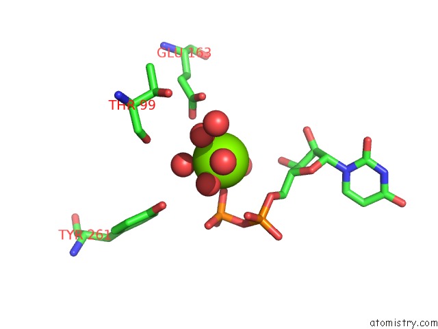
Mono view
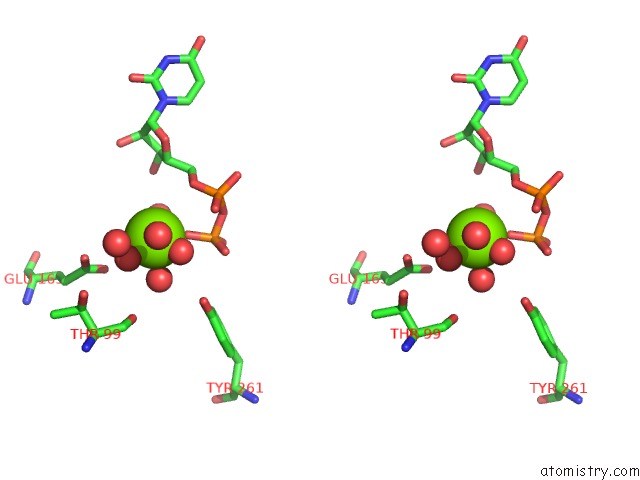
Stereo pair view
Mono view
Stereo pair view
A full contact list of Magnesium with other atoms in the Mg binding
site number 1 of T4 Phage Bgt in Complex with MG2+ : Form I within 5.0Å range:
|
Reference:
S.Morera,
L.Lariviere,
J.Kurzeck,
U.Aschke-Sonnenborn,
P.S.Freemont,
J.Janin,
W.Ruger.
High Resolution Crystal Structures of T4 Phage Beta-Glucosyltransferase: Induced Fit and Effect of Substrate and Metal Binding. J.Mol.Biol. V. 311 569 2001.
ISSN: ISSN 0022-2836
PubMed: 11493010
DOI: 10.1006/JMBI.2001.4905
Page generated: Sat Aug 9 23:22:48 2025
ISSN: ISSN 0022-2836
PubMed: 11493010
DOI: 10.1006/JMBI.2001.4905
Last articles
Mg in 6XHAMg in 6XGV
Mg in 6XG6
Mg in 6XEZ
Mg in 6XGU
Mg in 6XGO
Mg in 6XFO
Mg in 6XED
Mg in 6XET
Mg in 6XER