Magnesium »
PDB 1k9w-1kk3 »
1kil »
Magnesium in PDB 1kil: Three-Dimensional Structure of the Complexin/Snare Complex
Protein crystallography data
The structure of Three-Dimensional Structure of the Complexin/Snare Complex, PDB code: 1kil
was solved by
X.Chen,
D.Tomchick,
E.Kovrigin,
D.Arac,
M.Machius,
T.C.Sudhof,
J.Rizo,
with X-Ray Crystallography technique. A brief refinement statistics is given in the table below:
| Resolution Low / High (Å) | 32.23 / 2.30 |
| Space group | P 21 21 21 |
| Cell size a, b, c (Å), α, β, γ (°) | 40.489, 60.425, 159.787, 90.00, 90.00, 90.00 |
| R / Rfree (%) | 24.5 / 30.5 |
Magnesium Binding Sites:
The binding sites of Magnesium atom in the Three-Dimensional Structure of the Complexin/Snare Complex
(pdb code 1kil). This binding sites where shown within
5.0 Angstroms radius around Magnesium atom.
In total 2 binding sites of Magnesium where determined in the Three-Dimensional Structure of the Complexin/Snare Complex, PDB code: 1kil:
Jump to Magnesium binding site number: 1; 2;
In total 2 binding sites of Magnesium where determined in the Three-Dimensional Structure of the Complexin/Snare Complex, PDB code: 1kil:
Jump to Magnesium binding site number: 1; 2;
Magnesium binding site 1 out of 2 in 1kil
Go back to
Magnesium binding site 1 out
of 2 in the Three-Dimensional Structure of the Complexin/Snare Complex
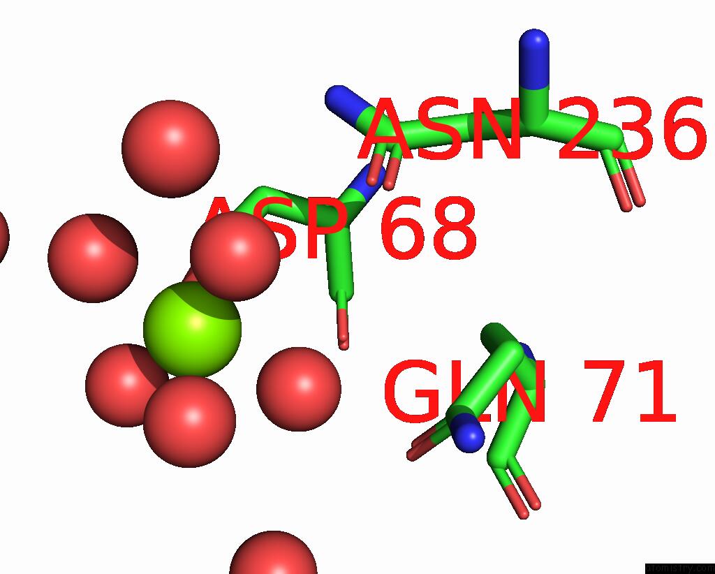
Mono view

Stereo pair view
Mono view
Stereo pair view
A full contact list of Magnesium with other atoms in the Mg binding
site number 1 of Three-Dimensional Structure of the Complexin/Snare Complex within 5.0Å range:
|
Magnesium binding site 2 out of 2 in 1kil
Go back to
Magnesium binding site 2 out
of 2 in the Three-Dimensional Structure of the Complexin/Snare Complex
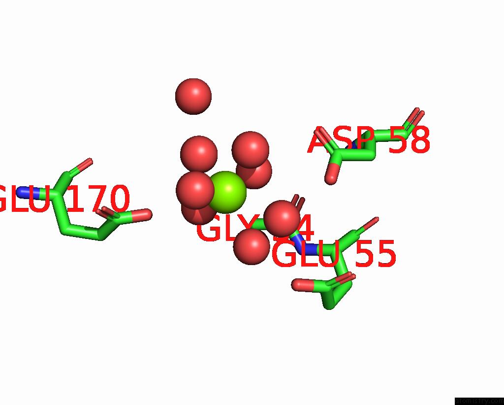
Mono view
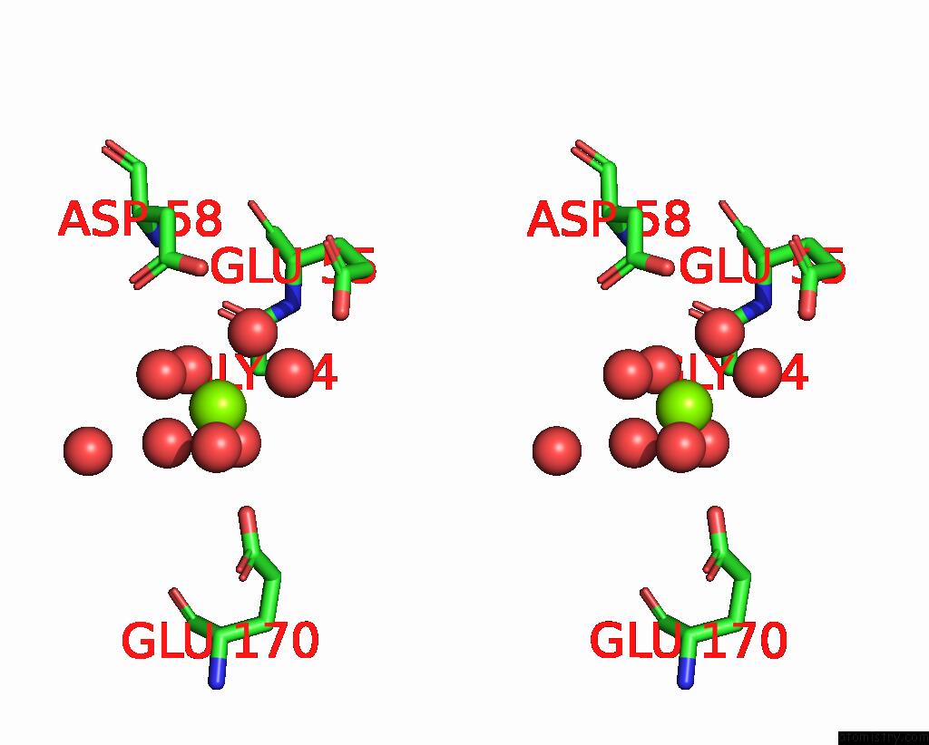
Stereo pair view
Mono view
Stereo pair view
A full contact list of Magnesium with other atoms in the Mg binding
site number 2 of Three-Dimensional Structure of the Complexin/Snare Complex within 5.0Å range:
|
Reference:
X.Chen,
D.R.Tomchick,
E.Kovrigin,
D.Arac,
M.Machius,
T.C.Sudhof,
J.Rizo.
Three-Dimensional Structure of the Complexin/Snare Complex. Neuron V. 33 397 2002.
ISSN: ISSN 0896-6273
PubMed: 11832227
DOI: 10.1016/S0896-6273(02)00583-4
Page generated: Sun Aug 10 00:11:01 2025
ISSN: ISSN 0896-6273
PubMed: 11832227
DOI: 10.1016/S0896-6273(02)00583-4
Last articles
Mg in 2UU7Mg in 2UAG
Mg in 2UKD
Mg in 2SHK
Mg in 2TPS
Mg in 2TRT
Mg in 2TRA
Mg in 2RMK
Mg in 2RUS
Mg in 2TCT