Magnesium »
PDB 1kk7-1l3p »
1l1r »
Magnesium in PDB 1l1r: Crystal Structure of Aprtase From Giardia Lamblia Complexed with 9- Deazaadenine, MG2+ and Prpp
Enzymatic activity of Crystal Structure of Aprtase From Giardia Lamblia Complexed with 9- Deazaadenine, MG2+ and Prpp
All present enzymatic activity of Crystal Structure of Aprtase From Giardia Lamblia Complexed with 9- Deazaadenine, MG2+ and Prpp:
2.4.2.7;
2.4.2.7;
Protein crystallography data
The structure of Crystal Structure of Aprtase From Giardia Lamblia Complexed with 9- Deazaadenine, MG2+ and Prpp, PDB code: 1l1r
was solved by
W.Shi,
A.E.Sarver,
C.C.Wang,
K.S.Tanaka,
S.C.Almo,
V.L.Schramm,
with X-Ray Crystallography technique. A brief refinement statistics is given in the table below:
| Resolution Low / High (Å) | 25.00 / 1.95 |
| Space group | P 31 2 1 |
| Cell size a, b, c (Å), α, β, γ (°) | 54.325, 54.325, 108.744, 90.00, 90.00, 120.00 |
| R / Rfree (%) | 21.8 / 26.2 |
Magnesium Binding Sites:
The binding sites of Magnesium atom in the Crystal Structure of Aprtase From Giardia Lamblia Complexed with 9- Deazaadenine, MG2+ and Prpp
(pdb code 1l1r). This binding sites where shown within
5.0 Angstroms radius around Magnesium atom.
In total only one binding site of Magnesium was determined in the Crystal Structure of Aprtase From Giardia Lamblia Complexed with 9- Deazaadenine, MG2+ and Prpp, PDB code: 1l1r:
In total only one binding site of Magnesium was determined in the Crystal Structure of Aprtase From Giardia Lamblia Complexed with 9- Deazaadenine, MG2+ and Prpp, PDB code: 1l1r:
Magnesium binding site 1 out of 1 in 1l1r
Go back to
Magnesium binding site 1 out
of 1 in the Crystal Structure of Aprtase From Giardia Lamblia Complexed with 9- Deazaadenine, MG2+ and Prpp
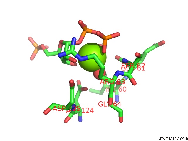
Mono view
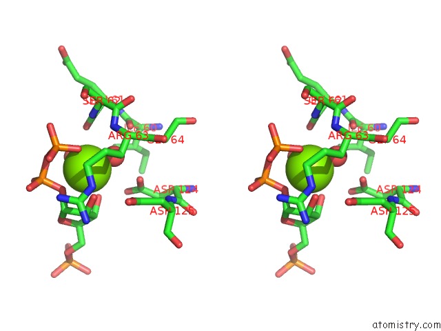
Stereo pair view
Mono view
Stereo pair view
A full contact list of Magnesium with other atoms in the Mg binding
site number 1 of Crystal Structure of Aprtase From Giardia Lamblia Complexed with 9- Deazaadenine, MG2+ and Prpp within 5.0Å range:
|
Reference:
W.Shi,
A.E.Sarver,
C.C.Wang,
K.S.Tanaka,
S.C.Almo,
V.L.Schramm.
Closed Site Complexes of Adenine Phosphoribosyltransferase From Giardia Lamblia Reveal A Mechanism of Ribosyl Migration. J.Biol.Chem. V. 277 39981 2002.
ISSN: ISSN 0021-9258
PubMed: 12171925
DOI: 10.1074/JBC.M205596200
Page generated: Sun Aug 10 00:30:00 2025
ISSN: ISSN 0021-9258
PubMed: 12171925
DOI: 10.1074/JBC.M205596200
Last articles
Mg in 3CCUMg in 3CCS
Mg in 3CCR
Mg in 3CCQ
Mg in 3CCM
Mg in 3CCL
Mg in 3CCJ
Mg in 3CC2
Mg in 3CCE
Mg in 3CC7